Op Exam 1 Combined
 Call Kai
 Learn
 Practice Test
 Spaced Repetition
 Match
 Flashcards
 Knowt Play
1/726

There's no tags or description
Looks like no tags are added yet.
727 Terms
Cariology
Study of dental caries and cariogenesis
Is caries all cavity?
No — non-cavitated lesions exist
Tooth worm theory
Old belief that a worm gnawed the tooth → caries thought infectious and transmissible
Keyes triad (1963)
Tooth (host) + bacteria + sugar substrate, with TIME in the center
What did the Keyes triad leave out?
The drop in pH (acid) that causes demineralization — the new paradigm
Does the Keyes triad still hold?
Yes
Infectious/transmissible era treatment
Chemicals or excision by drilling
Current caries paradigm
Ecological and non-communicable chronic disease — not transmissible, not infectious
Does caries pass between two touching teeth?
No — not patient to patient or tooth to tooth
Definition of dental caries
A BIOFILM-MEDIATED, DIET-MODULATED process that can lead to an IMBALANCE in the cycle of DEMINERALIZATION and REMINERALIZATION causing LOCALIZED DESTRUCTION of tooth structure
Biofilm mediated
Microscopic communities of bacteria in a slimy matrix (plaque) — gives bacteria a home
Diet modulated
With sugars (degradable carbohydrates), bacteria make acid and lower pH — frequency of exposure is critical
Extended low pH =
Microbiome in DYSBIOSIS
When does demineralization start winning?
When pH drops below the CRITICAL pH
Imbalance on the seesaw
De- and remineralization always occur; caries when demineralization wins
What drives remineralization?
Fluoride (including water fluoride), saliva, Ca²⁺/PO₄³⁻
Seesaw: disease indicators
White spot lesions; restorations < 3 years; enamel lesions; caries/dentin
Seesaw: risk factors
Pathogenic bacteria; absence of saliva; destructive lifestyle habits
Seesaw: protective factors
Saliva and sealants; antibacterials; fluoride/Ca²⁺/PO₄³⁻; effective lifestyle habits; risk-based reassessment
Risk factors share one thing:
They lower pH (low saliva/xerostomia, frequent sugar, poor hygiene)
Why is saliva protective?
It neutralizes acid (low flow/xerostomia raises risk)
Coffee with milk and sugar vs coffee alone
Sipping with sugar = frequent low pH (higher risk); coffee alone lowers pH less
Truck driver sipping soda all night
High caries risk at the PATIENT level
Rotated tooth that can't be cleaned
High caries risk at the TOOTH level
Tooth death spiral
Small filling → bigger filling → 1-, 2-, 3-surface → crown → root canal → extraction → implant
Why does the death spiral happen?
The DISEASE (risk) is never addressed — only lesions are drilled and restored
CariesCare International (CCI)
Practitioner-oriented guidelines, skeleton of the module (Martignon, Br Dent J 2019)
CariesCare 4-D in order
Determine, Detect, Decide, Do
Which D starts the cycle?
DETERMINE (risk) — not Detect
Four aims of CariesCare
Prevent new caries; prevent progression; preserve tooth structure; manage caries risk
1st D: Determine
Determine patient-level (and tooth-level) caries risk
2nd D: Detect
Detect and assess caries: staging (severity) and grading (activity)
3rd D: Decide
Personalized care plan at patient and tooth level
4th D: Do
Prevention and management of cavitated and non-cavitated lesions; then cycle restarts
Why is CariesCare a cycle?
Risk and disease are dynamic — reassess and keep lowering risk (risk-based recall interval)
Center of the 4-D gear diagram
Improving health outcomes
Patient-level Decide example
Truck driver sips ice water instead of soda
Tooth-level Decide example
Class I or II filling if the lesion demands it
Why classify caries? (slide 3)
Validated criteria for practitioners (caries control/management) and for researchers/public health planners (effects/need for interventions)
Classification is also a mode of…
Communication (e.g., 'caries in the distal pit of #2, deep, active')
Staging vs grading
Staging = severity (depth). Grading = activity (progressing or not)
Three criteria for classifying caries
Location, severity, activity (+ primary vs around restoration)
Only two classification systems you're responsible for
ICDAS and ADA CCS
Classification used at TUSDM
ADA Caries Classification System (ADA CCS)
ICDAS year and basis
based on SEVERITY (not activity); used worldwide
Coronal caries sites
Pit and fissure; proximal; smooth surface
Pit and fissure caries
Occlusal surface; easiest to see visually
Smooth surface caries location
Facial or lingual, most often near the gingival line (biofilm)
Proximal caries: where and how found
Just below the contact area; bitewing radiograph (not directly visible)
Primary caries
Caries with no existing restoration
CARS
Caries around restorations and sealants (older term: secondary caries)
Why not say 'secondary caries'?
Misnomer: you can't tell if it's a second primary lesion or truly secondary; sealants included
Three detection tools in the TUSDM clinic
Visual, bitewing radiographs, fiber-optic transillumination (FOTI)
Visual exam requirements
Clean (prophy/remove biofilm), dry, good light, loupes; mirror, reflected light, air-water syringe
Why 'bitewing'?
Its angle opens up the contacts
Three ways to look at severity
Histologically, visually, radiographically
Before a visual caries exam
Remove biofilm and dry the surface (5 seconds)
ONE exception to drying the tooth
ICDAS 4 (dentin shadow) is best seen slightly WET
ADA CCS ↔ ICDAS merge
Sound = 0; Initial = 1, 2; Moderate = 3, 4; Advanced = 5, 6
ADA CCS chart colors
Sound green/white; Initial yellow; Moderate orange; Advanced red
ICDAS 0
Sound: no change in enamel translucency after 5 s air drying
Non-carious enamel defects (opacity, hypoplasia, fluorosis, DI) are coded…
SOUND (ICDAS 0) — may still need restoration
Hypoplasia (ICDAS slide definition)
Reduced localized thickness of enamel, no dentin exposed
ICDAS 1
First visual change in enamel: nothing when wet
white/brown opacity after 5 s drying
limited to pit/fissure
outer 50% of enamel
ICDAS 1 histology
Outer 50% of enamel
ICDAS 2
Distinct visual change visible even when WET
inner 50% of enamel to outer 1/3 of dentin
no cavitation
ICDAS 2 histology
Inner 50% of enamel to outer 1/3 of dentin
ICDAS 1 vs 2 key question
'Can you see the white spot when it's wet, or only after you dry it?' Only after drying = 1; when wet = 2
ICDAS 3
Localized enamel breakdown (microcavitation) with NO visible dentin and NO shadow
middle 1/3 of dentin histologically
Microcavitation means…
Cavity just in enamel — you will not see dentin
ICDAS 4
Underlying dark shadow (grey/blue/brown) from dentin through apparently intact enamel, with or without breakdown; seen best WET; middle 1/3 dentin
ICDAS 5
Distinct cavity with visible dentin; inner 1/3 of dentin, close to pulp
ICDAS 6
Extensive distinct cavity with visible dentin (deep and wide); inner 1/3 of dentin
What separates Advanced from Moderate?
Visible dentin in a cavity (ICDAS 5–6)
ICDAS 3 vs 4
3: enamel microcavity, no shadow. 4: dentin shadow through intact enamel
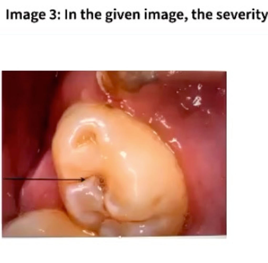
Slide 28 arrow lesion answer
ICDAS 2, Initial — enamel change, no dentin shadow, no microcavitation
Radiographic: Sound
No change
Radiographic: Initial
up to outer 1/3 of dentin
Radiographic: Moderate
D2 — middle 1/3 of dentin
Radiographic: Advanced
D3 — inner 1/3 of dentin
Moderate
shadowing + microcavitation

Advanced
dentin clearly visible (root lesion)

Initial
white chalky changes around grooves
Lecture 2 summary points
Classification is key to detecting, diagnosing, managing; consider caries in context of risk; tooth level: location, primary/secondary, severity, activity; ADA CCS used at TUSDM
Who gets root caries?
Elderly with gingival recession/exposed roots
decreased motor skills
less saliva
xerostomia drugs
systemic conditions
Why is root caries increasing?
Life expectancy up — people keep teeth longer
Tooth-level root caries factors
Recession from poor OH and attachment loss
irregular root surface
wide interproximal spaces protect biofilm
Where does root caries start?
Directly in DENTIN, after thin cementum is removed (e.g., traumatic brushing)
Critical pH: enamel vs dentin
Enamel 5.5; dentin 6.5
Why does root caries spread fast?
Higher critical pH, tubules, thin cementum, specific microbiota
Consequences of root caries
Sensitivity, pain, chewing difficulty → pulp inflammation, infection, tooth loss, poor nutrition/QOL
4 reasons root caries is alarming
Rapid progression; often asymptomatic; closer to pulp; harder to restore (saliva, bleeding)
Active root caries
Yellowish/slightly brown, undefined borders, soft/leathery, visible biofilm → restore
Inactive/arrested root caries
Dark brown/black, defined borders, smooth, bright, hard → monitor; restore only for esthetics
What arrests root caries?
Protective factors: better brushing, more fluoride, body reactions
Instrument to probe a suspected lesion
Rounded WHO probe (not explorer — avoids cavitating it)
CARS definition (Lecture 3)
Lesion at margins of existing restorations — just another primary lesion near a restoration/sealant
Procedural causes of CARS
Wrong matrix/wedge use, defective material placement, wrong adhesive/light-curing (marginal gaps)
Disease-level cause of CARS
Only the lesion was treated, not the patient's disease — risk stays high
CARS key statistic
8× more frequent than primary lesions in adults (esp. >50); greatest reason for restoration failure