Module 2 COnditioning and Learning
 Call Kai
 Learn
 Practice Test
 Spaced Repetition
 Match
 Flashcards
 Knowt Play
1/69

There's no tags or description
Looks like no tags are added yet.
70 Terms
Subdivisions
CNS, brain and spinal cord
forebrain, cerebrum, thalamus, hypothala.
midbrain
hindbrain, pons, medulla oblongata, cerebellum
PNS, nerves to muscle/sensory organs
Reflexes as innate behaviors
reflex as response to stimulus wo conscious thought, one type of innate behaviors
KNEE JERK/patellar reflex or withdrawal reflex hardwired into NS and present from birth
-autonomic/fast responses to certain stimuli, ensuring immediate responses wo the need for conscious though or prior learning
Reflexes mediated by neural circuits in spinal cord/brain
Spinal Reflexes: The spinal cord mediates relatively simple reflexes (e.g., grasp reflex, patellar reflex,
withdrawal reflex).
• Brainstem Reflexes: The brainstem is crucial for more complex reflexes, many of those involving the
head and face (e.g., salivary reflex, pupillary light reflex, startle reflex, postural reflexes for balance)
Complex innate behaviors (hibernation/migration) Mediated by…
complex innate mediated by forebrain. These behaviors are also innate and automatic and occur in response to stimuli, but they are not ‘fast’, like reflexes.
• Migration and hibernation behaviors in some animals are triggered by environmental cues
(temperature, daylight hours)
reflexes can be modified by..
experiences such as
Non-Associative Learning Processes:
• Habituation: A decrease in response to a repeated, harmless stimulus.
• Sensitization: An increased response to a stimulus following a strong or noxious event.
• Associative Learning:
• Conditioning: The process by which an innate reflex can be modified through associations
with other stimuli, such as in classical conditioning
Spinal Cord
is a major part of the central
nervous system (CNS) that extends from the
brainstem down through the vertebral column. It
serves as a communication highway between
the brain and the body, relaying sensory and
motor information via spinal nerves
-innate behaviors
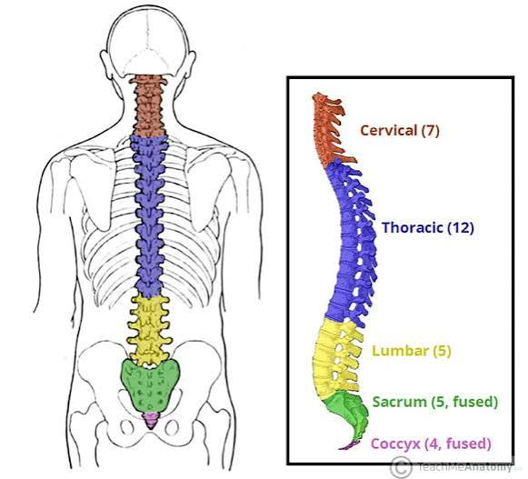
spinal cord divisions
small diameter, 16-18 in long
cervical, thoracic, lumbar, and sacral, each responsible for different body regions. Spinal nerves are pairs of nerves that originate from the spinal cord in these different segments.
• Damage to specific regions results in loss of function in the areas they control
Cross Section of Spinal Cord: Gray/ White matter
Gray Matter: Located in the center,
forming an "H" or butterfly shape, it contains neuronal cell bodies and is divided into dorsal horns– towards the back of the body (sensory processing) and ventral horns– towards the front of the body (motor neuron cell bodies)
White Matter: Surrounding the gray matter, it consists of axons organized into ascending (sensory) and descending (motor) pathways or tracts. These tracts carry signals to and from the brain and different parts of the body.

SC Cross Sec Cont: Yt matter and grey cell layers
GRAY MATTER:
• Divided into 10 layers with cells
• Dorsal horn: sensory (layers I-VI)
• Ventral horn: motor (layers VII-IX)
WHITE MATTER:
• Divided into ~ six columns
containing tracts
• Ascending tracts relay information
from the spinal cord to the brain
• Descending tracts carry
information from the brain to the
spinal cord
Key Neurons in Spinal Cord
Sensory neurons: Receive signals from the body (e.g., touch, pain, temperature) and transmit them to the
brain. They are part of the peripheral nervous system but connect with the spinal cord.
• Motor neurons: Send signals from the spinal cord to muscles, controlling movement.
• Interneurons: Excitatory or inhibitory (use GABA as neurotransmitter) and they connect sensory and
motor neurons, playing a crucial role in reflexes.

Sensory Neurons in SC cont
located outside the spinal cord
-no dendrites, single axon that splits into 2 branches, one goes to skin/muscles other goes to spinal cord.
-one collects periphery info and carries signal toward the soma, central branch takes signal into spinal cord via dorsal horn
-soma of sensory neurons located inside gnaglia, outside of SC, part of PNS
Motor Neurons in SC cont
are multipolar neurons
-extend long axon to muscles and initiate muscle contraction
-form synapses w muscle fibers, synapses known as neuromuscular junctions
-receive input from brain for voluntary movement and from spinal cord sensory neurons/interneurons for reflexes
visual of neurons

Neural Circuits behind spinal reflexs
2 main types: Monosynaptic and Polysynaptic
The monosynaptic component of the patellar reflex involves
a direct connection between the sensory neuron and motor
neuron in the spinal cord, resulting in a rapid, automatic
contraction of the quadriceps muscle in response to the tap
on the patellar tendon
polysynaptic circuit components
The polysynaptic components involve
interneurons and have 3 functions:
1) Relax antagonistic muscles
Polysynaptic components:
2) Coordinate the activity of
contralateral muscles
Polysynaptic components: 3) Send information to the forebrain, which mediates the
conscious perception of the stimulus that triggers the reflex
Central Pattern Generators
neuronal circuits that when active produce a rhythmic motor patterns like walking/breathing/swimming is abscence of descending inputs via brain
—not reflexes, both involve automatic actions, CPGs generate intrinsic rhytm act without needing ongoing sensory input
-reflexes triggered by certain stimulus
central PGs made of what circuits + exhibit 2 key properties
motor neurons and interneurons arranged in complex network within gray matter of SC
-controls repetitive behaviors like walking/breathing etc
exhibit
Rhythmogenesis: ability to produce rhythmic outputs.
• Presence of reciprocal inhibition, a neural circuit where the activation of one set of neurons
leads to the inhibition of an opposing set, allowing for alternating activation of muscle groups

Spinal Injury and Synaptic Plasticity
after injury neruons/axons limited regeneration
-synaptic plasticity possible, existing neurons can ,odify their connections/stregthen synapse in response to rehab
-increasing evidence magnetic/electrical stim promotes neural repair
using STDP and CPG for Spinal Cord Recovery + BSI
-They found that this stimulation protocol, based on spike-timing dependent plasticity (STDP) rules, efficiently enhanced synaptic plasticity in the spinal cord.
-In this study, the researchers utilized insights from central pattern generator (CPG) circuits and their role in the alternating activation of muscle groups to selectively stimulate the appropriate muscles when the animal initiated voluntary walking.
BSI= brain spine interface, wearable unit receives neural data predicts motor intention, sends updated stimulation commands
Brainstem location and composed of what subregions + function
base of the brain and connects brain to spinal cord
-midbrain: closest to forebrain
-pons
-medulla: closest to spinal cord
-does contain basilar artery and vertebral arteries
function= regulates essential autonomic funct ie heart rate, breathing, blood press. while also coordinating reflexes
-contains key neuromodulatory centers crucial for regualting array of funct like sleep wake cycles, attention, motivation
internal org of brainstem
The brainstem has a complex internal structure, with areas of white and gray matter intermingled. It contains:
1) Cranial nerve nuclei (which contain the somas of neurons that form the cranial nerves) and
2) Neuromodulatory centers
cranial nerves what they are and role in reflexes and vital functions
12 pairs of nerves that emerge directly from the forebrain and brainstem, responsible for transmitting sensory and motor info to and from the head and neck.
-Some cranial nerves are primarily sensory (e.g., the optic nerve for vision and the olfactory nerve for smell), while others are primarily motor (e.g., the facial nerve for facial movements). Some cranial nerves have mixed functions (sensory and motor), and some reflexes involve multiple cranial nerves, such as the pupillary light reflex and the gag reflex.
o They also help regulate vital functions like breathing, heart rate, and blood pressure.
Neuromodulatory Centers
Small clusters of neurons (‘nuclei’) that use neurotransmitters known as neuromodulators.
Neuromodulators are neurotransmitters that influence membrane potential and intracellular molecular cascades.
They may not contribute directly to encoding information, but they regulate the neurons that do by making them more or less likely to fire spikes.
• Cholinergic centers: Contain neurons that use acetylcholine as neurotransmitter: Attention
• Dopaminergic centers: Dopamine: reward and motivation (ventral tegmental area –VTA- in the midbrain)
• Serotonergic centers: Serotonin: Mood regulation and motivation (Raphe nuclei: in all sections of the brainstem
Attention Centers: Acetylcholine, basal forbreain, neocortical regions)
two main brain regions use acetylcholine as neurotransmitter and have widespread projections to other brain regions:
• Brainstem : project extensively to the thalamus
• Basal forebrain: projects extensively to the neocortex
—Basal forebrain cholinergic center is critical for focused, goal-directed attention
Example: Waking up in the morning
Example: Studying for an exam
—Brainstem cholinergic centers are involved in general arousal and alertness
Example: Waking up in the morning
Dopaminergic Centers memory and learning via sensory stimuli (VTA)
The ventral tegmental area (VTA) is located in the midbrain and contains dopaminergic neurons that send
their axons to forebrain structures involved in making decisions based on the potential value of stimuli.
• VTA neurons respond (fire spikes) to rewards, both innately rewarding stimuli (such as food, water, or social
interaction) and stimuli that have been learned to be associated with reward (such as a bell ringing before
food arrives in classical conditioning).
Serotonergic Centers support motivated behaviors
Serotonergic centers are known as the raphe nuclei, which are
distributed throughout all sections of the brainstem (midbrain, pons,
and medulla) and serve as the primary source of serotonin in the
brain. They project widely to various regions.
• The main role of serotonin is to promote motivation, but exactly how
serotonin affects motivation remains unclear.
• A lack of motivation is an important symptom of depression and
most medications for depression target serotonin to increase its level
in the brain.
expectancy theory of motivation + additional brain regions supporting motivation
suggests that motivation is driven by the expectation that efforts will
lead to rewarding outcomes. Serotonin plays a role in this process by influencing how individuals
evaluate effort and reward.
More serotonin could increase motivation by reducing the perceived cost of putting in more effort, or by
making people value the reward more
The anterior cingulate cortex promotes persistence toward goals.
-Its activation is correlated with increased perseveration and decreased apathy.
The cerebral cortex and thalamus important for..
encoding sensory info, for sensory content of memories
they interact to process increasingly complex info and relay it to other brain areas where mem traces can be formed
thalamus located where and does what for CC
in the forebrain deep in the center
-two thalami, one per hemisphere
-thalamus receives sensory input from outside, 15+ brain regions that perform diff functions,
-contains GLUTAMATERGIC excitatory neurons known as relay cells which project to cortex
2 main types of neurons thalamus
Thalamocortical/Relay cell Excitatory neurons (use
glutamate as neurotransmitter
2.Interneuron Inhibitory neurons (use GABA as neurotransmitter)
GABA interneurons concentrated in the thalamic reticular nucleus
-most thalamic intern. located in the TRN, thalamic reticular nucleus, which forms a layer that surrounds main thalamus
-interneurons in TRN establish inhibitory synapses w relay cells of thalamus
This circuit
between TRN and relay cells
can generate rhythmic
activity in the thalamus
TRN and sleep
TRN pacemaker of spindle oscillations, damage abolishes spindles
-projections from relay cells to cortex entrain cortical cells in oscillation
cerebral cortex subdivisions
Frontal (motor control, working memory, cognitive flexibility)
•Parietal (somatosensory)
•Temporal (audition, olfaction)
•Occipital (vision)
•Insula (taste, emotion)
•Limbic (motivation, learning, memory)
cortical lobes: grey is how many layers + pyramidal cells funct.
grey matter is in 6 layers
Pyram cells: Pyramidal cells are crucial for the encoding and processing of information in the brain, receive excitatory inputs
neocortex: interneurons
use GABA as NT, inhib, diff types and morphologies, most dont have spines on dendrites
-control level of activity of pyramidal cells and patter of activation
Hierarchical Model of Info to Neocortex + visual system
cortical areas extensively interconnected, w communication
-as info moves through cortical hierarchy, representations become increasingly complex and integrated
VS: neurons represent increasingly more complex info from edges/lines in primary visual cortex to objects and faces in higher order cortex areas
info transmission in thalamocortical circuits

neurons use APs to encode/ transmit info: neural rate code/receptive field
NRC= inividual neurons carry info about sensory stimuli using firing rate code, elevates firing rate when presented w prefered stim
receptive field: region of sensory space where presence of stimulus elicits response from neuron, neurons tuned to specific features of stimulus responding more strongly to some stimulus prop over others
Receptive field = where a neuron responds
Tuning = what features within that space the neuron prefers
EX. Neurons in the Primary visual cortex respond to edges and more strongly for edges of specific orientations
Fusiform face area
FFA: higher order cortical region in fusiform gyrus (bottom of temporal lobe) specialized in face perception
-responds preferentially to faces, more strongly in tact than scrambled faces and upright faces than to objects/animal faces
-is selective + has concept cells which responde specifically to one person or concept
concept cells in medial PFC experiment
spike activity of neurons in prefrontal cortex was quantified as animals viewed different exemplars of two categories (cats, dogs).
—Nearly one-third of neurons in prefrontal cortex were category- selective and responded specifically to one category but not the other
schemas and assimilation vs accomodation, cortical development w schema formation
Schema: A form of mental representation of complex knowledge. A schema is a mental model that the brain uses to help organize and interpret information.
assimilation, The process of incorporating new information into already existing schemas
Accommodation, Changing or altering existing schemas in light of new information. modifying existing schemas, or ideas, as a result of new information or new experiences
CD, neocortex integrates overlapping experiences for generalized representations and continues to adapt
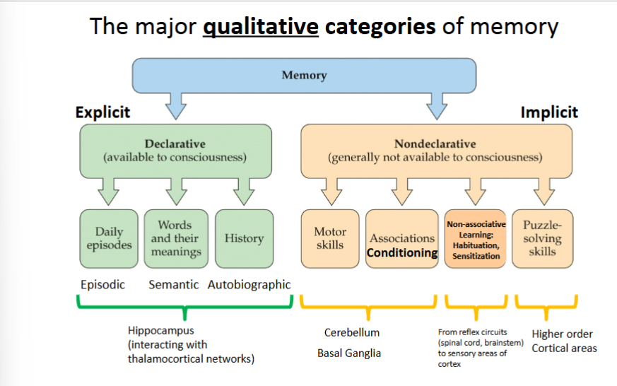
types of memory
HM patient demonstrates…
-HM unable to form new declarative memories since hippocampus removal
—motor skills and ability to learn intact, showing nondec mem relies on diff brain area
-mirror tracing experiment still performed but no declarative
Declarative memories rely on an intact hippocampus, and non-declarative memories must depend on non- hippocampal neural circuits
he had what amnesia, anterograde amnesia and retrograde amnesia
hippocampus located and part of what region
-part of cerebral cortex, within the temporal lobe
-considered evolutionarily older part of cortex,belongs to allocortex which has fewer layers compared to the 6 layered neocortex
-contains pyramidal cells (excitatory glutamate NT cells) and interneurons (GABA) interconnected
-considered essential for survival across evolution
Tri-synaptic circuit of the hippocampus
info in neocortex enters hippocampus via perforant pathway to dentate gyrus. from DG to CA3 to CA1 which is sent back to neocortex
-adaptations suggest they perform different functions and contribute differently to memory
Neurogenesis and pattern separation (Dentate Gyrus)
Pattern Separation: The ability to distinguish and store similar experiences as distinct, separate events, ensuring that individual episodes are not confused with one another.
—DG minimizes overlap between patterns of neural activity representing similar experiences
Neurogenesis is DG, over the period of a month, neural stem cells in DG undergo proliferation and differentiation until fully integrated in neural circuits of hippocampus
factors that regulate adult neurogenesis
molecular facotrs:
-NTs (serotonin)
-transcription factos (CREB which promotes growth/survival of neurons)
behavioral factors:
-exercise
-enriched environments
-diet (flavonoids)
-new neurons in this regio though to generate unique representations for similar inputs, preventing confusion
-helps each memory stores distinctly
Pattern completion and how it relates to connectivity (in CA3)
Pattern Completion: The ability to reconstruct a full memory from partial or incomplete cues, allowing for the recall of an entire episode based on limited information
—memory recall spike activity that encoded memory is restored and original activity pattern re-established
-reactivation process allows brain to reconstruct and reinstate spike activity in neurons
Recurrent Connections in CA3 pyramidal cells
-most synapses in ca3 are from recurrent connections

Representation of space (location) by hippocampal place cells
The hippocampus integrates where (spatial
context) and when (temporal context) an
experience occurred, forming a detailed
memory that we can later recall
-receptive field of hippoc. place cell is known as place field, specific to environment where neuron is active
-animal enters region, neuron active, helping brain map spatial locations
Amnesias
1- Anterograde amnesia: Inability to create new declarative memories
2- Retrograde amnesia: Inability to access memories from before the
damage to hippocampus (‘long-term’ or ‘remote’ memories).
—Graded Retrograde Amnesiafollowing HC damage: Graded retrograde amnesia refers to the loss of memories that were formed closer in time to the hippocampal damage, while older memories are spared. This temporal gradient suggests that declarative memories depend on the hippocampus initially but become gradually consolidated in the neocortex over time
standard mem consolidation theory vs multiple trace theory vs trace transformation theory
1- Standard Memory Consolidation Theory
proposes that memories gradually transfer from the
hippocampus to the neocortex.
-hippocampus is temporary role in mem retrieval
2- Multiple Trace Theory suggests that the
hippocampus remains involved in memory storage
indefinitely, with each retrieval creating new traces.
-hippocampus has continuing role in detailed episodic mem retrieval
3- Trace Transformation Theory proposes that
over time, detailed hippocampal memories are
transformed into more generalized, schematic
cortical representations
-detailed mems remain hippocampus dependent, generalized mems become independent of it
Complementary Learning Systems
Hippocampus
-rapid learning, crucial for new mems, acquires detailed info, mems can be unstable and fade
Neocortex
-learns more slow, integrates mems with existing knowledge, extracts gists of experiences, supports schemas and generalizations
The two systems complement each other: the hippocampus rapidly acquires new memories, while the neocortex gradually integrates those memories with older knowledge
Memory Generalization + Reconsolidation
is the process of identifying common features across individual experiences and applying that knowledge to new situations.
Examples from the lecture:
After visiting several grocery stores, you expect fruits and vegetables to be near the entrance.
Generalization allows the brain to learn concepts and rules rather than remembering every individual experience separately
Reconsolidation:
LT mem temp unstable and updated before stabilized again
—1 Retrieve an existing memory
2. Memory becomes temporarily unstable
3. New information can modify the memory
4. Memory is restabilized
Relevance for the treatment of memory disorders with neurostimulation methods
Transcranial electrical stimulation (tES): Uses electrical stimulation applied through electrodes on the scalp.
Transcranial magnetic stimulation (TMS): Uses magnetic pulses to influence brain activity.
A clinical study involving PTSD investigated combining prefrontal-cortex tES with virtual-reality exposure therapy. The lecture reports that this combination reduced physiological arousal and PTSD symptoms more than exposure therapy alone.
The potential application is that reactivated memories may be more open to modification, creating opportunities to improve treatments for memory-related disorders. These techniques are promising approaches, not guaranteed cures.
Brain Circuits for Sleep-Wake Control: role of hypothalamus, brainstem, thalamus
Hypothalamus — sleep-wake regulation
Acts as a master regulator of sleep and wakefulness.
Controls bodily functions such as temperature, hunger, and hormonal activity.
Influences brainstem neuromodulator systems
Brainstem — neuromodulator centers
Helps generate and regulate sleep stages.
Contains systems involving neurotransmitters such as acetylcholine and serotonin.
Interacts with the hypothalamus to regulate sleep and wakefulness.
Thalamus and cortex — sleep rhythms
Generate characteristic patterns of electrical activity during sleep.
Produce coordinated neural oscillations, including sleep spindles and slow oscillations.
Circadian Mechs via suprachiasmatic nucleus and pineal gland
Circadian rhythm: A roughly 24-hour cycle in physiology and behavior that helps coordinate sleep with the light-dark cycle.
Suprachiasmatic nucleus (SCN): Located in the hypothalamus; receives light information from the retina and acts as the brain's central circadian clock.
Pineal gland: Produces melatonin, a hormone that signals nighttime to the body.
Melatonin: Typically rises in the evening as darkness approaches, promoting sleepiness.
Cortisol: Typically peaks in the morning, supporting alertness.
SCN cont
light info from retina, SCN-central circadian clock, SCN controlled pathways regulate pineal melatonin production, daily timing of sleepiness and alertness
The SCN controls the timing of melatonin production; the pineal gland produces melatonin.
Sleep architecture: sleep stages
EEG: Electrical activity of the brain.
EOG: Eye movements.
EMG: Muscle activity and tone.
N1, Transition toward sleep; relatively low-voltage activity, Light sleep
N2, Sleep spindles; lighter NREM sleep, Clearly asleep
N3, High-amplitude, low-frequency slow waves, Deep, slow-wave sleep
REM, Low-voltage, relatively high-frequency activity resembling wakefulness, Rapid eye movements, vivid dreams, and muscle paralysis
NREM vs REM
NREM: Generally quieter neural activity, large-amplitude slow waves in deep sleep, and no REM-like muscle paralysis.
REM: Brain activity resembles wakefulness, dreams tend to be vivid and emotional, and skeletal muscle tone is largely suppressed despite occasional twitches.
NREM oscillations and memory consolidation
During NREM sleep, three important neural oscillations help coordinate communication between the neocortex, thalamus, and hippocampus
-slow oscillations, sleep spindles, hippocampal ripples
slow oscillations + sleep spindles
SO: Location: Neocortex
Stage: Most associated with N3, deep NREM sleep.
Cortical neurons alternate between:
Up state: Neurons are relatively active.
Down state: Neurons are relatively inactive.
These alternating states synchronize neural activity and help coordinate other sleep rhythms.
SS: Location: Thalamocortical circuits
Stage: N2, lighter NREM sleep.
Brief bursts of rhythmic electrical activity.
Approximately 10–15 cycles per second (Hz).
Typically last about 0.1–2 seconds, according to the lecture.
Generated by reciprocal circuits within the thalamus; thalamic relay cells help synchronize cortical activity.
Spindles are important for memory consolidation
hippocampal ripples + how all three work together
HR: Location: Hippocampus, particularly CA1
Stage: Occur during NREM sleep and also during awake immobility.
Brief events lasting approximately 100 milliseconds.
High-frequency activity, approximately 100–200 Hz.
Associated with reactivation of groups of neurons involved in episodic memory.
Can replay previously experienced spatial trajectories.
Important: The lecture states that ripples cannot be observed directly in a standard EEG.
How all work together: temporal coupling, the three rhythms occur in a coordinated sequence, allowing different brain regions to communicate
—ripples often occur nested within spindles, and spindles tend to occur during slow oscillations. This coordination provides a time window for the hippocampus, thalamus, and cortex to exchange information.
Synaptic homeostasis hypothesis (SHY)
Synaptic scaling is a homeostatic, negative-feedback process that adjusts synaptic strength in response to sustained changes in a neuron's activity. It helps keep neural activity within a functional range and prevents saturation.
-proposes sleep helps renormalize synaptic strength after changes that occur in wakefulness
Glymphatic system and metabolite clearance
The glymphatic system is a network associated with the movement of cerebrospinal fluid (CSF) through brain tissue and the clearance of metabolic waste from the central nervous system.
CSF: Cerebrospinal fluid is a clear, colorless fluid found in the brain and spinal cord.
Function: Helps clear metabolic waste from the central nervous system.
Fluid movement: The lecture identifies arterial pulsation as a primary driver of CSF flow in the glymphatic system.
Sleep: Glymphatic clearance is greater during sleep than during wakefulness.
N3 sleep: Deep NREM sleep is identified in the lecture as optimal for glymphatic function.
Clinical relevance: Disruption of glymphatic clearance may contribute to certain brain diseases, although the relationship is still being investigated.
Classical conditioning def
non declarative (implicit) learning where stimulus acquires capacity to evoke response initially evoked by another stimulus
-starts w an existing, innate response to a natural stimulus: reflx
ivan pavlov experiment/ appetitive conditioning
Begins with an innate (unlearned) reflex
– Unconditioned stimulus (US) [food]
– Unconditioned response (UR) [salivation]
• A neutral stimulus (CS) is then repeatedly and consistently presented before
the reflex is triggered, producing a new reflex.
– Conditioned stimulus (CS) [bell]
– Conditioned response (CR) [salivation]
Aversive conditioning ex
Odor Conditioning in Flies
• Exposure to shock (US) innately
produces escape/avoidance
behavior (UR).
• The CS is an odor, initially neutral.
• After CS is paired with US, CS
comes to produce avoidance (CR).
• Aversive conditioning: new
CSCR reflex helps avoid
noxious US
Eye blink conditioning
puff of air to eye(US) = blink (UR), tone plays (CR)
-form of aversive conditioning
-effectiveness of CS builds over many pairings w US and prepares for puff US
-CR takes place quickly after CS
Initially (day 1), the CS does not produce any response.
– After extensive training, the CR emerges as a slow squeezing shut of the eye prior to the US.
timing of stimuli important for conditioning :trace vs delay + examples
Delay conditioning (top): best learning
• Trace conditioning (bottom): For trace conditioning to happen the brain needs to form a mental representation of the conditioned stimulus after it has ended. This is called a stimulus trace.
Trace Ex. hungry when order taken when at first not
bell sounds leads to meeting friends before lunch =happy/hungry
just bell sounds can make feel happy or hungry
Delay Ex. notification bell creates anticipation