Articles about affective neuroscience, emotion and memory + presentations 9 and 10
 Call Kai
 Learn
 Practice Test
 Spaced Repetition
 Match
 Flashcards
 Knowt Play
1/19

Earn XP
Description and Tags
Dalgleish, T., Dunn, B. D., & Mobbs, D. (2009). Affective Neuroscience: Past, Present, and Future.. Emotion Review, 1(4), 355– 368. + LaBar, K. S., & Cabeza, R. (2006). Cognitive Neuroscience of Emotional Memory. Nature Reviews Neuroscience, 7(1), 54–64.
20 Terms
ARTICLE: “Affective Neuroscience: Past, Present, and Future”
Until further notice, the flashcards will be based on this article
Dalgleish, T., Dunn, B. D., & Mobbs, D. (2009). Affective Neuroscience: Past, Present, and Future.. Emotion Review, 1(4), 355– 368.
Give a historical overview of the pioneering work in affective neuroscience of James and Lange
Pioneering work!
The James-Lange Theory
Emotions are:
The experience (/interpretation) of bodily changes that occur in response to emotive cues in the world
So, different bodily changes code different emotions
Remain remarkably influential on contemporary thinking
Because of he emphasis it places on the embodiment of emotions, which today sits well with frameworks of embodied cognition
Give a historical overview of the pioneering work in affective neuroscience of Cannon and Bard
Early functional neuroanatomical models!
Critique of James-Lange - Cannon suggest:
(1) Autonomic activity cannot differentiate different emotional states
Findings today:
This suggestion has partly been debunked: it seems they can to a certain degree
(2) Emotional behavior in animals is not impaired when surgically separating the viscera from the brain
(viscera - part of limbic system - regulating the bodys automatic functions, linking physiological reactions to feelings and instincts (AI answer))
Findings today:
Partial disconnection of the brain from the body can actually sometimes reduce emotional intensity
Since it is not possible to totally isolate the brain from all aspects of bodily feedback - so (supporting James-Lange) emotional experience may be driven by remaining feedback in separation designs
(3) Bodily reactions are typically too slow to generate emotions
(4) Artificial hormonal activation of bodily activity is insufficient to generate emotion
Findings today:
Some artificial manipulations of organ activity
can induce emotions - e.g. inducing panic attacks
.
The Cannon-Bard Theory
Cannon developed the first neuroanatomical theory of emotion, later elaborated by Bard
Suggests:
Thalamus and hypothalamus are critically involved in the emotion response to stimuli
Evidence supporting hypothalamus’ role: important part of reward network (and therefore motivation)
These emotion responses are inhibited (i.e. controlled) by evolutionarily more recent neocortical regions - i.e. thalamic regions
Evidence supporting this:
Removal of the cortex in cats - freeing the thalamic circuitry from top-down control - allows uncontrolled emotion displays (e.g. anger attacks)
.
Schacter-Singer
The model accepted today - a hybrid position between James-Lange and Cannon-Bard
The model suggests:
The body contributes to a crude (i.e. rough) sense of emotional intensity
The crude sense is then cognitively appraised - leading to more nuanced emotional experience
Evidence for the model:
Studies showing that similar patterns of bodily arousal could be experienced as different emotions - e.g. anger or happiness - depending on the social and cognitive context
Give a historical overview of the pioneering work in affective neuroscience of Hess
Hypothalamic stimulation studies
Cannon-Bard theory was influenced by Walter Hess’ studies on hypothalamus
Hess implanted electrodes into the hypothalamic region of cats
Electrical stimulation of one part of the hypothalamus led to an “affective defence reaction” - associated with increased heart rate, alertness, and a propensity to attack
Indications of Hess’ findings:
Stimulation of certain brain regions can induce animals to act in different ways/recognizable emotional responses - e.g. angry, fearful, curious, lethargic
Give a historical overview of the pioneering work in affective neuroscience of Papez
Development of increasingly sophisticated neuroanatomical frameworks
The Papez Circuit
An anatomically broader - compared with Cannon-Bard - scheme for the central neural circuitry of emotion
Suggesting:
Sensory input into the thalamus diverge into an upstream “thought” and a downstream “feeling” pathway
Thought stream —> link the thalamus to the sensory cortices
Especially to the cingulate region
Stream continues: —> cingulum pathway —> hippocampus —> fornix —> mammillary bodies of hypothalamus —> mammillothalamic tract —> back again to anterior thalamus
Here sensations are turned into perceptions, thoughts, and memories
Feeling stream —> transmitted from the thalamus directly to the mammillary bodies
Generating emotions - with downward projections to the bodily systems - and then upwards to the cingulate cortex via he anterior thalamus
Emotional feelings are:
A function of cingulate activity generated through either of the two streams
Echoing Cannon-Bard:
Top-down cortical regulation of emotional responses is permitted by downward projections from the cingulate to the the hypothalamus
Today’s views on The Papez Circuit
Many of the pathways he proposed exist! - but less evidence that all regions specified are central to emotion
Give a historical overview of the pioneering work in affective neuroscience of MacLean
Development of increasingly sophisticated neuroanatomical frameworks
MacLean’s Limbic System
A more broadly supported anatomical model of the brain regions involved in emotion and the dominant conceptualization of the “emotional brain” today
Built on:
Papez’s and Cannon-Bard
Studies on monkeys by Kluver an Bucy - “Kluver-Bucy syndrome“
Suggesting: key role for temporal lobe structures in normal emotional behavior
Later work showed that damage to the
amygdala accounts for important aspects of the emotional changes
The brain as a triune architecture with three interacting systems
(1) evolutionary ancient reptilian brain
Brain areas:
Striatal complex + basal ganglia
Seat of primitive emotions - e.g. aggression + fear
(2) “old” mammalian brain (aka the “visceral brain” / the limbic system)
Brain areas:
Hypothalamus, thalamus, hippocampus and cingulate cortex, amygdala, prefrontal cortex + other
Augments (förstärkar) primitive reptilian emotional responses - e.g. aggression
Elaborates social emotions
(3) “new” mammalian brain
Brain areas:
Neocortex
The interface of emotion with cognition
The seat of top-down control over emotional responses originating within other systems
Emotion experiences are generated from:
Integration - in system (2) (specifically the hippocampus) - of sensations from the world with information from the body
Messages about bodily changes - caused by external events - return to the brain and are integrated with ongoing perceptions of the world
Describe Zajonc-Ledoux theory of emotion
Compare the Z-L theory to the Schacter-Singer theory
Core of the Z-L theory:
Emotional responsen can occur automatically and instantly at the same time
Instantly: without conscious cognitive interpretation or thinking first
.
The “low” road / direct pathway
Route:
Senses —> Thalamus —> Amygdala
Any sudden, alarming sensory input can trigger the instant survival loop - since the thalamus acts as the main relay station for all senses (except for smell)
Speed:
Extremely fast - “quick and dirty“
Function:
Triggers immediate survival responses for simple emotions
(e.g., fear of a snake).
.
The “high” road / indirect pathway
Route:
Senses —> Thalamus —> Sensory Cortex —> Amygdala
Speed:
Function:
Describe the current state of research on the amygdalas contribution to emotion
Amygdala
Position in brain:
Medial temporal lobe
Rests on the anterior tip of hippocampus
Central research findings
Kluver-Bucy syndrome
Many of the tendencies in the syndrome can be sufficiently induced by bilateral lesions of the amygdala + disrupting the social behavior of monkeys
Fear and emotional learning
Critical for fear conditioning and for linking stimuli with emotionally significant outcomes

E.g S.M.
In contrast: damage to hippocampus, with intact amygdala, can be fear conditioned with no explicit memory of the conditioning procedure
E.g. Amygdala-damage and recognition of fear in facial expressions
Emotional expression and social signals of emotion
Amygdala damage can impair the recognition and expression of fear, particularly in response to fearful facial expressions.
E.g. S.M.
fMRI when presented with fearful faces or fear communicated in other ways (e.g. vocally or bodily movements) show selective brain activation in the amygdala
Even when not aware that the fearful face was presented or when presented to blindsight patients
Can be modulated by top-down control on attention
Emotional memory
Amygdala activity can enhance the encoding and consolidation of emotionally significant memories
Amygdala damage:
Does not show usual enhanced memory for emotional aspects of stories
Take-home message
The amygdala is not simply a “fear centre”!
It helps detect and evaluate the emotional significance of stimuli + coordinate appropriate learning and responses


Describe the current state of research on the PFC’s contribution to emotion
Phineas Gage
PFC lesion —> radical changes in personality and emotional behavior
.
Guiding behavior
Integrates emotional information with cognitive processes to guide behavior
Involved in learning the emotional and motivational value of stimuli
PFC regions work together with the amygdala to learn and represent relationships between new stimuli and primary reinforcer (e.g. food, drink, sex)
.
Decision-making:
The vmPFC uses emotion-related signals from past experience (“somatic markers”) to anticipate the likely consequences of different choices
Somatic marker hypothesis (SMH)
Somatic marker:
Physiological reactions which tag previous emotionally significant events
Can be reactivated by cues, providing a signal delineating which current events have had emotion-related consequences in the past
Allow decisions to be made in situations where a logical analysis of the available choices proves insufficient
Helps anticipate outcomes and guide decisions
Patient EVR - lesion in vmPFC
Unable to utilize somatic markers
Tries and fails to deal with complex situations of uncertainty using logical reasoning alone
Iowa Gambling Task
Lesion - did not get “hunches” about which cards to choose
Non-lesion - unknowingly became sensitive to the delayed rewards and could process their somatic markers that had developed early in the game
S.M. patient and amygdala
The amygdala seems necessary for initially seating up the emotional response that the vmPFC can regenerate, but it doesn’t seem necessary once the somatic markers have been established.

.
Emotion regulation:
PFC regions contribute to evaluating, and regulating emotional responses
Emotion regulation = Processes that influence:
(1) which emotion one has
(2) when one has them
(3) how one experiences and expresses these
emotion
Multiple points in emotion generative process can be regulated, affecting the emotion

Reappraising how we think about or evaluate a situation
One of most effective means of regulating our emotions
Less useful when emotions are very intense (more useful to redeploy our attention)
Less useful when the situation can be modified
Associated with activation in PFC
Increased PFC activity when participants regulated negative emotions - both up- and –down - compared with when just told to look at images
.
Take-home message:
The PFC allows emotional information to be used flexibly:
emotions can guide decisions, while cognitive processes can also regulate emotional responses
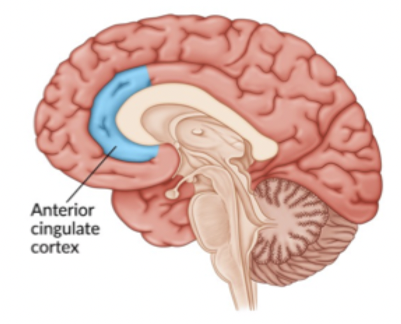
Describe the current state of research on the anterior cingulate cortex’s (ACC) contribution to emotion
ACC sits at an important interface between cognition and emotional control - contributing to:
Monitoring emotionally significant information
Activates to more subjective elements of pain including empathy for pain and social ostracism
Conflict and error monitoring
ACC is involved in the monitoring of conflict between the current functional state of the organism and any new information that has potential affective or motivational consequences
When such conflicts are detected, the ACC projects information about the conflict to areas of PFC where adjudications among response options can occur
Selecting and adjusting behavior
Regulating emotional responses
Integrating emotion, motivation and cognitive control
Describe the current state of research on the insula’s contribution to emotion
Strongly involved in interoception
Interoception = perception and representation of signals from inside the body
E.g. if asked to judge whether ones own heartbeat is simultaneous to a tone
Helps integrate bodily signals with information form external world
Provides a link between physiological changes in the body and our subjective emotional experience
.

Insula is involved in:
Awareness of bodily states
E.g. heartbeat, breathing and visceral sensations
Emotional experience
By integrating bodily signals with other information
Involved in the generation of a risk prediction error - guiding decision-making in risky situations and facilitating learning about uncertain rewards
Bodily feedback in decision-making proposed by the somatic marker hypothesis
Disgust
One of the emotions consistently associated with the insula
Empathy and pain
Including responses to our own and others’ bodily/emotional states
ARTICLE: “Cognitive neuroscience of emotional memory”
Until further notice, the flashcards will be based on this article:
LaBar, K. S., & Cabeza, R. (2006). Cognitive Neuroscience of Emotional Memory. Nature Reviews Neuroscience, 7(1), 54–64.
Flashbulb memories
Definition of flashbulb memory
A highly vivid memory for how one learned about an emotionally engaging and surprising event
Involving memory for the source of the event information - as opposed for the event itself
E.g. memory for:
Where you were
What you were doing
Who told you something
How it affected you
What the aftermath was
Emotion-induced memory enhancement - and the role of amygdala
Consequences of amygdala damage on emotional episodic memory?
Unilateral damage to amygdala:
Does not show enhanced arousal-mediated memory consolidation for emotionally arousing words
But instead show parallel forgetting rates for arousing and neutral words from immediate to 1-h retention intervals
Bilateral damage to amygdala:
Show impairments in long-term (1 h to 1 month) recall or recognition of emotional words, pictures and stories1
.
Weapon focus / gist memory effect
When emotionally aroused:
Focusing of attention on central gist information at the expense of peripheral details for complex events
e.g. complex events: emotional narratives or social encounters
Attentional focusing ensures that emotionally salient features of complex events are preferentially retained in memory - an evolutionary advance
Amygdala lesion
Do not focus on “central gist” of information - when memory is testen for audiovisual narratives that describe emotionally arousing events
Valence is the crucial factor in engaging amygdala during emotional memory tasks
In some cases patients do preferentially remember words that are effectively valent, but low in arousal - relative to neutral ones
Memory modulation hypothesis
States that:
Greater long-term memory for emotional than neutral events reflects the neuromodulatory influence of the amygdala on consolidation processes in MTL (medial temporal lobe) memory regions through engagement of stress hormones
E.g. supported by findings from research on rodents
Emotional arousal does not simply strengthen memory - it modulates which information gets prioritized for LTM storage
.
How amygdala modulates consolidation of memories during emotional arousing events:
1. When experiencing an emotionally arousing event, hormones (e.g. adrenaline and cortisol) are released
2. The hormonal signals influence the amygdala - by acting on the norepinephrine receptors - increasing its activity
3. The amygdala then modulates memory-related brain regions - e.g. hippocampus and sensory cortex - enhancing processes involved in memory and consolidation
The amygdala acts as a modulator - helping to strengthen the consolidation of memories formed during emotionally arousing events
E.g. Evidence showing effects of activation vs inhibited activation of amygdala on encoding and consolidation of emotional memories - important for the memory enhancing effects of emotion
Healthy adults given a placebo 1h before the story, remember the emotionally arousing portion of the story better than the neutral portions 1 week later
Proppranolol, blocks the adrenergic receptors, thereby reducing the effects of adrenaline and noradrenaline. Its administration during encoding abolishes the retention advantage (for emotionally arousing portion of the story) in healthy adults
Two patients with selective amygdala damage (SM and BP) also lack the retention advantage

The role of amygdala during memory retrieval
Resent research in rodents suggests that the amygdala also contributes to the reconsolidation of emotional memory traces following their retrieval - not only encoding and consolidation stages of memory
Dolcos et al. (2005)
Participants viewed neutral and emotional pictures - then returning 1 year later to perform a memory test:
Emotional pictures were remembered better than neutral ones - correlating with activity in amygdala and MTL memory regions
But only when participants indicated that they remembered (recollected) seeing the pictures 1 year ago
Recollection = retrieval of qualitative information (e.g. place time, perceptual, emotional details) about an event
Recollection is especially enhanced behaviourally by emotion
Familiarity = Feeling of having previously encountered something without the retrieval of any associated contextual details - (i.e. “knowing”)
Greater retrieval success activity was found in the amygdala, hippocampus and entorhinal cortex for emotional relative to neutral pictures.
In amygdala and hippocampus the activity was enhanced for emotional items recognized with a sense of recollection rather than familiarity
Fear conditioning - the role of the amygdala
How fear conditioning usually is measured in humans
Skin conductance response (SCRs)
Potentiation of eyeblink startle reflexes
.
Neuroscientific research pointing to the importance of the amygdala in fear conditioning
In rodents:
Lesions of the basolateral and central nuclei of the amygdala prevent the acquisition of fear to discrete cues and environmental contexts
During fear conditioning, electrophysiological changes occur in the lateral amygdala before they do in other brain regions
To extinguish fear behaviours, the ventromedial PFC suppresses amygdala function by engaging a network of inhibitory interneurons
Double dissociation regarding the contributions of the amygdala and hippocampus to conditioned emotional learning in humans
Damage to the amygdala:
Consistently impairs fear conditioning and fear-potentiated startle responses
(similar to those observed in non-human animals)
By contrast, amnesic patients with damage restricted to the hippocampus show the opposite dissociation:
They can acquire conditioned fear on simple tasks but cannot verbalize the appropriate stimulus relationships
Important distinction between conditioned fear response and verbalizing stimulus relationships
Unlike rodents, humans acquire conscious knowledge of the stimulus relationships, which can modify learning under some circumstances
The evidence therefore show how declarative and non-declarative aspects of simple forms of fear learning are neurally dissociable
Contextual reinstatement of fear - the role of hippocampus
The contextual reinstatement of extinguished fear - hippocampal dependent
(Although the initial acquisition of simple fear conditioning to sensory cues depends on the amygdala and not the hippocampus)
Rodent lesion studies
Shown that: the integrity of the hippocampus is important for the contextual recovery of extinguished fear
Amnesic patients with selective hippocampal damage
Do not show contextual fear reinstatement, despite acquiring fear normally
In contrast to non-amnesic persons!
Normally conditioned fear responses that have been extinguished can be recovered in a context-dependent manner
Post Traumatic Stress Disorder (PTSD) and emotion induced memory enhancement
Diagnostic criteria
Exposure to a traumatic event
Persistent re-experiencing of the traumatic event
Persistent avoidance of reminders of the event
Persistent symptoms of heightened arousal
Duration of symptoms must be more than 1 month
Significant impairment in social, occupational, or other important areas of functioning
.
Reduced hippocampal volume correlating with months of combat exposure and development of PTSD

.
PTSD and generalization (worsened memory discrimination)
Memory discrimination serves to disambiguate new experiences from stored memories
Upon experiencing a situation that shares features of a traumatic event:
Healthy individuals (non-PTSD):
Able to discriminate a novel experience from a fearful one and successfully encode the novel experience as novel and safe
PTSD individuals:
Excessive generalization manifests as a failure to discriminate the novel, safe ecperience from the previously stored emotional memory
Leading to excessive arousal and fear responses
.
Psychopharmacological studies providing greater specificity about what phase of memory is affected by emotion
Memory phases: encoding, consolidation, storage, retrieval
Neurohormonal memory modulation in rodents:
Post-training infusion of β-adrenergic receptor antagonists into he basolateral amygdala blocks the memory-enhancing effects of adrenaline
While infusion of β-adrenergic receptor agonists facilitates memory consolidation
.
Neuohormonal memory modulation in PTSD
Beta-blockers are currently being evaluated as potential agents for secondary prevention of PTSD
Administration of the β-adrenergic receptor antagonist propranolol seems to selectively reduce memory consolidation for emotionally arousing material
i.e. similarly to the rodent study above
Adaptive forgetting and Inhibitory control - connect to PTSD
Adaptive forgetting and Inhibitory control
May promote resilience by limiting the involuntary retrieval and emotional impact of trauma-related information
Allowing flexible control over attention and memory
Evidence - think/no-think - can we inhibit a memory?
First, Training faze:
Learn to associate 40 pairs of pictures (a face and an item) to a precision level of 95%
Second, experiment faze:
Baseline:
8 pairs that was previously trained on, will not be shown during this faze - is used as a control to see what you remember naturally without manipulation
Think:
16 faces with green shown around them: instruction is to “think of the associated picture”
No-think:
16 faces with red shown around them: instruction is to “not think of the associated picture”
Third, test:
Tested for all 40 pairs of pictures
Results:
Actively “think”ing gave better memory results than baseline
Actively inhibiting (no-think) gave worse memory results than baseline
PFC inhibiting hippocampus when asked not to think about something —> weakening hippocampus’ ability to recall the memory later
.
Inhibitory control and PTSD
Waldhauser et al. (2018) - trauma exposed refugees in Think/No-Think task
With PTSD showed poorer memory suppression than without PTSD
With PTSD - failed to downregulate gamma activity associated with the reactivation of sensory memory traces
.
Mary et al., (2020) - Paris terrorist attack survivors with and without PTSD
Without PTSD - had stronger PF control system (especially in right anterior dorsolateral PFC) - inhibiting memory-related brain activity
With PTSD had reduced PF control
.
Both studies show that PTSD may involve a general deficit in inhibitory control over memory
Contributing to difficulties in suppressing unwanted thoughts and memories