L7 - Plasma Proteins
 Call Kai
 Learn
 Practice Test
 Spaced Repetition
 Match
 Flashcards
 Knowt Play
1/47

There's no tags or description
Looks like no tags are added yet.
48 Terms
What are the Main Components of Plasma?
Plasma contains:
Water
Biochemical constituents, including proteins, electrolytes, enzymes, and metabolites.
What is the Difference between Plasma and Serum?
Plasma:
Obtained from anticoagulated blood(e.g., EDTA purple-top tube)
Contains all proteins, including fibrinogen and clotting factors
Plasma total protein is typically slightly higher than serum total protein
Serum:
Obtained blood after blood clots (e.g., red-top tube)
Lacks fibrinogen and clotting factors consumed during clot formation
Use the same sample type when monitoring protein trends

What are the Main Plasma Protein Groups, their Sites of Synthesis, and their Functions?
Albumin:
Synthesized by hepatocytes
Maintains oncotic pressure and transports substances such as bilirubin
Gamma globulins (immunoglobulins):
Synthesized by plasma cells
Function as antibodies in adaptive immunity
Alpha and beta globulins:
Primarily synthesized by hepatocytes
Include acute phase, transport, complement, clotting, and lipoproteins
Why is Albumin Important for Maintaining Oncotic Pressure?
Albumin is the most abundant individual plasma protein and helps retain water within blood vessels
Severe hypoalbuminemia can decrease oncotic pressure, resulting in edema or body cavity effusions
How are Plasma Proteins Measured Using Refractometry?
Refractometry measures the refractive index of plasma, which correlates with dissolved solute concentration, primarily proteins
It is used to estimate total plasma protein (TPP), with fibrinogen measurable after heat precipitation
What Factors can Interfere with Refractometric Protein Measurements?
These substances affect light refraction independently of protein concentration.
High glucose, sodium, or urea concentrations can falsely increase TPP
Hemolysis and lipemia can interfere with readings
How are Serum Total Protein, Albumin, and Globulins Measured?
Total protein: Biuret method
Albumin: Bromocresol green method
May vary by species, particularly birds, and can be affected by bilirubin or certain drugs
Globulins: Calculated as (total protein − albumin)
Albumin: Globulin (A/G) ratio: Used to identify disproportionate protein changes
What is Serum Protein Electrophoresis (SPE), and What Does It Measure?
SPE separates serum proteins into albumin, alpha-1, alpha-2, beta, and gamma fractions based on their electrophoretic properties
SPE helps identify which protein fractions are increased or decreased
Albumin: Largest peak in healthy animals
Alpha and beta fractions: Primarily acute phase and other non-immunoglobulin proteins
Gamma fraction: Primarily immunoglobulins

*What Terminology Describes Increased and Decreased Protein Concentrations?
Hyperproteinemia / Hypoproteinemia:
Increased / decreased total protein
Panhyperproteinemia / Panhypoproteinemia:
Both albumin and globulins increased / decreased
Hyperalbuminemia / Hypoalbuminemia:
Increased / decreased albumin
Hyperglobulinemia / Hypoglobulinemia:
Increased / decreased globulins
Hyperfibrinogenemia / Hypofibrinogenemia:
Increased / decreased fibrinogen
Hypergammaglobulinemia / Hypogammaglobulinemia:
Increased / decreased gamma globulins
What is the Difference between Relative and True (Absolute) Protein Changes?
Relative changes:
Protein mass remains unchanged, BUT plasma water volume changes
Dehydration → Apparent increased protein concentration
Overhydration → Apparent decreased protein concentration
True changes:
Actual protein mass changes due to:
Altered synthesis
Increased loss
Changes in intake/utilization
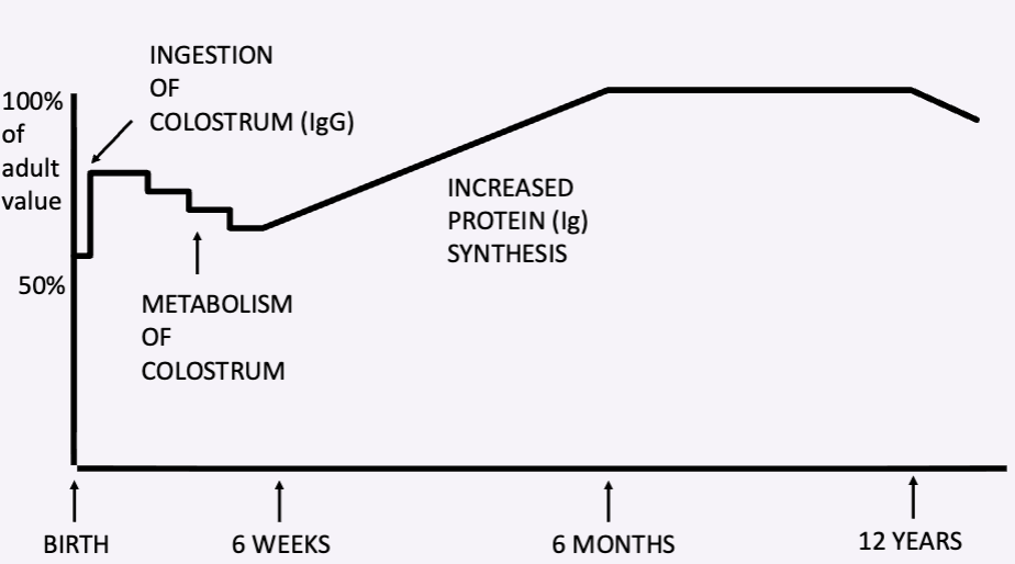
How does Age Affect Total Protein Concentrations?
Neonates have lower total protein concentrations than adults
Colostrum ingestion causes a temporary increase in immunoglobulins, especially IgG
Maternal immunoglobulins gradually decline as endogenous protein and antibody synthesis increases
Failure of passive transfer prevents the expected post-colostrum increase in proteins
Age-specific reference intervals are important when interpreting protein values.
What is the Primary Mechanism of Hyperalbuminemia?
Dehydration (relative increase) is the only clinically relevant cause of hyperalbuminemia
The liver does not normally overproduce albumin
What Laboratory Pattern Suggests Dehydration?
CBC
Hematocrit (HCT) may also increase
Increase plasma proteins
CHEM
Increased total protein
Increased albumin
Normal or proportionately increased globulins
Clinical findings
Tacky mucous membranes, decreased skin turgor, and sunken eyes support dehydration
What are the Major Mechanisms of Hyperglobulinemia?
Relative increase: Dehydration
True increase:
Increased gamma globulin production due to chronic inflammation or lymphoid neoplasia
Increased alpha and beta globulin production during an acute phase response

What is the Difference between Polyclonal and Monoclonal Gammopathy?
Polyclonal gammopathy:
Broad-based gamma peak on SPE caused by increased production of multiple immunoglobulins
Commonly due to chronic inflammation
Monoclonal gammopathy:
Narrow-based gamma peak on SPE caused by a abnormal immunoglobulin population
Commonly associated with lymphoid neoplasia

What are the Major Causes of Polyclonal and Monoclonal Gammopathies?
Polyclonal:
Chronic inflammation and persistent antigenic stimulation, including infectious diseases such as feline infectious peritonitis (FIP)
Monoclonal:
Lymphoid neoplasia, particularly plasma cell myeloma
Non-neoplastic monoclonal gammopathies may also occur with infections, especially canine ehrlichiosis and leishmaniasis
Why can Hyperglobulinemia be accompanied by Hypoalbuminemia?
During inflammation, albumin synthesis may decrease because albumin is a negative acute phase protein
The lecture also describes a compensatory reduction in albumin synthesis during marked hyperglobulinemia to help balance oncotic pressure.
*What Protein Patterns Help Distinguish Dehydration from Inflammation or Neoplasia?
Interpret these patterns alongside clinical findings and hematocrit:
Increased albumin with normal or increased globulins
→ Dehydration
Increased globulins with normal or decreased albumin
→ Chronic inflammation, lymphoid neoplasia, or certain infections
What is the Acute Phase Response, and How Does It Affect Plasma Proteins?
Inflammation stimulates cytokine-mediated changes in hepatic protein synthesis
Positive acute phase proteins increase
Negative acute phase proteins decrease
Alpha and beta globulin fractions may increase on SPE
Most acute phase proteins are present in relatively low concentrations, so their increases may have little effect on total protein

What are the Major Positive and Negative Acute Phase Proteins?
Positive (increase):
Fibrinogen
C-reactive protein
Serum amyloid A
Haptoglobin
Alpha-1-acid glycoprotein
Ceruloplasmin
Alpha-2-macroglobulin
Negative (decrease):
Albumin
Transferrin

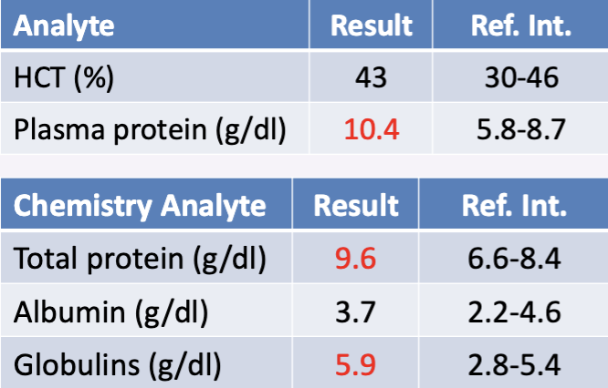
PRACTICE QUESTION: A 9-month-old filly has a cough and nasal discharge
ANSWER → Hyperproteinemia due to hyperglobulinemia
Likely chronic inflammation due to upper airway infection
Why is Fibrinogen Clinically Important in Large Animals?
Fibrinogen is the most abundant acute phase protein and increases during acute or chronic inflammation
Its concentration can significantly increase total plasma protein, making it a useful inflammatory marker in large animals

What is the Protein:Fibrinogen Ratio, and How is It Interpreted?
Protein:Fibrinogen Ratio = Total plasma protein (g/dL) / Fibrinogen (g/dL)
Ratio ≥ 15: Proportionate increase, suggesting dehydration
Ratio < 15: Disproportionate fibrinogen increase, suggesting inflammation
Fibrinogen must be converted from mg/dL to g/dL before calculation

PRACTICE QUESTION: A 9-month-old filly has a cough and nasal discharge
ANSWER → Hyperproteinemia due to hyperglobulinemia, most likely from chronic inflammation associated with an upper respiratory infection
Plasma protein: 10.4 g/dL
Fibrinogen: 900 mg/dL; Convert fibrinogen: 900 mg/dL = 0.9 g/dL
10.4/0.9 = 11.5
The ratio is below 15, indicating a disproportionate increase in fibrinogen
Albumin and HCT are within reference intervals, making dehydration less likely as the primary explanation


PRACTICE QUESTION: A 2 year old quarter horse with colic is moderately dehydrated on examination
ANSWER → Dehydration
HCT: 48% (30–46)
Plasma protein: 9.2 g/dL (5.8–8.7)
Fibrinogen: 600 mg/dL (100–400); Convert fibrinogen: 600 mg/dL = 0.6 g/dL
9.2/0.6 = 15.3
The ratio is ≥ 15, indicating a proportionate increase in plasma protein relative to fibrinogen, consistent with the clinical findings
What are the Major Mechanisms of Hypoalbuminemia?
Decreased synthesis:
Hepatic insufficiency
Acute phase response
Compensatory response to marked hyperglobulinemia
Increased loss:
Renal loss through glomerular disease
Gastrointestinal loss (protein-losing enteropathy, diarrhea, malabsorption)
Blood loss, exudates, or vasculitis
Decreased intake or increased utilization:
Malnutrition or cachexia
What are the Major Mechanisms of Hypoglobulinemia?
Decreased synthesis:
Immunodeficiencies causing hypogammaglobulinemia
Hepatic insufficiency (decreased alpha and beta globulins)
Increased loss:
Gastrointestinal (protein-losing enteropathy, diarrhea, malabsorption)
Blood loss, Hemorrhage or exudates
Decreased intake:
Failure of passive transfer in neonates
What are the Major Patterns of Hypoproteinemia and their Possible Causes?
Panhypoproteinemia (decreased albumin and globulins):
Hemorrhage, gastrointestinal protein loss, or dilution
Hypoalbuminemia with normal or increased globulins:
Renal protein loss, decreased albumin synthesis, or inflammation
Hypoglobulinemia with normal or increased albumin:
Immunodeficiency or failure of passive transfer

PRACTICE QUESTION: A 6-year-old Siberian Husky was hit by a car yesterday and today collapsed
ANSWER → Hemorrhagic blood loss
Decreased HCT indicates anemia
Decreased albumin and globulins indicate panhypoproteinemia
Together with the history of trauma, these findings strongly suggest hemorrhage

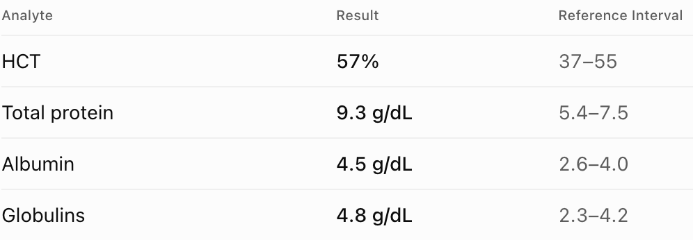
Case 1 — A Dog with Vomiting
A 5-year-old dog presents with vomiting, tacky mucous membranes, and decreased skin turgor.
QUESTION: What is the most likely cause of the increased total protein?
ANSWER → Dehydration (relative hyperproteinemia)
Albumin and globulins are both increased
HCT and physical examination support reduced plasma water volume

Case 2 — A Cat with Chronic Inflammation
A cat has persistent fever and weight loss.
QUESTION: Which protein group explains the hyperproteinemia, and what mechanism is most likely?
ANSWER → Hyperglobulinemia due to increased immunoglobulin production, consistent with chronic inflammation
Albumin is mildly decreased, which can occur because albumin is a negative acute phase protein

Case 3 — A Dog with Protein Loss
A dog has marked proteinuria, with no evidence of dehydration.
QUESTION: What is the most likely mechanism of hypoalbuminemia?
ANSWER → Renal protein loss through the glomeruli
Albumin is markedly decreased while globulins remain within reference limits, supporting selective albumin loss

Case 4 — A Dog with Chronic Diarrhea
A dog has chronic diarrhea and weight loss.
QUESTION: What is the most likely cause of the hypoproteinemia?
ANSWER → Protein-losing enteropathy
Both albumin and globulins are decreased (panhypoproteinemia), consistent with gastrointestinal protein loss

Case 5 — A Horse with Fever
An adult horse has fever, nasal discharge, and inflammatory changes on its CBC.
QUESTION: Calculate the protein:fibrinogen ratio. Does it support inflammation or dehydration?
ANSWER → Since the ratio is below 15, it supports inflammation
1000 mg/dL = 1.0 g/dL
Ratio = 9.0 / 1.0 = 9.0
Case 6 — A Dehydrated Horse
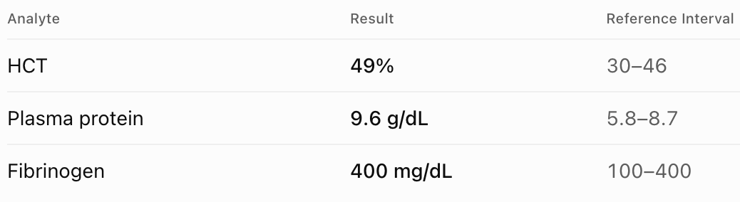
A horse has colic, decreased skin turgor, and dry mucous membranes.
QUESTION: Calculate the protein:fibrinogen ratio and identify the likely mechanism.
ANSWER → The ratio is at least 15, and the increased HCT and dehydration signs support a relative increase due to dehydration
400 mg/dL = 0.4 g/dL
Ratio = 9.6 / 0.4 = 24

Case 7 — A Dog with a Plasma Cell Tumor
A dog has marked hyperglobulinemia. Serum protein electrophoresis shows a tall, narrow gamma peak.
QUESTION: What type of gammopathy is present, and which disease category is strongly suspected?
ANSWER → Monoclonal gammopathy, raising concern for lymphoid neoplasia such as plasma cell myeloma
Certain infections can also produce a monoclonal pattern, so the finding alone is not diagnostic

Case 8 — A Dog After Fluid Therapy
A hospitalized dog receives excessive IV fluids. There is no evidence of hemorrhage or gastrointestinal protein loss.
QUESTION: What is the most likely reason for the low total protein and HCT?
ANSWER → Overhydration causing hemodilution
The plasma water volume increases, making protein and red cell concentrations appear lower without necessarily reducing their total mass
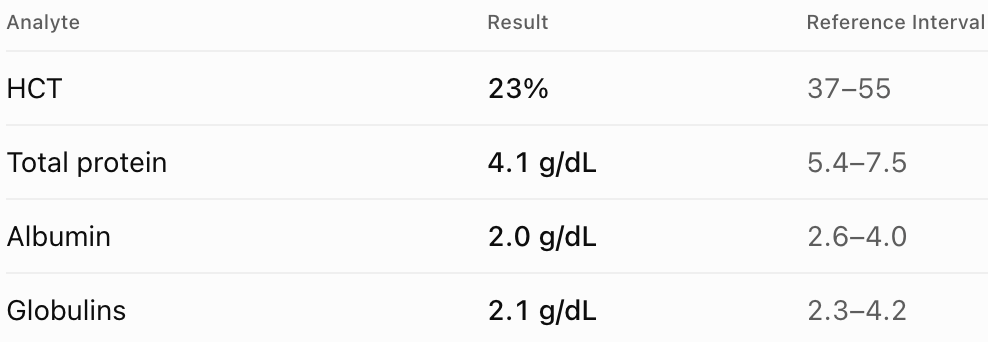
Case 9 — A Dog with Acute Blood Loss
A dog presents after trauma with weakness and evidence of internal bleeding.
QUESTION: What is the most likely explanation for the combined abnormalities?
ANSWER → Hemorrhagic blood loss
Decreased HCT plus decreased albumin and globulins indicates loss of red cells and plasma proteins
The timing and fluid shifts affect when these changes become measurable.

Case 10 — A Neonatal Foal
A newborn foal did not nurse adequately after birth. Its serum total protein and immunoglobulin concentrations remain low.
QUESTION: What mechanism best explains these findings, and why is age important?
ANSWER → Failure of passive transfer due to inadequate colostrum intake
Neonates normally acquire maternal IgG through colostrum, causing an early increase in immunoglobulin concentration
Age-specific reference intervals are necessary
Table 1. Major Plasma Proteins — Synthesis & Function
Protein | Site of Synthesis | Main Function |
Albumin | Hepatocytes | Maintains oncotic pressure; transports bilirubin and other substances |
Gamma globulins (Ig) | Plasma cells | Antibodies; adaptive immunity |
Alpha & beta globulins | Primarily hepatocytes | Acute phase response, transport, complement, clotting |
Fibrinogen | Hepatocytes | Coagulation; positive acute phase protein |
Table 2. Plasma vs. Serum & Laboratory Methods
Feature | Plasma | Serum |
Collection | Anticoagulated blood (EDTA) | Clotted blood (no anticoagulant) |
Tube | Purple-top | Red-top |
Fibrinogen / clotting factors | Present | Consumed during clotting |
Total protein measurement | Refractometry | Biuret method |
Albumin measurement | Not routinely measured by refractometry | Bromocresol green |
Additional measurements | Fibrinogen, protein:fibrinogen ratio | Calculated globulins, A/G ratio |
Important: Plasma total protein is usually slightly higher than serum total protein because plasma contains fibrinogen and clotting factors
Avoid switching between plasma and serum measurements when monitoring trends
Table 3. Causes of Increased vs. Decreased Plasma Proteins
Protein | Increased (Hyper-) | Decreased (Hypo-) |
Total protein | Dehydration, increased globulin production | Overhydration, hemorrhage, protein loss |
Albumin | Dehydration (relative) | Decreased synthesis, renal/GI loss, hemorrhage, inflammation |
Globulins | Dehydration, chronic inflammation, lymphoid neoplasia | Immunodeficiency, protein loss, failure of passive transfer |
Gamma globulins | Chronic antigenic stimulation, lymphoid neoplasia | Immunodeficiency, failure of passive transfer |
Fibrinogen | Acute/chronic inflammation, dehydration (relative) | Consumption or impaired production |
Key distinction:
Relative changes = Change in plasma water volume.
Absolute changes = Change in actual protein mass.
Table 3A. Causes of Increased Protein Concentrations (Hyperproteinemia)
Protein | Mechanism | Causes |
Total protein ↑ | Relative increase | Dehydration |
True increase | Increased globulin production | |
Albumin ↑ | Relative increase ONLY | Dehydration |
Globulins ↑ | Relative increase | Dehydration |
Increased production | Chronic inflammation, lymphoid neoplasia | |
Gamma globulins ↑ | Increased antibody production | Chronic inflammation (polyclonal), lymphoid neoplasia (monoclonal) |
Alpha & beta globulins ↑ | Increased production | Acute phase response |
Fibrinogen ↑ | Increased production | Acute or chronic inflammation |
Remember: Hyperalbuminemia is caused by dehydration, NOT increased albumin synthesis.
Table 3B. Causes of Decreased Protein Concentrations (Hypoproteinemia)
Protein | Mechanism | Causes |
Total protein ↓ | Relative decrease | Overhydration |
True decrease | Decreased albumin and/or globulin concentrations | |
Albumin ↓ | Decreased synthesis | Hepatic insufficiency, acute phase response, compensatory response to hyperglobulinemia |
Increased loss | Hemorrhage, renal disease, gastrointestinal disease, exudates | |
Decreased intake / increased utilization | Malnutrition, cachexia | |
Globulins ↓ | Decreased synthesis | Immunodeficiency, hepatic insufficiency |
Increased loss | Hemorrhage, gastrointestinal disease, exudates | |
Decreased intake | Failure of passive transfer in neonates |
Table 4. Laboratory Protein Patterns & Clinical Interpretation
Laboratory Pattern | Most Likely Interpretation |
↑ Albumin + ↑ Globulins + ↑ HCT | Dehydration |
↑ Albumin + normal globulins | Dehydration |
↑ Globulins + normal/↓ albumin | Chronic inflammation, infection, or lymphoid neoplasia |
↑ Gamma globulins + broad SPE peak | Polyclonal gammopathy; commonly chronic inflammation |
↑ Gamma globulins + narrow SPE peak | Monoclonal gammopathy; commonly lymphoid neoplasia, occasionally infection |
↑ Fibrinogen + protein:fibrinogen ratio <15 | Inflammation |
↑ Total plasma protein + ratio ≥15 | Proportionate increase; supports dehydration |
↓ Albumin + ↓ Globulins + ↓ HCT | Hemorrhage |
↓ Albumin + normal/↑ Globulins | Renal protein loss, decreased synthesis, or inflammation |
↓ Albumin + ↓ Globulins | Hemorrhage, GI protein loss, or dilution |
Exam tip: Always interpret protein values together with HCT, clinical signs, and the individual albumin and globulin concentrations. Total protein alone cannot identify the underlying mechanism.
Table 5. Acute Phase Proteins & Serum Protein Electrophoresis
Protein / Pattern | Change | Clinical Significance |
Positive acute phase proteins | ↑ Fibrinogen, CRP, SAA, haptoglobin, etc. | Acute or chronic inflammation |
Negative acute phase proteins | ↓Albumin, transferrin | Inflammatory response |
Alpha / beta SPE fractions | ↑ | Increased acute phase protein production |
Broad gamma peak | ↑ | Polyclonal gammopathy; commonly chronic inflammation |
Narrow gamma peak | ↑ | Monoclonal gammopathy; commonly lymphoid neoplasia, but some infections |
Protein:Fibrinogen Ratio — Quick Reference

Ratio | Interpretation |
≥ 15 | Proportionate increase; supports dehydration |
<15 | Disproportionate fibrinogen increase; supports inflammation |
This ratio is particularly useful in large animals.
The ONLY Table I Want You to Memorize
Disease / Condition | Albumin | Globulins | WHY? |
Dehydration | ↑ | ↑ | Less water |
Chronic inflammation | ↓ or normal | ↑ | More antibodies |
Lymphoid neoplasia | ↓ or normal | ↑ | Abnormal immunoglobulin production |
Hemorrhage | ↓ | ↓ | Losing blood and proteins |
Kidney disease | ↓ | Normal | Albumin lost in urine |
GI disease | ↓ | ↓ | Proteins lost through intestines |
Check total protein: Is it increased or decreased?
Check albumin and globulins: Which one is causing the change?
Check HCT and clinical signs: Does dehydration, dilution, or hemorrhage fit?
Identify the mechanism: Water change, increased/decreased production, or protein loss.
For large animals: If fibrinogen is provided, calculate the protein:fibrinogen ratio.