Lecture 6 - Cardiac Imaging 1
 Call Kai
 Learn
Learn Practice Test
 Spaced Repetition
 Match
Match Flashcards
 Knowt Play
1/100

There's no tags or description
Looks like no tags are added yet.
Name | Mastery | Learn | Test | Matching | Spaced | Call with Kai | Chat |
|---|
No analytics yet
Send a link to your students to track their progress
101 Terms
What is the leading cause of death worldwide
Cardiovascular disease
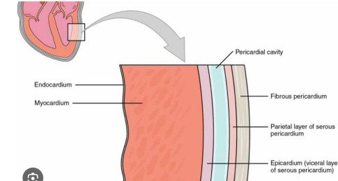
What’re the layers of the heart
Pericardium → Epicardium → Myocardium → Endocardium
Role of pericardium
Protective
Fluid containing sac around heart
Role of epicardium
Outer surface of the heart
Role of myocardium
Muscular layer
Responsible for contraction
Role of endocardium
Smooth inner lining of the chambers and valves
Ensures blood an flow through heart without friction
Name the atrioventricular valves
Tricuspid
Mitral
Name the semilunar valves
Pulmonary
Aortic
Flow of mitral valve and what phase of cardiac cycle
LA → LV
Diastole
Flow of tricuspid valve and what phase of cardiac cycle
RA → RV
Diastole
Flow of pulmonary valve and what phase of cardiac cycle
RV → pulmonary trunk
Systole
Flow of aortic valve and what phase of cardiac cycle
LV → aorta
Systole
What is the difference between diastole and systole
Diastole: Heart muscles relax and and ventricles fill with blood
Systole: Heart muscles contract and pump blood to body
Explain why there is a difference in pressure between the RV and LV
LV pumps blood into the high resistance systemic circulation
RV pumps blood into the low resistance pulmonary circulation
Both hearts pump the same amount of blood
Describe the pulmonary circulation
RV → pulmonary trunk → pulmonary arteries → lungs → pulmonary veins → LA
Low pressure
What kind of blood do the pulmonary arteries carry
Deoxygenated
What kind of blood do the pulmonary veins carry
Oxygenated
Describe the systemic circulation
LA → mitral valve → LV → aorta → systemic arteries → tissues → veins → IVC/IVC → RA
High pressure
What is cardiac hypertrophy
Abnormal thickening of cardiac walls

What is the cardiac output formula
CO = HR x SV
CO: cardiac output (mL/min)
HR: heart rate (beats/min)
SV: stroke volume (mL/beat)
What is the stroke volume formula
SV = EDV - ESV
SV: stroke volume
EDV: end diastolic volume
ESV: end systolic volume
What is the typical resting adult CO
5-6 L/min
What’re the branches of the aorta
Brachiocephalic trunk
Left common carotid
Left subclavian artery
Where do the pulmonary veins attach to the heart
Posterior aspect of the LA
Which vessel carries the least amount of oxygen
SVC/IVC
Which vessel carries the most amount of oxygen
Pulmonary veins
What is a CTPA and what is it used for
Pulmonary angiogram
Uses IV contrast to display the vessels supplying the lungs
Done to diagnose PE
What is CCTA and what is it used for
Coronary CT angiogram
To see if the coronary lumens patent
Where is the trigger ROI placed for a CTPA
Pulmonary trunk
Where is the trigger ROI placed for a CCTA
Ascending aorta

Name the vessel
Right coronary artery (RCA)

Name the vessel
Left circumflex artery (LCX)
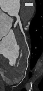
Name the vessel
Left anterior descending (LAD)
CTPA or CCTA contrast timing earlier
CTPA
Contrast reaches lungs and pulmonary vessels before coronary arteries
Where does the coronary tree lie in relation to the aortic valve
Superior
What plane is used to image coronary vessels
Epicardial fat
Origin of right coronary artery
Right aortic sinus
What does the RCA supply
RA
RV
SA node
AV node
Origin of left coronary artery
Left aortic sinus
Branches of the left coronary artery
Left anterior descending
Left circumflex
What does the LAD artery supply
Anterior LV
What does the LCX artery supply
Lateral LV
Why does CCTA need high resolution scanning
Coronary arteries are only 2-5mm in diamter
Why is diastole preferred for cardiac imaging
Ventricles are full
AV valves open
Semilunar valves closed

What does the P wave represent
Atrial depolarisation
When the atria are contracting to push blood into the ventricles

What does the QRS complex represent
Ventricular repolarization
R wave is used as the trigger for ECG gated imaging
Ventricles contract to eject blood into the pulmonary trunk and aorta

What does the T wave represent
Ventricular repolarisation
Ventricles relax and prepare to fill with blood
Describe the route of electrical signal in the heart
SA node → AV node → bundle of His → Purkinje fibers
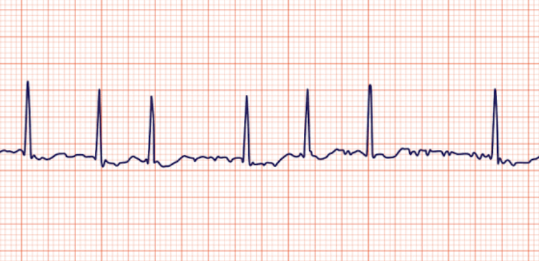
Normal or nah
No P wave
Atrial fibrillation

Normal or nah
Ventricular fibrillation
Chaotic and irregular
What is eh ideal HR for CCTA
60 bpm
What is atherosclerosis
Inflammatory disease that causes build up of fat to block the lungs
Describe the inflammatory process of atherosclerosis
Endothelial dysfunction (inner layer of arteries)
↓
LDL (low density lipoprotein) enters vessel intima
↓
LDL oxidises
↓
Monocytes enter
↓
Inflammatory mediators released
↓
Plaque grows
↓
Fibrous cap weakens
↓
Plaque rupture
↓
Thrombus
↓
Myocardial infarction
What’re the AHA plaque stages
1-2: Adaptive intimal thickening fatty streak, below CCTA spatial resolution
3: Pathological intimal thickening
4: Atheroma with necrotic lipid core (can be seen on CCTA)
5: Fibroatheroma (calcified)
What is positive remodeling
Outward growing of plaque
Vessel expands as plaque grows outwards, away from lumen
High risk feature

What is negative remodeling
Inward growing of plaque
Vessel shrinks inwards

What're the high risk plaque features on CCTA
Low attenuation plaque (lipid rich core)
Spotty calcium (small calcifications)
Positive remodeling
Napkin ring sign (high attenuation rim with low attenuation core)
If plaque shows up very bright on CCTA what kind of plaque is this
Calcified

What is going on here
Nothing

What is going on here
Negative remodeling

What is going on here
Negative remodeling
Soft lipid plaque
What is the FAI
Fat attenuation intext
Evaluates the perivascular inflammation around the coronary arteries
How does inflammation change fat
Normal lipid rich fat has strong negative HU
Inflammation changes adjacent fat so it becomes more positive (moves towards 0)
-60 HU vs -90 HU, which is more inflamed
-60 HU due to the FAI
What is the high risk cut off for FAI
FAI ≥ −70.1 HU = high-risk perivascular inflammation (moving towards 0 = higher risk)
What is CAC
Coronary artery calcium scoring
measures atherosclerotic calcified burden not the degree of coronary narrowing
Describe prospective gating
Tube current only on during a chosen cardiac phase, mid-late diastole
Scanner waits for R wave

What’re the disadvantages of prospective gating
Slow HR
Regular rhythm required
Predictable RR interval
Describe retrospective ECG gating
Scanner acquires data throughout the cardiac cycle
ECG trace is recorded and images can later be reconstructed at different phases
What’re the advantages of using retrospective ECG gating
High HR
Irregular rhythm
Functional assessment
Reconstructing multiple phases
Explain what partial-volume averaging is and how it relates to slice thickness
Image artifact that happens when a single voxel contains a mix of tissue types
Having thicker slices leads to partial-volume averaging and can obscure pathology
What is the slice thickness for CCTA
0.5-0.75mm recons
What is the slice thickness for calcium scoring
2.5-3mm
Purpose of beta-blockers
Control HR/rhythm
Decreases HR
Regularizes rhythm
Lengthens diastole
Improves motion free imaging window
What is the role of GTN
Coronary vasodilation
Increases coronary vessel caliber
Improves lumen visualization (bigger coronary lumen)
Does not control HR
Process of CCTA
Preparation → HR target (tall R wave) → GTN → acquisition → contrast → recon
What’re the contraindications for GTN
Hypotensive
Server aortic stenosis
Recent PDE-5 inhibitor (Viagra)
What is the HU of calcium
≥130 HU
What’re the levels of Agatston scoring
0: No detectable calcium
1-99: Mild
100-299: Moderate
≥300–400: High burden
High calcium score ≠ proof of severe stenosis
What’re the post processing ways for CCTA
cMPR/CPR
Orthogonal MPR
MIP
Volume rendering
What is cMPR/CPR
Curved multiplanar/planar reconstruction
Follows the coronary centerline and unrolls the artery
Useful for seeing a long coronary segment
What is orthogonal MPR
Cross section perpendicular to vessel centerline
Can help measure lumen diameter, vessel wall, stenosis
What is stenosis
Narrowing of the lumen
What is MIP
Highlights highest attenuating structures
Useful for calcium
Contrast filled vessels
Can conceal soft plaque
What is volume rendering
Useful for overview of anatomy
Not used for stenosis grading
HU measurements
What is a myocardial perfusion used for
Evaluates contrast distribution through the myocardium
Uses stress agent (adenosine)
Anatomy and physiology
Why is cardiac MRI used
Non-ionizing radiation
issue characterization
Follow ups
Congenital heart disease
Gives good values for ejection rate, stroke volume, mass
When would you use CMR vs CCTA
CMR: muscle related clinical question
CCTA: coronary artery disease
What is the magnetohydrodynamic effect
The current of blood flow behaves like conductive fluid while interacting with the magnet
Interferes with the ECG signal and produces abnormal T wave
Scanner can confuse R with T wave

Cine on CMR = ?
Retrospective gating
What is steady state free precession (cine) CMR
Blood = bright
Myocardium = dark
Used for:
Wall motion
Ventricular volumes
Valve motion Jets
What is T1/T2 black blood CMR
Blood appears dark
T1:
Anatomy
Fat infiltration
T2:
Oedema
Myocardial infarction
What is late gadolinium enhancement used for
10-15min after GAD
Scar tissue
Infarction
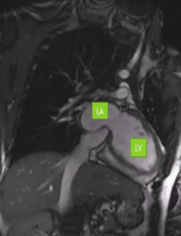
Describe the vertical long axis view
2 chamber view
LA and LV
LV → mitral valve → LA
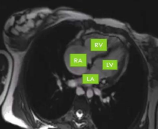
Describe the 4 chamber view (horizontal long axis view)
RA
RV
LA
LV
Mitral valve
Tricuspid valve
Describe the short axis view
Base → apex
Volume
Wall motion

Outflow tract views
LVOT: Aortic outflow
RVOT: Pulmonary outflow
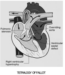
What is tetralogy fallot
Ventricular spetal defect
Overriding aorta
Pulmonary stenosis
RV hypertrophy
What is TGA
Transposition of the great arteries
When the aorta arises from RV
When the pulmonary artery arises from LV

What is ebstein anomaly
Abnormal tricuspid valve
Causes:
Apical displacement
Arterialization of part of the RV
Enlarged RA
