BIO202: Lab Practical #2
 Call Kai
 Learn
 Practice Test
 Spaced Repetition
 Match
 Flashcards
 Knowt Play
1/160

There's no tags or description
Looks like no tags are added yet.
161 Terms

Identify the indicated vessel.
right external jugular vein

Identify the indicated vessel.
right femoral vein

Identify the indicated vessel.
abdominal aorta

Identify the indicated vessel.
left popliteal artery

Identify the indicated vessel.
Right basilic vein
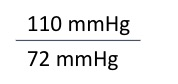
Using the blood pressure reading above, indicate if the blood pressure of the individual is normal or hypertension.
normal

Identify the indicated vessel.
inferior vena cava

Identify the specific vessel indicated at the pointer.
aorta

Identify the indicated vessel.
right internal thoracic artery

Identify the indicated vessel.
right axillary artery

Identify the indicated vessel.
right small saphenous vein
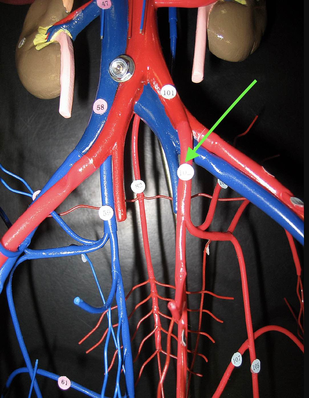
Identify the indicated vessel.
left internal iliac artery

Identify the indicated vessel.
right cephalic vein

Identify the indicated layer of the vessel wall.
tunica media

Identify the indicated vessel.
Left axillary artery
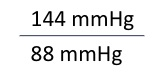
Using the blood pressure reading above, indicate which value represents the patient's systolic blood pressure?
144 mmHg

Identify the indicated vessel.
superior vena cava

Identify the indicated layer of the vessel wall.
tunica intima

Identify the indicated vessel.
thoracic aorta
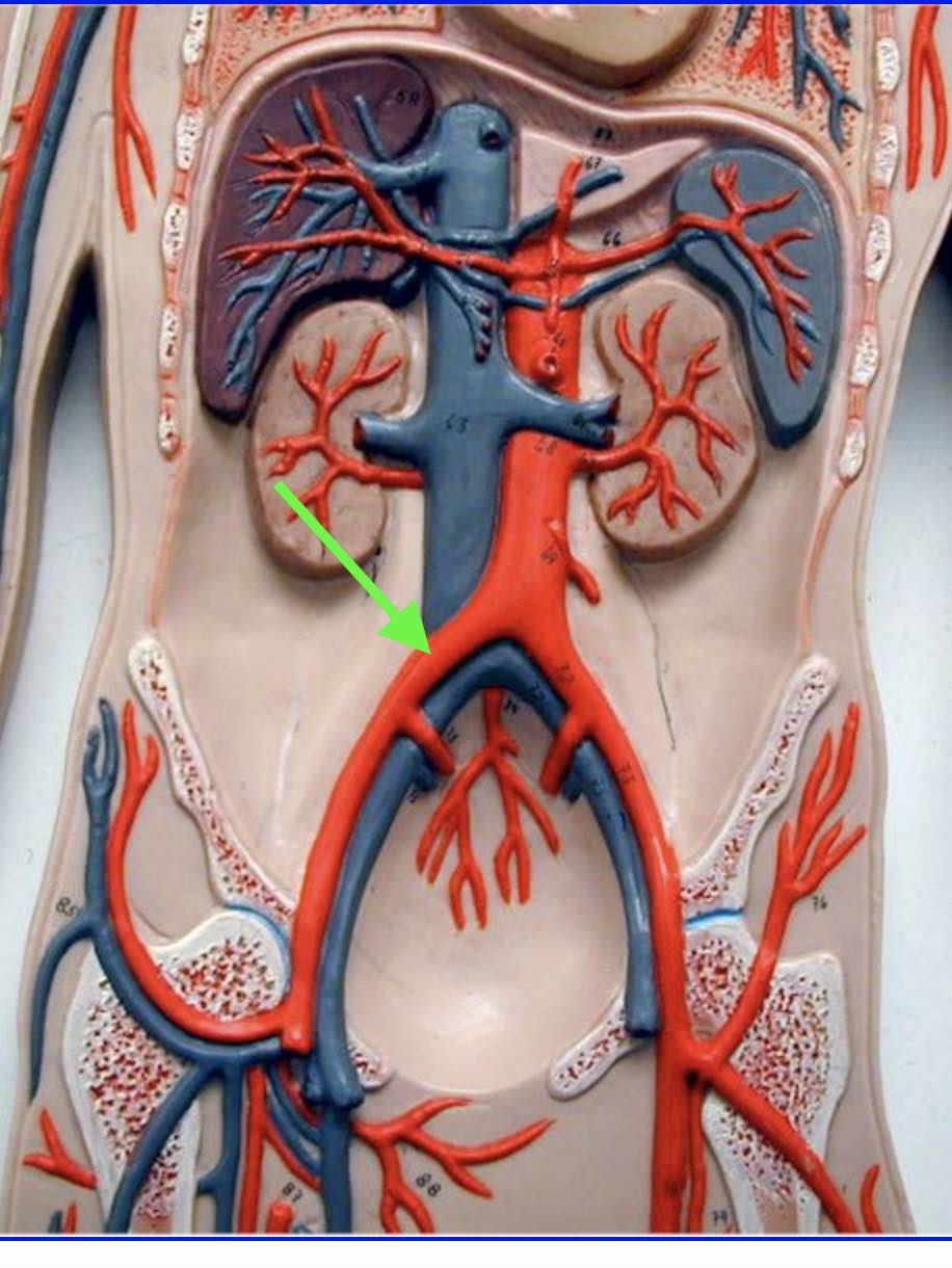
Identify the indicated vessel.
right common iliac artery

Identify the indicated vessel.
left anterior tibial artery

Identify the indicated vessel.
Left brachial artery

Using the blood pressure reading above, indicate if the blood pressure of the individual is normal or hypertension.
hypertension

Identify the indicated vessel.
right median cubital vein

Identify the indicated vessel.
left brachiocephalic vein

Identify the indicated vessel.
left common carotid artery

Identify the indicated vessel.
right internal iliac vein
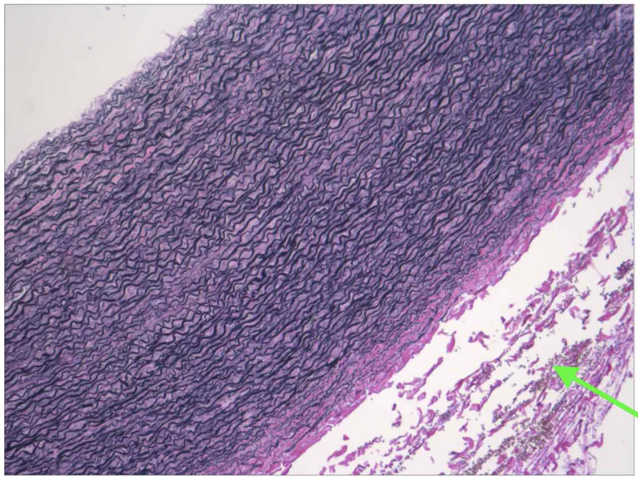
Identify the indicated layer of the vessel wall.
tunica externa
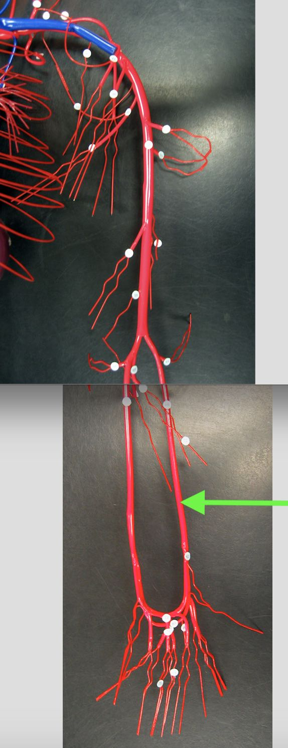
Identify the indicated vessel.
Left radial artery

Identify the indicated vessel.
right great saphenous vein

Identify the indicated vessel.
left common iliac vein
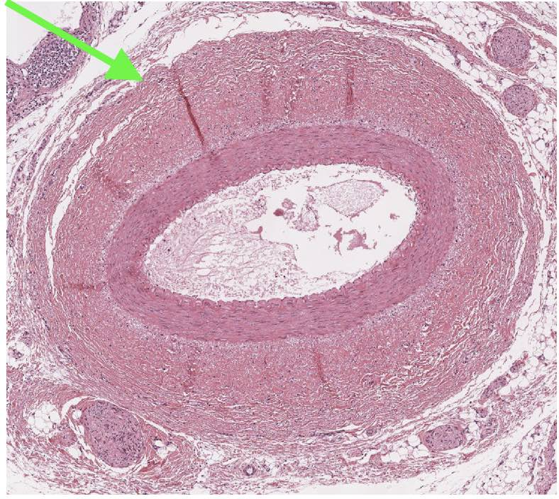
Identify the type of vessel indicated at the pointer.
artery

Identify the indicated vessel.
Left ulnar artery

Identify the indicated vessel.
right subclavian vein

Identify the indicated vessel.
left peroneal artery

Identify the indicated vessel.
right common iliac vein
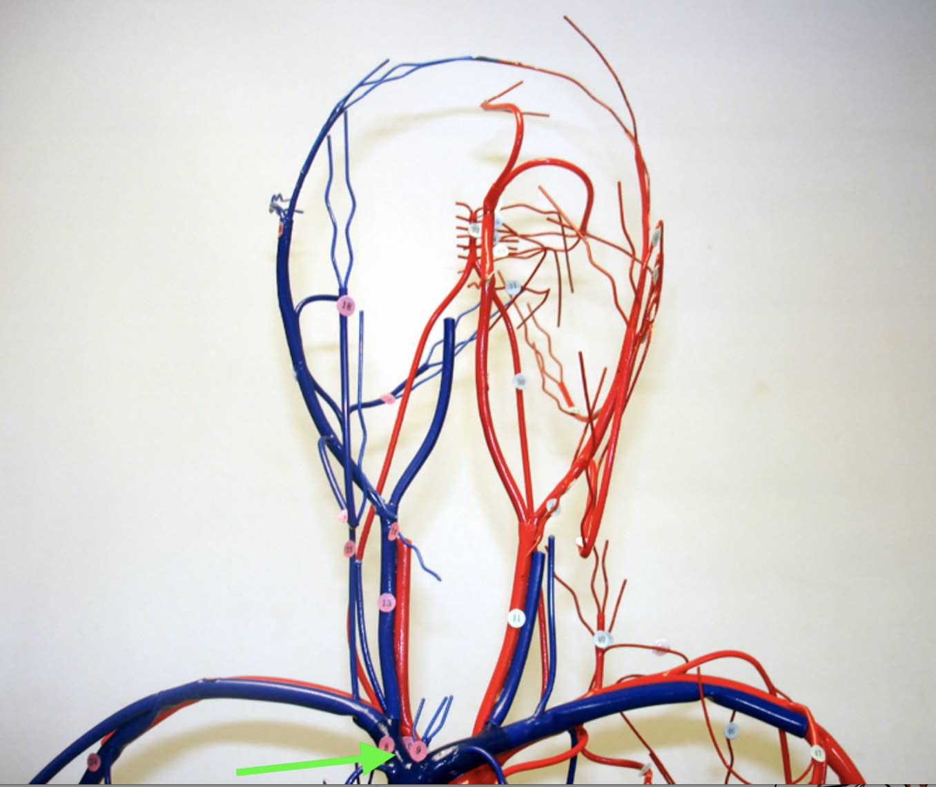
Identify the indicated vessel.
right brachiocephalic vein

Identify the structure(s) indicated at the pointer.
elastic fibers
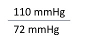
Using the blood pressure reading above, indicate which value represents the patient's diastolic blood pressure?
72 mmHg

Identify the indicated vessel.
right external iliac vein

Identify the indicated vessel.
left internal jugular vein
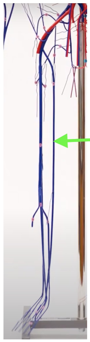
Identify the indicated vessel.
right great saphenous vein
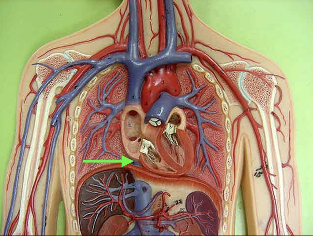
Identify the indicated vessel.
inferior vena cava

Identify the indicated layer of the vessel wall.
tunica media
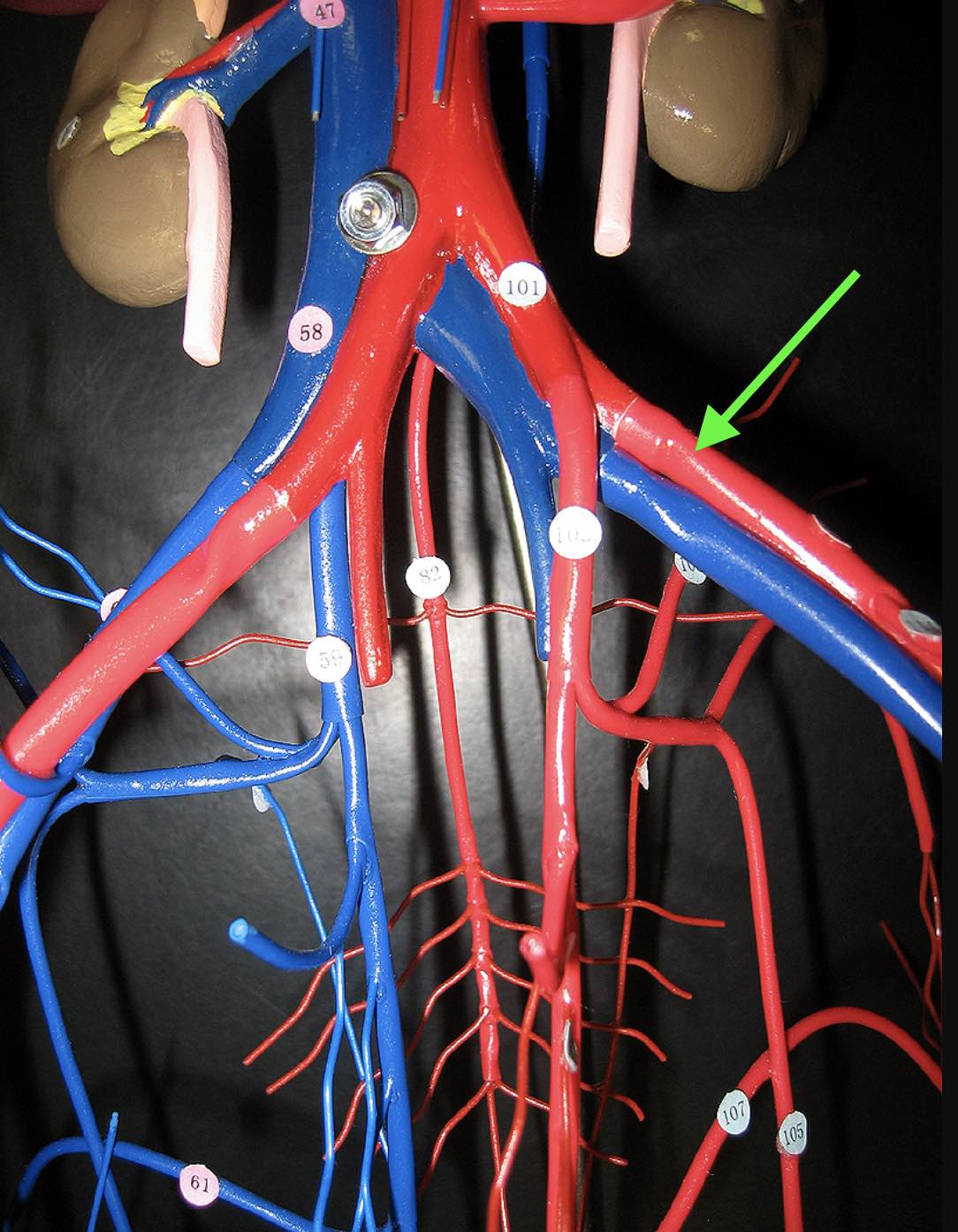
Identify the indicated vessel.
Left external iliac artery

Identify the indicated layer of the vessel wall.
tunica externa

Identify the indicated vessel.
left internal iliac vein

Identify the indicated vessel.
left subclavian vein
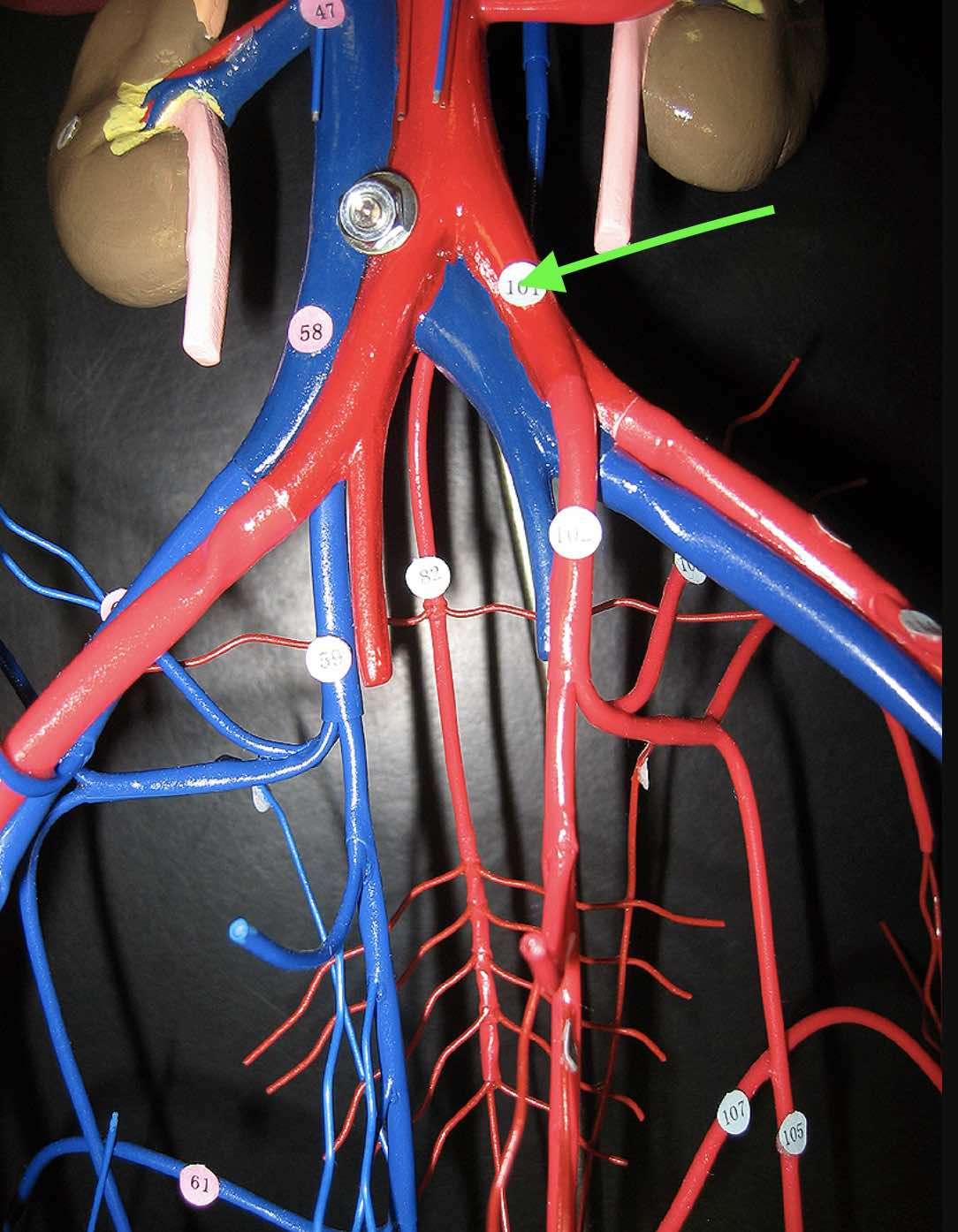
Identify the indicated vessel.
left common iliac artery

Identify the indicated vessel.
right internal iliac artery
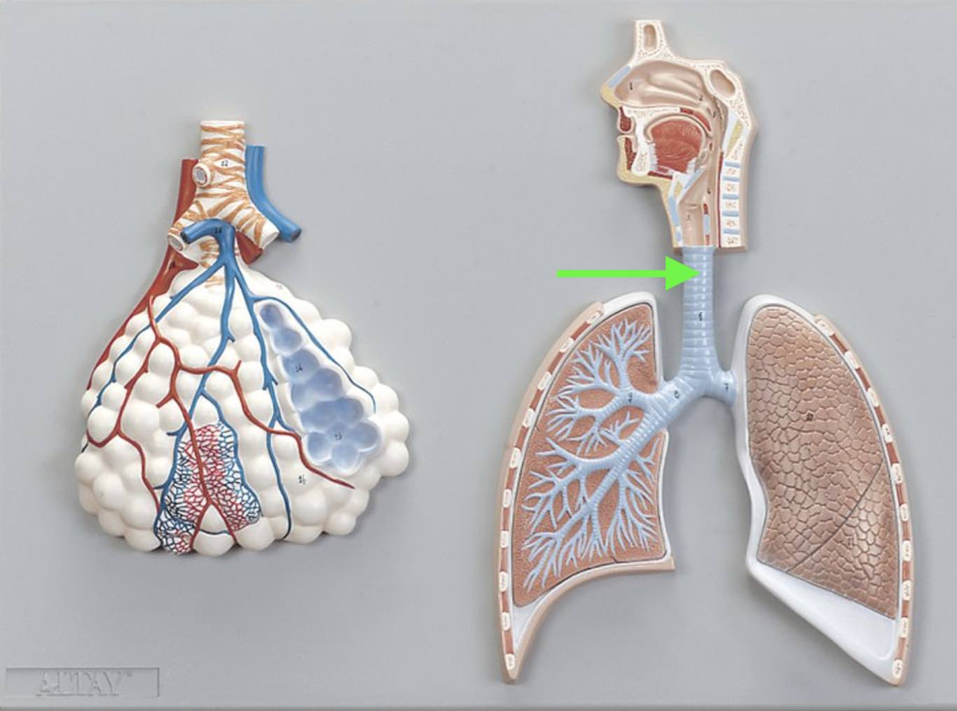
Identify the indicated structure.
trachea

Identify the indicated structure.
alveolus

Identify the disorder indicated in the image.
emphysema

Identify the indicated muscle.
diaphragm

Identify the indicated lung volume.
tidal volume

Identify the indicated structure.
lingual tonsils

Identify the indicated structure.
nasopharynx

Identify the indicated structure.
epiglottis
Identify the lung volume defined as "the volume of air remaining in the lungs after maximal exhalation."
residual volume

Identify the indicated muscle.
diaphragm
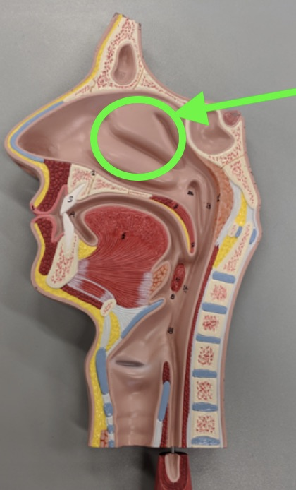
Identify the indicated structure.
nasal cavity

Identify the indicated structure.
left primary bronchus
Identify the lung volume defined as "the volume in the lungs at maximal inflation."
total lung capacity

Identify the indicated structure.
soft palate

Identify the indicated muscle.
external intercostal muscles

Identify the indicated structure.
thyroid cartilage

Identify the indicated lung volume.
total lung capacity

Identify the indicated structure.
parietal pleura
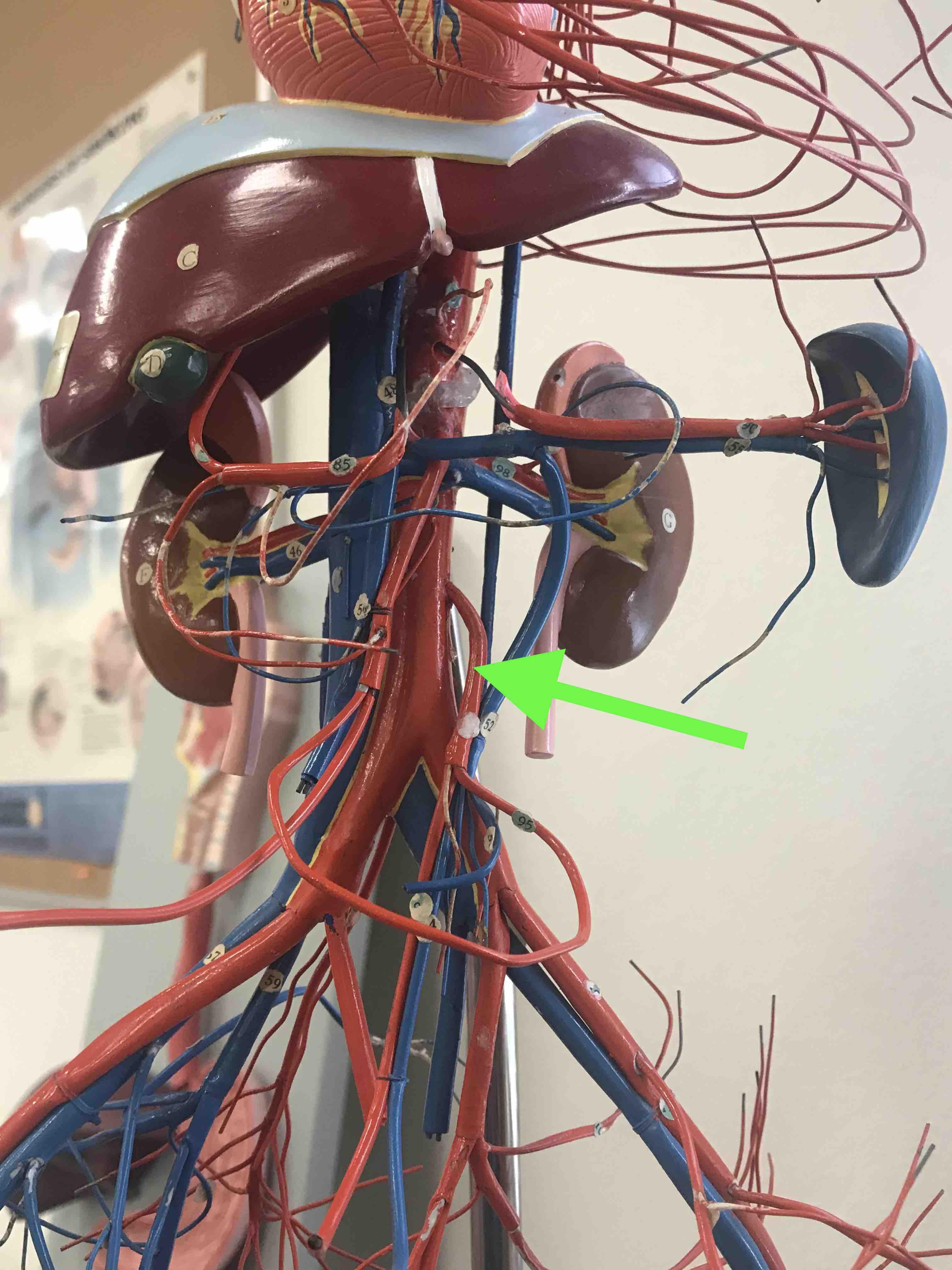
Identify the indicated vessel.
inferior mesenteric artery

Identify the indicated vessel.
splenic vein

Identify the indicated organ.
liver
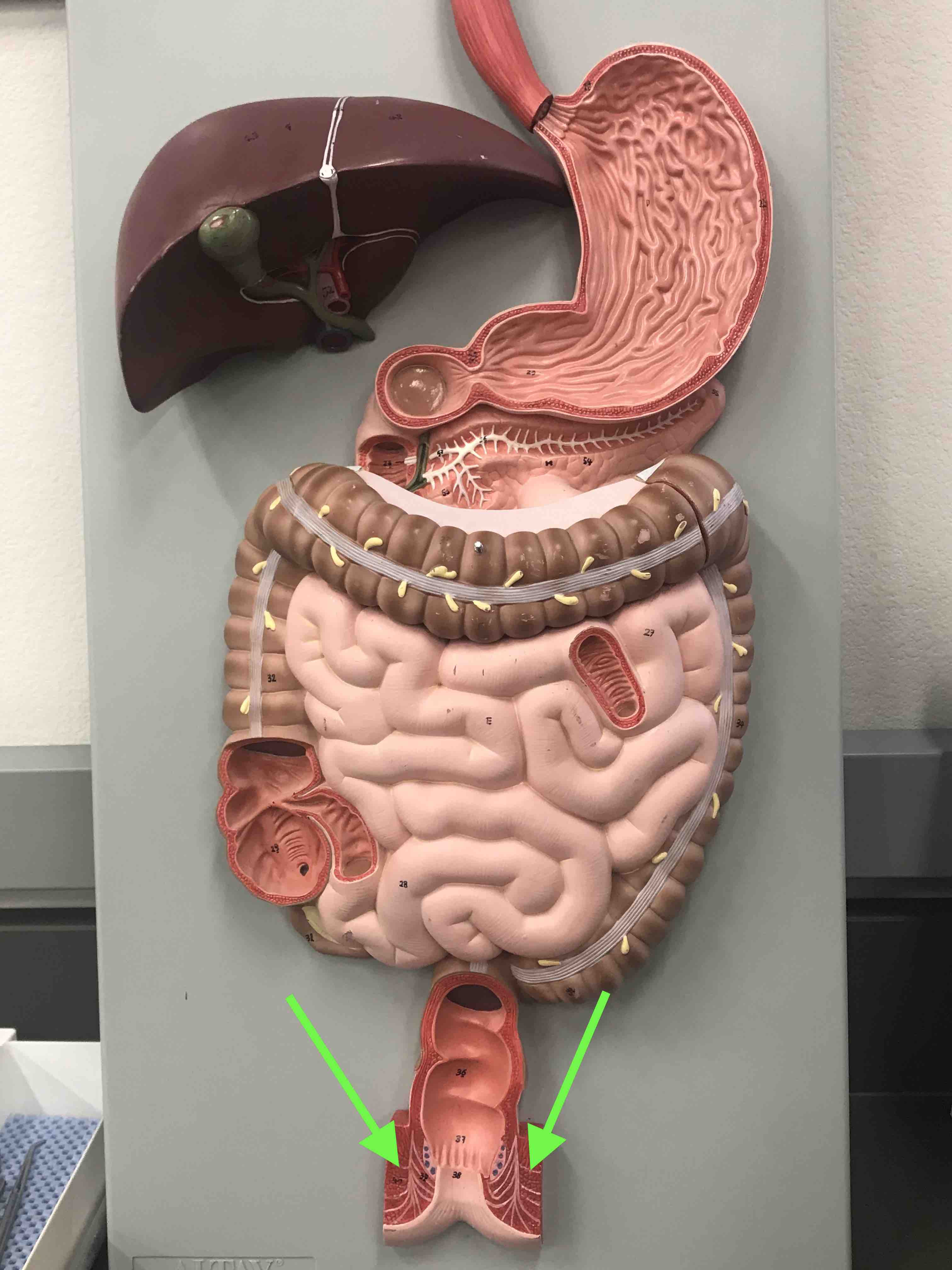
Identify the indicated structure.
external anal sphincter

Identify the indicated structures.
salivary glands

Identify the indicated organ.
liver

Identify the indicated region.
duodenum

Identify the indicated cells you are viewing at the pointer.
acini cells

Identify the indicated cells you are viewing at the pointer.
goblet cells

Identify the indicated structure.
pyloric sphincter

Identify the indicated organ.
stomach

Identify the organ you are viewing on the slide.
large intestine

Identify the indicated organ.
gall bladder

Identify the indicated structure.
ileocecal sphincter

Identify the indicated structure.
submandibular glands

Identify the indicated vessel.
hepatic portal vein

Identify the indicated vessel.
splenic artery

Identify the indicated structure.
alveolus

Identify the indicated structure.
bronchioles

Identify the indicated structure.
diaphragm

Identify the indicated region.
laryngopharynx

Identify the indicated organ.
small intestine
What lung volume is defined as the total volume of air moved into or out of the lungs during quiet breathing?
tidal volume

Identify the indicated structure.
cardia

Identify the indicated lung volume.
expiratory reserve volume

Identify the indicated structure.
transverse colon

Identify the indicated organ.
tongue

Identify the indicated structure.
quadrate lobe
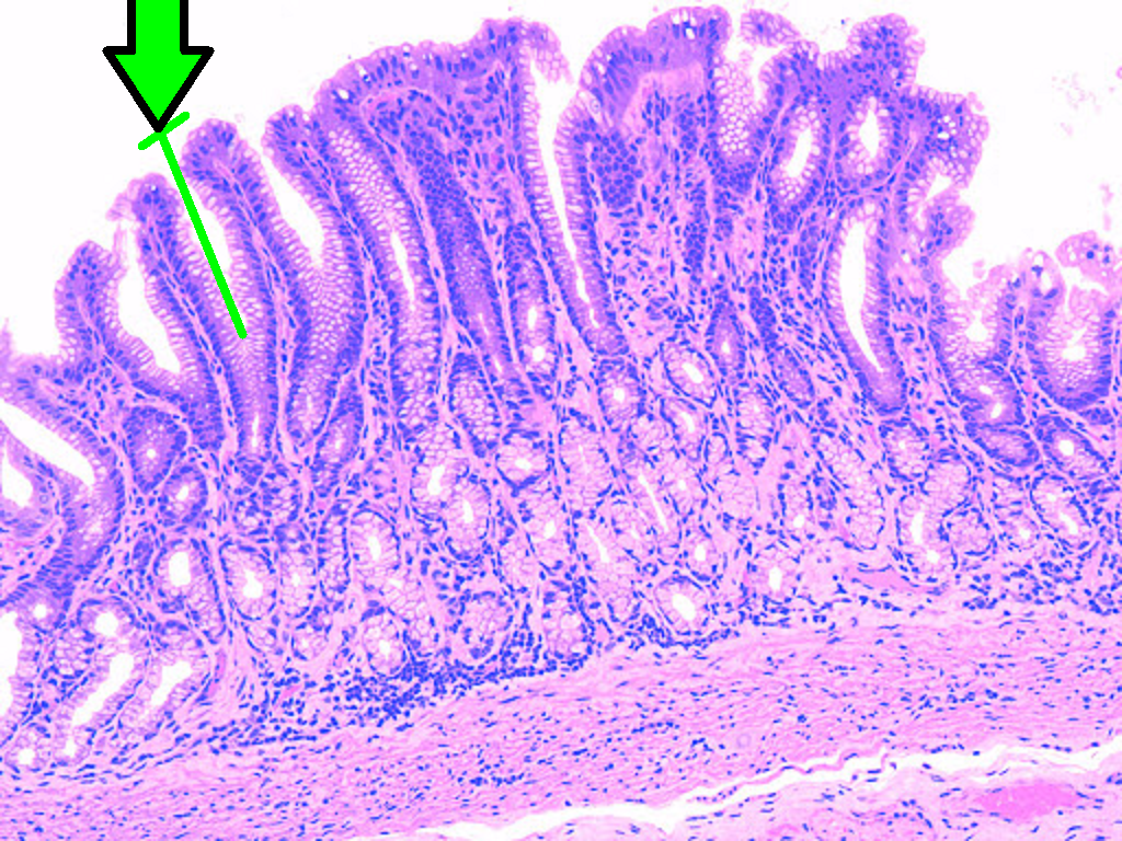
Identify the indicated structure.
gastric pit
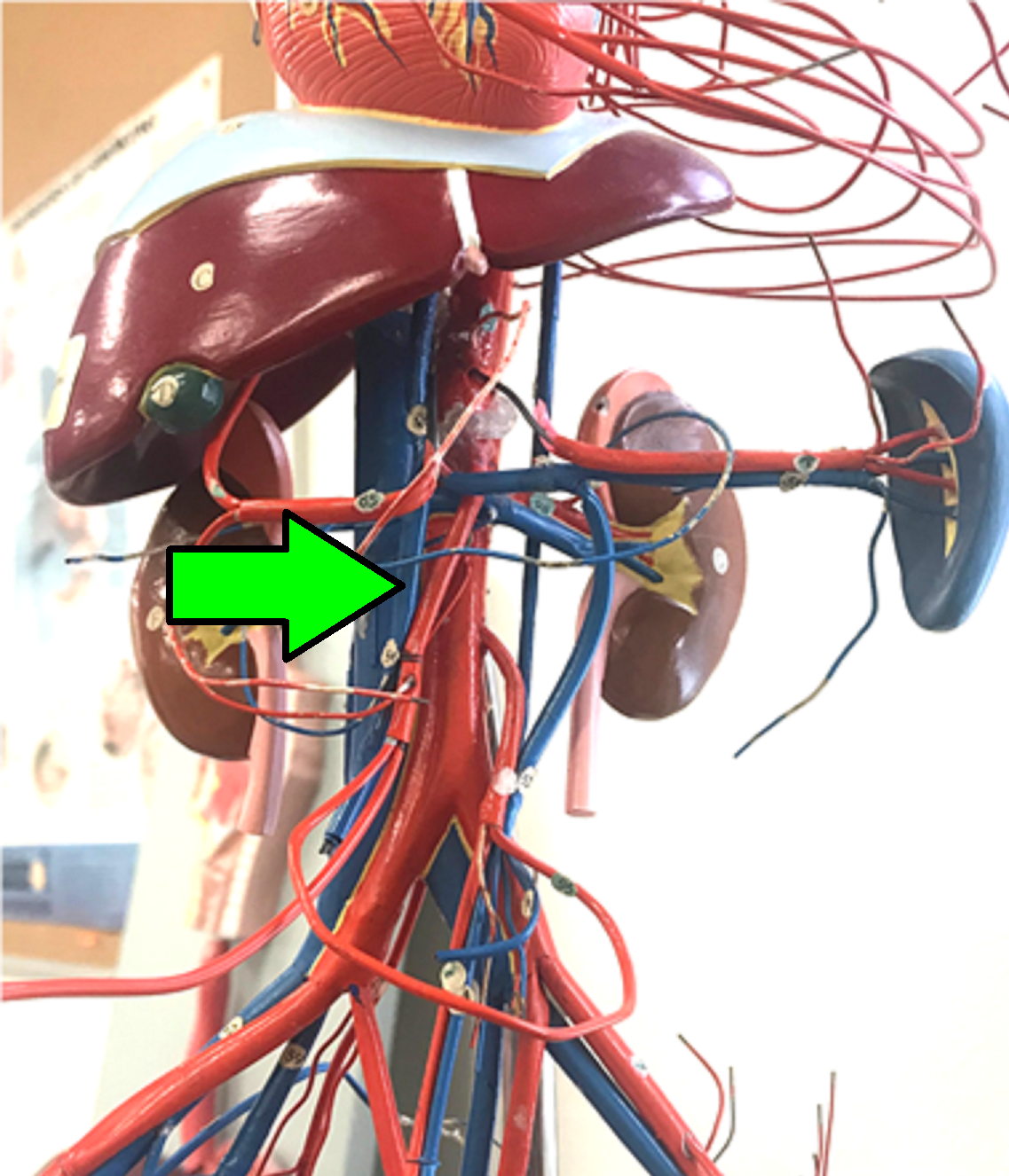
Identify the indicated structure.
superior mesenteric vein

Identify the indicated vessel.
right popliteal vein

Identify the indicated layer of the vessel wall.
tunica intima