electrophoresis
 Call Kai
 Learn
 Practice Test
 Spaced Repetition
 Match
 Flashcards
 Knowt Play
1/91

There's no tags or description
Looks like no tags are added yet.
92 Terms
what is electrohporesis
technique used for separation and purification of ions, proteins, and other molcules
what is electrophoresis most commonly used for in the clinical laboratory
to separate the serum proteins, isoenzymes, lipoproteins, and hemoglboins in various body fluids
what does electrophoresis involves in the movement of
charged particles when an external electrical current is produced in liquid evironment
how is the electric field applied to solution
oppositely charged electrodes place in the solution.
cations moves towards
negatively charged electrode
anions move towards
positively charged electrodes
what are organic molecules?
amphoteric
charge in solution and magnitiude of charge can be altered by varying the pH
what is amphoteric
can be either positive or negative
what factors affect migration?
migration of charge
size of molecule
propertiesof support medium
strength of electric field
endosmosis
ionic strength of the buffer
temperature
migration factor:
migrationof charge
greater net charge = faster molecule moves
less net charge = slower molecule moves
migration factor:
size of molecule
large = slower
small = faster
migration factor:
properties of support medium
pores, filter the separtation not only by charge but by size
increase viscosity = slower migration
decrease viscosity = faster migration
migration factor:
strength of electric field
greater voltage applied = faster migration
migration factor:
enodomosis (limiting factor)
support medium will absorb some ion in buffer causing ion cloud
can slow stop or push migration backwards
migration factor:
ionic strength of buffer
lower buffer = faster migration
increase strength = slow migration
mirgraton factor:
temperature
increase temperature = faster migration
but too high of tempature can denature the protein
what are the components of electrophoresis
sample applicator
support medium
buffer system
electrophoresis chamber
electrodes
timer
power supply
stains
what does the buffer system do in components of electrophoresis
set the pH for migration to occur in
what are the functions of buffers?
fix and maintain a constant pH that determines the net charge on the molcules and controls the direction of migration
conduct current
what is the ideal pH for protein?
alkaline pH = protein takes a negative charge and migrates towards anode (positive charge)
8.6
what are the important consideration for buffers?
buffer must not reactwith sample
pH chosen should charge but not denature molecule
ionic strength of buffer must be considered
what does a slower separation result in?
result in higher resolution more distinct band separation
what are the 2 most commonly used buffers?
barbital buffer
tris-boric acid-EDTA buffers
what types of support media does electrophoresis use?
membrane or gel
when does endosmosis occur
when support medium becomes negatively charged b/c of the adsorption of hydroxyl ions from the buffer.
how does the ion cloud happen?
the negatively charged surface attracts positive ions from the buffer. ion cloud forms
depending on the strength of the charge on the molecule and the magnitude of the endomosis, the proteins can be slowed remain immobile or be pushed towards the cathode
forms friction and inhibitrs normal migration
what is wick flow?
the upward movement of buffer through both immersed ends of the gel membrane to replace lost moisture from heat generation.
what does the flow of wick flow add to?
adds distance to molcule moving towards the center of the membrane and slows molecules moving aways from the center.
what is the net result of wick flow?
results in a reduced separation and lead to compression of the final band patterns
not clean band. all glob together in the middle
what are the types of support medias?
agarose gel
cellulose acetate
polyacrylamide gel
starch gel
what is the most widely used support media?
agarose gel
what is agarose gel made up of?
composed of agrose and agaropectin
what is agarose gel free of and so what does not occur?
ionizable groups
so little endosmosis occurs
what does agarose gel give better results than cellulose acetate?
sharper results
what sample size will agarose gel accept?
will accept larger sample size
what is an advantage of agarose gel?
optically clear, used for densitometry immediately (measurement of band)
what is densitometry?
use to measure the amount of compound in each band
what needs to happen to media if it is cloudy or turbid?
needs a clearing step with a solvent before measuring bands
what is cellulose acetate composed of?
cellulose and acetic anhydride
how does cellulose acetate start as?
brittle, film of interlocking cellulose acetate fibers
what must cellulose acetate be done first before starting electrophoresis?
presoak in buffer to make membrane pliable
how are the bands in cellulose acetate?
protein absorption is minimal, and sharp, well-defined bands are obtained
what is the strength and pore size of cellulose acetate?
tends to be strong, and have uniform pore size
what is the color of cellulose acetate media?
opaque, so solvent must be used to clear media for densitometry, when solvent is used, it partially dissolves the cellulose acetate fibers leaving the stained fraction on the transparent background
what is polyacrylamide gel limited to?
limited to separation of alkaline phosphatase isoenzymes
how is polyacrylamide gel formed?
by the reaction of “activated” acrylamide molecules that produce long polymer chains.
what does polyacrylamide gel chain require?
requires that the chains be cross-link with one another
what happens if the concentration of acrylamide is adjusted?
the pore size can be varied which determines the extent of cross-linkage
what happen when the pore size is large in polyacrylamide gel?
almost all serum protein will migrate unhindered, and separation will be according to charge
what happens when pore size is smaller in polyacrylamide gel?
moleculare sieving becomes a factor as larger molecules are slowed down and separation is not only by charge but by size
what is polyacrylamide gel considered as now?
carcinogen
what is starch gel composed of?
composed of granules containing amylose and amylopectin
when a concentrated suspension of the gransules is heated a viscous solution is formed and becomes a gel upon cooling
starch gel separates molecules based on what?
based on size and charge
why is starch gel not consistant?
due to the wide range of pore sizes, consistency is difficult to achieve
harder to obrain reproducible results
what stains are used in electrophoresis?
ponceau S
Coomassie Brilliant Blue
Oil Red O
Schiff’s reagent
SyBr
coupled rxn
what is the most common stain for electrophoresis?
Ponceau S
what medium is Ponceau S used for
cellulose acetate x protein separation
bright red
what media is coomassie brilliant blue used for?
polyacrylamide gel x alkaline phosphatase isoenzymes
bright blue
what is Oil Red O
is a fat stain, fatty acid will absorb it
red color
phospholipids, lipoproteins will absorb stain
what is Schiff’s reagent for?
lipoprotein and glycoproteins
what is SyBr
used for separation of DNA and RNA
common colors are gold but also green
how are coupled reactions used in electrophoresis
it generates a product that can be detected by fluorescence or by its color
amount of fluorescence or amount of color is proportional to how much in the band
what is the most common specimen for electrophoresis?
serum and plasma - most common
following urine and CSF
what other body fluids are used in electrophoresis?
pleura, pericardium, peritoneum, synovial fluid, and tears
what may a sample be required to before electrophoresis?
pretreatment depending on concentration of molecules to be separated
possible interfering substances and support medium used and information to be collected
insouble sample need to pretreat to make soluble
what pretreatment does fluids like urine and csf need?
small qty of protein, need to concentrate protein
what pretreatment does fluids like synovial fluid?
often vry thick so needs to be thinned
what pretreatment does fluids like insoluble or aggresgating molcule
need to make soluble
source of error:
problems with sample application
over fill well causes distorted band
not enough causes incomplete migration
source of error:
buffer stability
expired contaminated causes shorten migration
refigeration to decrease bacterial growth and increase shelf stability
source of error:
support media preparation and/or contamination
making own media = increase error avoid fingerprints before it can dry
presoak, put media in slowly so no air bubbles can distort band
wet media can cause distorted band
source of error:
stain issue
leaching, coming off the protein = stain is old and needs replacement
what are the 2 negative phase reactant?
albumin and Transferrin
where do most of our antibodies lie in?
gamma region (largest %)
where are IgA, IgM, and IgG located on?
small % on the right/far side of beta region

normal

increase gamma spike (IgM)
multiple myeloma
monoconal spike/gamopathy
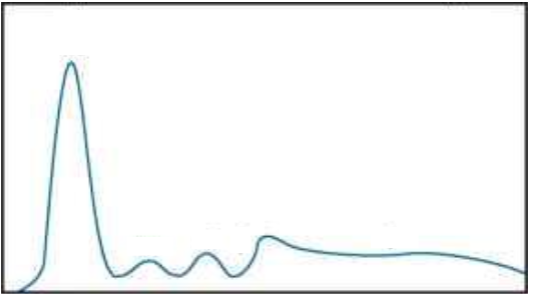
increased IgA
beta-gamma bridge
cirrous
CRP elevated located between beta and gamma region no separation

alpha 1 lower, alpha 2 higher, beta and gamma lower
nephrotic syndrome
alpha 2 have most positve acute phase reactents

elevation in alpha 1 and 2
inflammation
acute phase reaction or immediate phase reaction
recent infection because gamma not elevated (immune not involved yet)

immune reaction

on the right
reduced gamma region
immunocompromised, unable to produce antibodies
what are some examples of immunocompromised
treated for cancer, chemotherapy or drug that wipes out immune system
transplants, anti-rejection med
steroids lowers immune system
AIDS
what is the immunocompromised pattern also called?
hypogammaglobulinamia
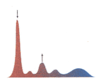
acute phase reactant
immedate reaction

immune response
rounded gamma

immunocompromised,
no gamma region

cirrous
beta gamma bridge

multiple myeloma
more spikey

nephrotic syndrome
decrease albumin and gamma
increase alpha 2
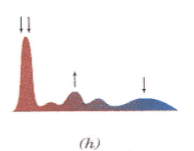
Protein-losing enteropathy - PLE
losing protein in feces
similar to nephrotic
how can you tell the difference between PLE and nephrotic syndrome on electrophoresis
nephrotic - inflammatory condition, higher alpha 2, lower gamma