Vet Med Term - Ch 7
 Call Kai
 Learn
 Practice Test
 Spaced Repetition
 Match
 Flashcards
 Knowt Play
1/146

There's no tags or description
Looks like no tags are added yet.
147 Terms
Functions of urinary system
Removes waste from the body by constantly filtering blood
Maintains proper balance of water, electrolytes, and acids in body fluids and removes excess fluids from body
Urea
Major waste product of protein metabolism
this is filtered by kidney and used in some diagnostic tests to determine kidney health
Homeostasis
Stable internal environment
involves continually adjusting to conditions to maintain a relatively constant internal environment
Home/o
Unchanging

Structures of urinary system
pair of kidneys
Pair of ureters
Urinary bladder
Urethra
Urine is
Formed in kidneys, flows through ureters to urinary bladder, and flow through urethra and outside body

Kidneys
Located Retroperitoneally
one sits on each side of vertebral column below diaphragm
Retroperitoneally
Located behind lining of abdominal cavity or outside peritoneal cavity
Ren/o, Nephr/o
Kidney

Each kidney has 2 layers that surround renal pelvis
Cortex
Medulla

Cortex
Outer layer of kidney
Cortic/o
Outer region

Medulla
Inner layer of kidney
Medull//o
Middle or inner portion

Nephron
Functional unit of kidney

Nephron consists of
glomerulus
Bowman’s capsule (glomerular capsule)
Proximal convoluted tubule
Loop of Henle
Distal convoluted tubule
Collecting duct
Nephron forms urine process by
filtration (glomerulus)
Reabsorption (proximal convoluted tubule, loop of Henle, collecting tube)
Secretion (distal convoluted tubule)
Glomerul/o
Wind into a ball
Glomeruli
Plural form of glomerulus

Glomerulus
Cluster of capillaries that filter blood

Bowman’s capsule (glomerular capsule)
Cup-shaped structure that contains the glomerulus
Renal capsule
The glomerulus and Bowman’s capsule together

Proximal convoluted tubules
Hollow tubes located between Bowman’s capsule and loops of Henle that are involved in reabsorption

Loop of Henle
U-shaped turn in the convolute tubules of the kidney located between the proximal and distal convoluted tubules that is involved in reabsorption; has ascending and descending loop

Distal convoluted tubules
Hollow tubes located between the loops of Henle and the collecting tubules that are involved in secretion
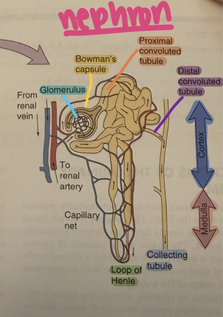
Collecting tubules
Hollow tubes that carry urine from the cortex to the renal pelvis
Renal pelvis
Area of kidney where nephrons collect urine before entering ureters
Pyel/o
Renal pelvis
Renal pelvis collects urine from
Calyces

Calyces (singular = calyx)
Irregular cup like shapes that collect urine from kidney

Hilus
Concave depression that serves as point of attachment of renal blood vessels, nerves, and ureter
located on medial surface of kidney
Gives some kidneys their bean shape

Ureters
Pair of narrow tubes that carry urine from kidneys to urinary bladder
Ureter/o
Ureter
Ureters enter urinary bladder at the
Trigone
Trigone
Triangular portion at base of organ where the 3 angles are marked by the 2 ureteral openings and one urethral opening

Urinary bladder
Singular hollow muscular organ that holds urine
elastic and size depends on amount of urine it’s holding
Cyst/o
Urinary bladder
Flow into urinary bladder
Enters from ureters at angle that serves as a natural value to control backflow
Flow out of urinary bladder
To urethra is controlled by sphincters
Sphincters
Ringlike muscles that close a passageway

Urethra
Tube extending from urinary bladder to outside of body
Urethr/o
Urethra
Urethral meatus (urinary meatus)
External opening of urethra
Meat/o
Opening
Urethra - females
Transports urine from urinary bladder to outside of body
Urethra - males
Transports urine from urinary bladder and repro fluids from repro organs out of body
Urination, excretion, voiding, elimination, micturition
Act of excreting urine
Erythropoietin
Hormone produced by the kidney that stimulates red blood cell production in the bone marrow
Antidiuretic hormone (ADH)
Hormone released by the posterior pituitary gland that suppresses urine formation by reabsorbing more water
Aldosterone
Hormone secreted by the adrenal cortex that regulated electrolyte balance via that reabsorption of sodium
Urine
End product of renal filtration of wastes
Uropoiesis
Process of urine production
-poiesis
Formation
Normal urine color for most species
Clear and pale yellow
-chrome
Color
Some species may have normal urine that is
turbid
Brown
White
Another color
Turbid
Cloudy
Sometimes urine color depends on
Diet or reproductive cycle
Urine color when dehydrated
More concentrated and deeper shade of yellow
Urine has a pH which depends on
Species and diet
Herbivores have a basic urine with
Higher pH
Carnivores have acidic urine with
Lower pH
Cystocentesis
Surgical puncture of urinary bladder, usually to collect urine
performed with needle and syringe
Cystography
Radiographic study of urinary bladder after contrast material has been placed in urinary bladder via a urethral catheter
contrast is water-soluble
Single contrast
When one contrast is used
Double contrast
When more than one contrast material is used
Cystogram
Radiographic film of urinary bladder after contrast has been placed via urethral catheter
Retrograde
Used to describe path that contrast takes (if it goes in reverse order of how urine normally flows)
Cystoscopy
Visual examination of urinary bladder using a fiberoptic instrument - cystoscope
Intravenous pyelogram (IVP)
Radiographic study of kidneys (especially renal pelvis) and ureters in which a dye is injected into a vein to define structures more clearly
Pneumocystography
Radiographic study of urinary bladder after air has been placed in bladder via a urethral catheter
Radiography
Imaging of internal structures that is created by exposing specialized film to x-ray
Scout film
Plain x-ray made without use of contrast
Retrograde pyelogram
Radiographic study of kidneys and ureters in which contrast is placed directly in urinary bladder
Albuminuria
Presence of the major blood protein in urine
Anuria
Complete suppression of urine production
Bacteriuria
Presence of bacteria in urine
Crystalluria
Urine with naturally produced angular solid of definitive form (crystals)
Dysuria
Difficult or painful urination
Glucosuria
Glucose (sugar) in urine
Glycosuria
Glucose (sugar) in urine
Hematuria
Blood in urine
Ketonuria
Presence of ketones in urine
ketones are produced during increased fat metabolism
Nocturia
Excessive urination at night
Oliguria
Scanty or little urine
Pollakiuria
Frequent urination
Polyuria
Excessive urination
Proteinuria
Presence of proteins in urine
Pyuria
Pus in urine
Stranguria
Slow or painful urination
Urinalysis (UA)
Examination of urine components
3 parts
1. Macroscopic analysis (gross exam)
2. Chemical analysis
3. Microscopic analysis (urine sediment exam)
Macroscopic analysis (gross exam)
Visual observation of urine in which things like color, transparency, odor, and foam are recorded
Chemical analysis
Applying urine to a reagent strip impregnated with chemicals that react with substances in urine and change colors
the colors are compared to a set of standards to determine whether or not there are abnormal levels
pH, leukocytes, erythrocytes, protein, glucose, specific gravity, etc
Microscopic analysis (urine sediment exam)
Detect presence of cellular material in urine that has not been centrifuged and is viewed under microscope
Urinary catheter (cath)
Insertion of tube through urethra into urinary bladder (usually to collect urine)
Catheter
Hollow tube that is inserted into a body aviary to inject or remove fluid
Azotemia
Presence of urea or other nitrogenous elements in blood
Azot/o
Urea or nitrogen
Calculus
Abnormal mineral deposit
urine may contain crystals that remain in solution in urine
Stones, calculi, and liths are formed when crystals precipitate and form solids
Lith/o
Stone or calculus