Genome Maintenance and Regulation
 Call Kai
 Learn
Learn Practice Test
 Spaced Repetition
 Match
Match Flashcards
 Knowt Play
1/99

There's no tags or description
Looks like no tags are added yet.
Name | Mastery | Learn | Test | Matching | Spaced | Call with Kai | Chat |
|---|
No analytics yet
Send a link to your students to track their progress
100 Terms
Why is regulation needed? x4
Efficient economy: make things in the right amount, no buildup of unnecessary proteins
Environmental responsiveness: respond to changes in the environment
Internal state responsiveness: respond to changes in cellular conditions, need to detect if there is a buildup of unnecessary proteins
Coordination: express groups of genes that contribute to the same process together
RNA Polymerase holoenzyme structure
2x alpha subunits - act as scaffold for holoenzyme assembly, also contain C-terminal domains (⍺CTD) which contacts UP element and activators
1x beta subunit and 1x beta prime subunit - forms crab claw and catalytic core. Beta subunit used for nucleotide addition, β’ has Mg2+ for phosphodiester bond chemistry
1x omega subunit - Assembly chaperone - helps β’ fold properly and is the smallest subunit
1x sigma factor - there are several types, attaches to core enzyme to create the holoenzyme (“whole enzyme”). This factor recognises and binds to DNA (essential for specific DNA coding)

Holoenzyme DNA recognition
RNA polymerase needs to find a signal in the DNA to initiate transcription, sigma factor recognised specific DNA sequences
e.g. House-keeping sigma factor (sigma70) binding: recognised two sites, -10 and -35, with optimal spacer of 17bp between them BUT it can bind many variants of the -10 and -35 sites. Holoenzyme binds different binding sites less strongly, leading to different expression levels. Mutations have different effects, so that a single mutant can have a bigger effect than a double/triple
Promoters that bind only the holoenzyme are called constitutive (always switched on)
Sequence-dependent of sigma factor binding allows making proteins in the right amounts

Transcription initiation and elongation mechanism
1. Closed complex formation — Holoenzyme locates the promoter. UP element binds αCTDs and adds further nuance to control of expression levels.
2. Open complex formation (isomerisation) — The DNA duplex is locally melted, unwinding roughly 13 bp around the transcription start site. This creates the transcription bubble — a short stretch of single-stranded DNA exposing the template strand.
3. Abortive initiation — RNAP begins synthesising short RNA transcripts (only a few nucleotides long at a time), but repeatedly releases them without leaving the promoter.
4. Promoter escape — Once RNAP successfully synthesises a transcript >~10nt, it undergoes a conformational change, releases σ, and moves off.
5. Elongation – enzyme moves along the DNA, maintaining the bubble but also annealing the DNA back. Inside the bubble, RNA is bound to DNA, but separates due to the structure of RNAP – rudder and lid domains force RNA to peel off.
Transcription factors
Constitutive promoters are always in one state and always turned on
Second state requires a second molecule
Proteins that bind DNA
They bind specific sequences called operators, usually located near RNAP binding site
Just like RNAP, they can tolerate variation in the operator, which determines how strongly TF binds
Functionally, they can be repressor, activators, or both - depends on the binding site location
Bacterial transcription factors
Most are dimers
They bind via a defined structural motif (e.g. helix-turn-helix)
Operators are partially symmetrical due to dimeric nature
Repressor transcription factor
• Reduces RNAP binding
• Most commonly through direct hindrance or loop formation
Activator transcription factor
• Enhances RNAP binding
• Recruit RNAP usually through direct contact with αCTDs
Regulation of the Lac operon
Lac promoter (PLAC) controls expression of an operon: set of genes expressed from the same promoter - DNA sequence where transcription begins
Three LacI operators binds to one proximal and two distal sites, then loops to provide stable repression as individually it is weak
Lac repressor (LacI) forms a dimer, is a regulatory domain that binds to allolactose from lactose metabolism, inducing conformational change and preventing DNA binding
Where does allolcatose come from?
Downstream product of lactose metabolism
lacZ - beta galactosidase: cleaves lactose into glucose and galactose - when this doesn’t work perfectly, allolactose is produced. This mechanism doesn’t always work as promoters are leaky, the extent of leakiness varies between promoters
lacY - permease: transports lactose into cell
lacA - transacetylase : detoxification enzyme, minor role
What are operons, and how does LacI differ?
Lac genes form an operon: Group of genes expressed from the same promoter, allows coordinating expression and ensuring proteins with related function are co-expressed
LacI is expressed as a single gene, its promoter is weak, efficient for proteins with independent actions, across multiple functions
Autoregulation
Autoregulation is an example of how the structure of the gene regulatory network - what transcription factors controls the expression of what genes/operons - is in itself a method of coordination and regulation
Negative auto regulation
Regulator represses to its own promoter
Fast response times and more precise expression
Positive auto regulation
Regulator activates to its own promoter
Bistability - once locked into a state, hard to go back
What two conditions does the lac operon respond to?
The presence of lactose and the absence of glucose
What is the internal signal for glucose status?
cAMP
What enzyme synthesises cAMP from ATP?
Adenylate cyclase
How does glucose affect adenylate cyclase activity and cAMP levels?
High glucose → adenylate cyclase inhibited → low cAMP.
Low glucose → adenylate cyclase active → high cAMP.
Role of CRP (cAMP receptor protein) in the lac operon
CRP (cAMP Receptor Protein) is the activator that detects the cAMP signal.
How is CRP activated?
CRP is inactive alone and becomes active when cAMP binds to it
How does activated CRP increase lac operon transcription?
It recruits RNA polymerase (RNAP) through direct contact with the αCTDs.
What signals does Lac operon respond to?

AND gate
Output when two conditions are true (glucose absent AND lactose present in lac operon) so transcription occurs
OR gate
Output when one of two conditions is true (x OR y present) so transcription occurs - e.g. unlike lac operon
Global control
Operons allows coordinating expression of groups of genes, but this is still a limited number
Sometimes, you need an even broader response:
SOS response to DNA damage
Heat shock response
Stringent response
Osmotic stress response
Iron limitation response
3 common ways to achieve global regulation
Global regulators
Two-component systems
Sigma factors
Global control: Global regulators
One regulator (or a group of regulators) that act on multiple genes/operons simultaneously.
Many promoters contain binding sites - coordinate response
Allows fast responses to Environmental or Internal changes
A group of genes under the control of the same transcription factor is called a regulon
Global regulator example
e.g. CRP (cAMP receptor protein)
Many promoters contain binding sites, and it binds them in three different ways
*CRP binding site is turquoise in diagram

Global control: two-component systems
Two-component systems (TCS) take a more direct route to environmental responsiveness - doesn’t require a small molecule intermediate (ligand)
Instead, it has a dedicated protein architecture built specifically to sense an external (or internal) signal and transduce it directly into a transcriptional response, without needing a small-molecule intermediate.
Precise and coordinated response to environmental stimuli
Allows fast responses to Environmental(and less so, Internal) changes
Global control: two-component systems - Sensor histidine kinase
Membrane-embedded, with an extracellular/periplasmic sensor domain and a cytoplasmic kinase domain. Detects a specific signal (a change in osmolarity, a nutrient level, a quorum signal, etc.)
Global control: two-component systems - Autophosphorylation
Upon sensing the signal, the histidine kinase phosphorylates itself on a conserved histidine residue, using ATP
Global control: two-component systems - Phosphotransfer
The phosphoryl group is transferred from the histidine kinease’s histidine to a conserved aspartate residue on a partner protein, the response regulator.
Global control: two-component systems - Response regulator activation
Phosphorylation of the Response Regulator triggers a conformational change that activates its DNA-binding domain, letting it act as a transcription factor — activating or repressing target genes, often across a whole regulon
What defines a two component system?
Histidine → Aspartate phototransfer
Phosphorylation of histidine and one aspartate in the response molecule receiving the phosphate
Global control: alternative signma factors
Alternative sigma factors swap out RNA polymerase's specificity subunit, redirecting the enzyme to recognize an entirely different set of promoters, genome-wide, all at once.
Most direct route to global coordinated activation
Allows fast responses to Environmental or Internal changes
Each sigma factor has its own DNA recognition sites (i.e., -10 and -35 sites are different)
E. Coli sigma factors

Global control: alternative sigma factors - Competition for limited core enzyme mechanism
Only one sigma factor can occupy core enzyme at a time, and core enzyme is present in limited amounts — so different sigma factors compete for access to core. Expression level of the sigma factors determine which promoters get transcribed.
Global control: alternative sigma factors - Anti-sigma factors
Some alternative sigma factors are always expressed, but held inactive by a dedicated anti-sigma factor that sequesters them until the appropriate signal arrives.
(e.g., σ²⁴ is held inactive by the anti-sigma factor RseA until envelope stress triggers its regulated proteolysis, releasing σ²⁴).
Global control: alternative sigma factors - Regulated synthesis/degradation
Sigma factor σ³⁸ is controlled at the level of protein stability (always generated consistently, in small amounts) but rapidly degraded under normal conditions, stabilized under stress, giving a fast on/off switch without needing new transcription of the sigma gene itself.
Global control: overlapping mechanisms
Global transcriptional control is achieve through multiple mechanisms
But they overlap in function and what they respond to: there is redundancy but also nuance
Redundancy, as several systems ensure coordinated response, when it really matters
Nuance, as different mechanisms can overlap only partially
Global control: Overlapping mechanisms SOS response example
Trigger: DNA damage or stalled replication generates single-stranded DNA (ssDNA). RecA binds ssDNA and becomes activated (RecA*).
Derepression mechanism: RecA* acts as a coprotease, stimulating LexA (the SOS master repressor) to autocleave – irreversible process. LexA normally binds SOS box in operators at >40 promoters across the genome, repressing them. As LexA is cleaved and its levels fall, repression is lost.
Temporal response/escalation programme: SOS boxes vary in LexA-binding affinity. Weak-affinity operators derepress first, so when DNA damage is low → early genes are expressed. Higher-affinity operators need more extensive LexA depletion → late genes only derepress under severe damage.
1. 2. 3. Order of escalation:
Enhanced DNA Repair - turn on genes to repair
Cell division arrest - buys time for repair, prevents faulty cells from being made
Error-prone translesion - damaging, mutagenic last resort that ignores the lesion but loses DNA code
Turning off: once damage is repaired, ssDNA disappears, RecA* deactivates, LexA is no longer cleaved, and LexA (itself under negative autoregulatory control) reaccumulates and re-represses the regulon.
Transcriptional termination
Transcription is controlled not only at the start, but at the end as well
RNA polymerase has a low likelihood of spontaneous drop-off (stopping transcription)
But genome has ways of encoding stop signs: Intrinsic and Rho-dependent termination
Intrinsic termination
Intrinsic termination exploits the difference between DNA and RNA
Specifically, RNA is single stranded - it has exposed residues so can bind/fold onto itself
Two sequence elements:
1. GC-rich inverted repeat, which, once transcribed into RNA
2. Immediately following the hairpin, a run of ~7-8 U residues in the RNA
Intrinsic termination Mechanism
As RNAP transcribes, RNA folds into the hairpin inside the enzyme's RNA exit channel — direct physical disruption.
Hairpin's formation mechanically slows down and destabilises the elongation complex.
The U-tract makes dissociation easier: an RNA:DNA hybrid made of rU:dA base pairs is the weakest of all possible RNA:DNA hybrids

Rho-dependent Termination
Intrinsic termination is self-reliant; Rho-dependent require a protein, Rho
Rho is a hexameric ring-shaped ATPase, a helicase-translocase.
Two key activities of Rho:
• RNA binding — recognizes and loads onto rut sites (Rho utilization sites) sequences on the nascent RNA
• Typically 70-80 nt long, located upstream of the termination point.
• Rut sites have no strict consensus sequence — they're defined by being cytosine-rich, largely unstructured
• ATP-dependent translocation — once loaded onto the rut site, Rho uses ATP hydrolysis to translocate along the RNA in the 5′→3′ direction, chasing after the transcribing RNAP.

Rho-dependent Termination Mechanism
• Rho loads onto the nascent RNA at the rut site and begins translocating along it, 5′→3′, powered by ATP hydrolysis.
• If RNAP is transcribing at full speed, Rho typically can't catch up. But if RNAP pauses — which happens naturally at certain sequences, and is also promoted by specific pause sites near termination regions — Rho has time to catch up to the elongation complex.
• Once Rho reaches RNAP, it unwinds the RNA:DNA hybrid within the enzyme (using its helicase activity), physically destabilizing the elongation complex and causing release of RNAP, RNA, and DNA
Why is it important to have both intrinsic and rho dependent intrinsic termination?
Allows different levels of control:
Intrinsic is fast and contributes to genomic regulation by indicating transcript ends;
Rho-dependent is quality control by stopping RNAP when it transcribes what it shouldn’t.

Termination as form of transcriptional control
Not all terminators are the same - their efficiency can vary
Inefficient terminator = transcriptional read-through - terminator wasn’t working when it should’ve
Read-through provides another level of control to transcription, linking adjacent operons in a ratio of expression
RNA based control
Not all transcriptional regulation happens on promoters via protein-DNA binding
Regulation also happens after the transcript starts being made, but still during transcription. This is possible because nascent RNA can be identified and bound to
RNA-based control is different from post-transcriptional and translational regulation, which occur after transcript is fully made.
Two major mechanisms in prokaryotes: attenuation and riboswitches
Attenuation
Exploits a unique feature of bacteria - transcription-coupled translation.
Because bacteria have no nucleus, ribosomes can attach to nascent RNA as it is being transcribed and translate it on the go
Attenuation uses the position of the translating ribosome as real-time sensor of amino acid availability
Key idea: If amino acid level is high, translation is fast. If amino acid level is low, translation is slow
Allows responding to Internal signals
Attenuation: trp operon example
trp operon (tryptophan biosynthesis genes) - you want to
make amino acid synthesis genes when amino acid is low!
• Operon has upstream leader sequence, containing two consecutive tryptophan codons.
• Leader region can fold into one of two alternative RNA secondary structures, depending on ribosome position: a terminator hairpin or an antiterminator hairpin (allows RNAP to continue transcribing).
• High tryptophan means ribosome translates through the Trp codons quickly, its position favors the terminator hairpin forming → transcription stops → no wasteful synthesis of enzymes for an amino acid the cell already has.
• Low tryptophan leads to ribosome stalling, favouring the antiterminator hairpin → RNAP reads through → structural genes get transcribed → more Trp gets made.

Riboswitches
Using the nascent RNA transcript itself as the regulatory sensor, but no ribosome involvement
Instead, the mRNA directly binds a small-molecule ligand and changes conformation in response
Allows responding to Environmental and Internal signals
Riboswitches Structure
All riboswitches have the same basic architecture, located in the 5′ untranslated region of the mRNA, upstream of the operon it controls:
1.Aptamer domain — a highly structured RNA region that folds into a specific three-dimensional shape capable of binding a target ligand with high affinity and specificity;
2.Expression platform —adjacent, overlapping region that physically implements the regulatory decision. It can fold into one of two alternative structures, and which one forms depends on whether the ligand is bound.

Riboswitch mechanism
Controls formation of intrinsic terminators to achieve control:
• Ligand absent: aptamer domain remains unbound/flexible, the expression platform folds into an antiterminator structure, RNAP reads through, downstream genes are transcribed.
• Ligand bound: binding stabilizes a specific aptamer conformation, which sequesters nucleotides that would otherwise form the antiterminator — forcing the expression platform instead into a terminator hairpin causing premature termination - genes are not transcribed.
Riboswitches overwhelmingly control genes involved in synthesizing or transporting the ligand they sense — negative loop (product senses itself, shuts down its own further production)

Patterns of gene expression during normal development
Normal development:
Begins at fertilization – the zygote is totiopotent
During development – stem cells become lineage restricted and acquire: pluripotent, then multipotent and eventually unipotent capabilities
This process correlates with their ability to express proteins of a specific cell lineage
The human body has ~250 distinct differentiated cell lineages
Differentiated cells are defined by the functions they perform – which in turn are determined by the specialised proteins that they are programmed to express
Refer to technologies for monitoring patterns of gene expression such as:
• Southern blots
• PCR (i.e. RT-PCR)
• Microarrays
• RNA seq S
Microarray analysis
Prepared and labelled with fluorescent tags:
- e.g. green probe (B-cell) and red probe (T-cell)
Probes are hybridised to DNA target sequences bound in a know pattern (i.e. grid-based) on the microarray surface
After hybridisation, samples are washed and fluorescence indicates bound probe quantity
Expression of specific loci is defined and loci clustered to monitor cell-type specific (a) or condition specific (b) patterns of expression
RNA seq (Illumina HiSeq technology) provides a state-of-the-art alternative which delivers improved quantification and can monitor all classes of RNA

Eukaryotic gene expression
Chromatin Remodeling (Chromatin – Substrate):
Genomic DNA is packaged around octameric histone cores into nucleosomes, forming compact chromatin fibers.
Chromatin Relaxation: Epigenetic modifications (such as histone acetylation) relax condensed chromatin into accessible euchromatin so transcription machinery can access the gene template.
Transcription and RNA Processing (Pre-mRNA):
RNA Polymerase II uses the exposed DNA template strand to transcribe precursor messenger RNA (pre-mRNA).
Co-transcriptional processing adds:
A 5' Cap (7-methylguanosine cap) at the 5' end.
A Poly-A tail (AAAA) at the 3' end.
Splicing (mRNA):
Pre-mRNA contains coding regions (exons) interleaved with non-coding regions (introns).
The spliceosome removes the introns and ligates the exons together to form mature mRNA.
Nuclear Export (Transport):
Mature, spliced mRNA bound by export factors is transported through the nuclear pore complex out of the nucleus and into the cytoplasm.
Translation (Protein Synthesis):
In the cytoplasm, ribosomes bind to the 5' cap of the mRNA.
The ribosome reads the mRNA codons and synthesizes a growing polypeptide chain, yielding a functional protein
Class II Gene regulatory region: Promoter region
Located immediately upstream of and around the transcription start site.
Upstream Sequence Element: Binding sites for regulatory transcription factors near the core promoter.
TATA Box (
TATAAA): Consensus sequence located approximately 25 base pairs upstream of the start site.Initiator (
Inr) Element: Overlaps the transcription start site (+1 position)
Class II Gene regulatory region: Enhancer region
Located variable distances away (0 to 10kb or more upstream or downstream)
Assembly of RNA Polymerase II initiation complex
TFIID = TATA Binding Protein (TBP) + TBP-Associated Factors (TAFs) (there are 10-12 TAFs)
TBP provides the sequence-specific protein-DNA interaction (similar role in eukaryotic cells to bacterial factor)
TBP recruits other factors in TFIID to the promoter region and also helps with transcription stimulation
TBP measures distance from TATA element to start site (i.e. Inr)
TBP helps recruit TFIIA and TFIIB to determine the positioning of RNA polymerase
TBP binds TATAA and bends the DNA at approx. 90deg - allows recruitment of other general transcription factors to the promoter
TBP + TFIIA + TFIIB = recruit RNA polymerase II

RNA Polymerase II structure
10 subunits
Catalytic activity
TFIIF
Stabilizes RNA polymerase interaction with TBP-TFIIB
Blocks non-specific binding of polymerase II to DNA
Attracts TFIIE and TFIIH
TFIIE
Attracts and regulated TFIIH
TFIIH
Contains ATP-dependent helicase to unwind DNA
Polymerase II C-terminal kinase that activates polymerase and releases it from the promoter
RNA Polymerase II initiation - promoter clearance
Phosphorylation of Pol II CTD (C-Terminal Domain) by TFIIH
TFII (D, A and B) become dispensable for elongation
TFII (E and H-kinase) become dispensable for elongation
Other proteins associate with TFIIH to form a repairosome
Defects in this function are responsible for Xeroderma Pigmentosum

Stages of transcription by RNA Pol II
Pre-initiation Complex (PIC Assembly):
General transcription factors (TBP/TFIID, TFIIE, TFIIH, etc.) and the Mediator complex assemble with unphosphorylated RNAPII at the promoter region (TATA box).
Initiation and Promoter Clearance (1→2 Transition):
Serine-5 Phosphorylation (Ser5-P): Upon initiation, the CTD tail of RNAPII is phosphorylated at Serine 5 (mediated by the CDK7 kinase subunit of TFIIH).
This phosphorylation breaks interactions with the Mediator complex, allowing RNAPII to clear the promoter.
Promoter-Proximal Pausing and Elongation (2 → 3 Transition):
Pausing & Capping: Shortly after promoter clearance, RNAPII pauses (20-60bp downstream) until the 5' capping complex is recruited to cap the newly synthesized 5' end of the nascent RNA (m7GpppX).
Serine-2 Phosphorylation (Ser2-P): Transition into productive, elongation-competent synthesis requires phosphorylation at Serine 2 on the CTD by CDK9 (part of P-TEFb). Without this, transcription may abort.
Termination of transcription
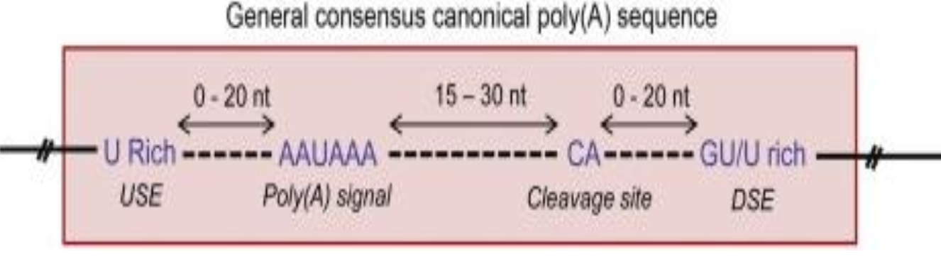
• RNA polymerase II arrives at the poly-A signal (PAS) 3’-TTATTT-5’
• Adds 5’ -AAUAAA-3’ to the mRNA.
• Cleavage site is 15-30nt downstream of the PAS.
• RNA polymerase II starts adding A bases (poly-A tail) to the mRNA 3’ end.
• More efficient if there is also a U-rich upstream signal element (USE) and GU-rich downstream signal element (DSE).
Determining the DNA sequence bound by a protein when the DNA sequence is unknown
Generate random sequence oligonucleotides (20bp long)
Anneal oligonucleotides to generate all DNA combinations
Stick the protein to a column
Run the DNA over the column (protein binds to target DNA)
Separate bund DNA and seuqence
Use alignment to determine the consensus sequences (logo plot)
EMSA (electrophoretic mobility shift assay) to determine the DNA sequence bound by a protein
Purify the protein and add it to known DNA sequence that has been 5’- end radiolabelled
Run the protein-DNA on a native gel
DNA bound to the protein will be in a higher molecular weight
DNAse I foot printing to determine the DNA sequence bound by a protein
Synthesise a known DNA sequence and radiojlabel 5’ end
Add pure protein and then add DNAse I are a conc where it will randomly cut once per strand
Isolate the DNA and run on a gle. Regions where the DNA was bound to protein will have been protected (not cut) so toy get a footprint on the gel. The footprint indicated which bases were bound to protein
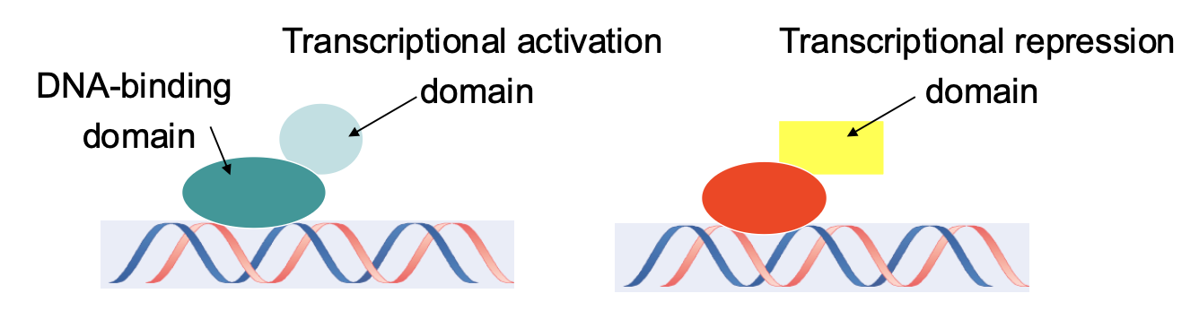
Modular structure of Transcription Factors
TFs contain DNA-binding domains & transcriptional regulation domains
Some have activation domains and others have repression domains
Some have domains to interact with other TFs
TFs are grouped into families on the basis of their DNA-binding domains
DNA-binding domain structures x4
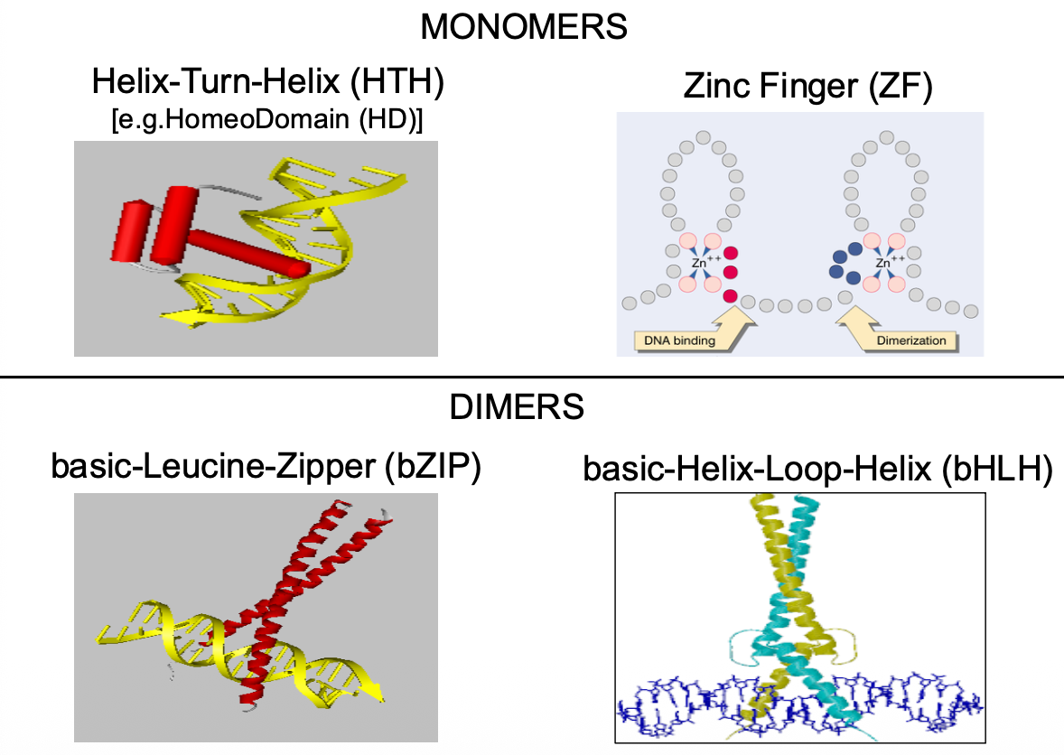
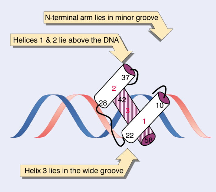
Helix-Turn-Helix motif
Helix 3 binds major groove
Helices 1&2 stabilize structure
N-terminal arm contacts minor groove
Zinc finger structure
Form helical and sheet structure
Zinc fingers classes x3
Class 1 – C2-H2 – e.g. TFIIIA
Class 2 – C2-C2 – e.g. steroid receptors (note: usually simplified to C4)
Class 3 – C6 – 2xZn – e.g. GAL4
C4 zinc fingers
A nuclear hormone receptor family
Large superfamily of ligand-dependent transcription factors/receptors
Nuclear Oestrogen Receptors are ERα and ERβ
Can bind DNA as homodimer (αα or ββ) or heterodimer (αβ)
bZip motif (Leucine Zipper) structure
The Leucine zipper is an amphipathic helix
Leucines at every 7th position form a hydrophobic side of the helix
The Leucine zipper is an amphipathic helix
Leucines at every 7th position form a hydrophobic side of the helix
• The hydrophobic helices bind to one another
• The foot of each helix is rich in basic residues
• These basic residues then bind to the DNA
• DNA-specificity depends on the bZip dimer
bHLH stricture
• Similar structure to bZip
• Helix-Loop-Helix domains interact
The basic-regions can then bind to DNA
• They target the DNA-sequence CAnnTG (a.k.a. E-box)
• Many are heterodimers that use a common Type-1 partner
• DNA-specificity is due to the Type-2 variable partner
Activation Domains
- Activation domains share loose similarity
- Fall into a number of classes
(1) ACIDIC (Asp [D] and Glu [E] rich)
(2) GLUTAMINE RICH (Gln [Q])
(3) PROLINE RICH (Pro [P])
- Interact with GTFs/TAFs
![<p>- Activation domains share loose similarity</p><p class="p1"><span style="line-height: normal;">- Fall into a number of classes</span></p><p class="p1"><span style="line-height: normal;">(1) ACIDIC (Asp [D] and Glu [E] rich)</span></p><p class="p1">(2) GLUTAMINE RICH (Gln [Q])</p><p class="p1">(3) PROLINE RICH (Pro [P])</p><p class="p1"><span style="line-height: normal;">- Interact with GTFs/TAFs</span></p>](https://assets.knowt.com/user-attachments/b4e281b7-9eea-40ff-b083-0c010c929f80.png)
Specific vs general transcription factors
CLOCK-BMAL1 Complex: Form heterodimers of basic helix-loop-helix (bHLH-PAS) transcription factors that bind specifically to E-box enhancer elements (
CACGTG) in target circadian genes.Transactivation: The glutamine-rich (polyQ) activation domain of CLOCK-BMAL1 recruits and interacts with general transcription factors (TFII complex and RNA Polymerase II at the TATA box) to stimulate gene transcription.
General Principle: Sequence-specific TFs bind unique DNA motifs via specific DNA-binding domains (e.g., bHLH, zinc finger) and use activation domains (acidic-rich, glutamine-rich, or proline-rich) to engage basal machinery for tissue- or signal-specific regulation.
Chromatin Barrier Problem
Nucleosome Obstacle: DNA in cells is not linear or naked; it is tightly wrapped around histone octamers to form nucleosomes (black ovals).
Core Problem: The physical presence of nucleosomes blocks RNA Polymerase II from moving along the DNA template during elongation.
Central Question: How does the transcriptional machinery overcome this dense chromatin barrier to allow active RNA synthesis? (This sets up chromatin remodeling and histone modification mechanisms).
Chromatin structure
DNA wrapped twice around histones - forms the nucleosome
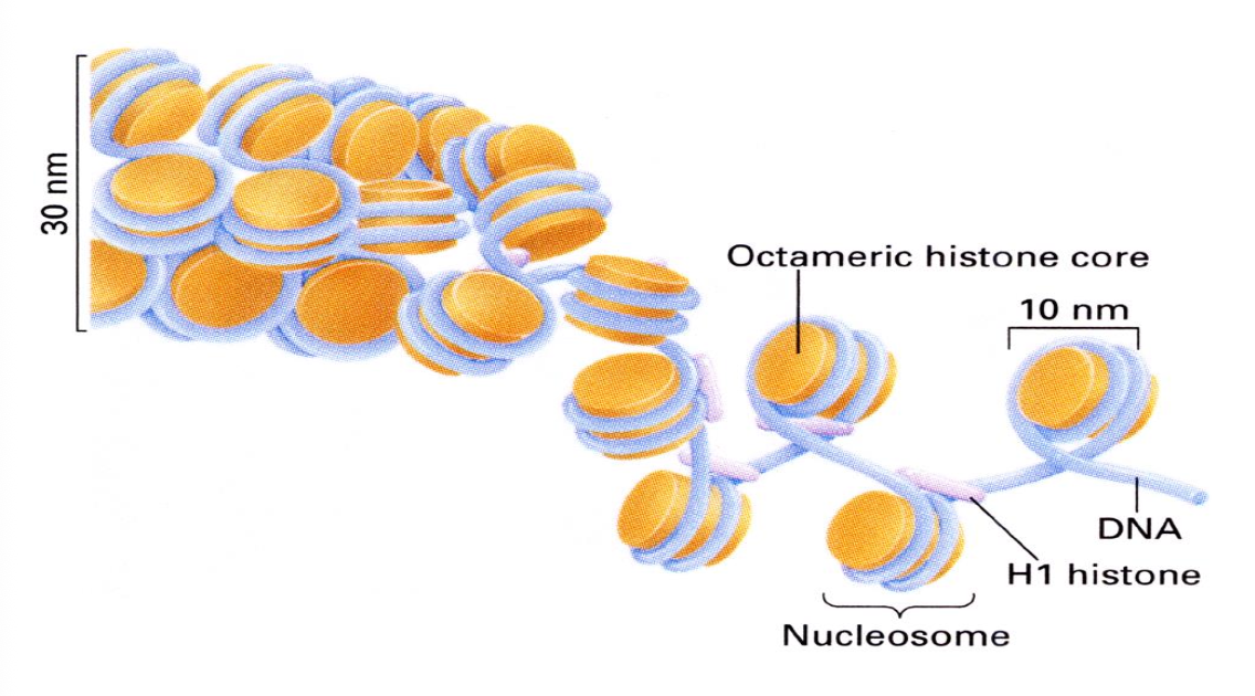
Nucleosome structure
8 Core Histones (2 of each)
• ~2 Turns of DNA
• 1 Linker Histone (H1) attached on the outside
Core tetramer of H3 and H4 bind the DNA and bend it
• The two H2A-H2B dimers then join
• The N-terminal tails are exposed and contain highly charged basic residues
• These tails are crucial for subsequent chromatin modelling
3 alpha helices fold domain

Histone post-transcriptional modifications x6
(1) Acetylation - of Lysine (K)
(2) Methylation - of Lysine (K) and Arginine (R)
(3) Phosphorylation - of Serine (S), Threonine (T) and Tyrosine (Y)
(4) Citrullination - of Arginine (R)
(5) Ubiquitination - of Lysine (K)
(6) Sumoylation - of Lysine (K)
Acetylation modification in lysine
H2A, H2B, H3 and H4
Methylation modification of lysine and arginine
H3 and H4
Post-translational modification of histones
Occurs in situ (i.e. whilst histones are bound to DNA) and correlates with the functional status of the relevant genomic locus
• These changes define an epigenetic ‘histone code’ and corresponding functional ‘landscape’
• The code defines the binding potential of chromatin associated factors
Lysine acetylation
- Acetylation is a dynamic process
• Histone Acetyltransferases (HATs) add an acetyl group to Lysine - stars transcription
• Histone Deacetylases (HDACs) remove the acetyl group from Lysine - stops transcription
Acetylation influences the structure of chromatin and TF access
Acetylation is associated with transcriptional activation
Acetylation = Less charge = Less cross-binding of Histone proteins = Relaxed state - DNA more accessible
Chromatin effects and transcription factors
- CLOCK-BMAL1
- bHLH-PAS transcription factors that form heterodimers
- Bind E-box enhancer sequences in circadian (i.e. biological-clock) genes
- Glutamine-rich activation domain (polyQ) interacts with the general factors
- Polymerase-II is stimulated to transcribe the gene
Histone acetylation and transcriptional activation
- CLOCK contains HAT-activity and can acetylate H3 and H4
- This opens up the core tetramer making the DNA accessible for transcription
- Hence, CLOCK relaxes the chromatin in addition to recruiting TFII+pol-II
- CLOCK keeps the basal promoter region open for TFII+Pol-II accessibility
• p300/CBP can interact with a variety of transcriptional regulators such as pCAF (a histone acetyltransferase) and TBP (which recognizes the promoter [TATAA])
• During transcription, they are all assembled at the promoter region.
• Histone acetylation by p300/CBP/pCAF and TAFII250 (also known, more simply, as TAF1) facilitates assembly of the pre-initiation complex to drive transcription
![<p>- CLOCK contains HAT-activity and can acetylate H3 and H4</p><p class="p1">- This opens up the core tetramer making the DNA accessible for transcription</p><p class="p1">- Hence, CLOCK relaxes the chromatin in addition to recruiting TFII+pol-II</p><p class="p1">- CLOCK keeps the basal promoter region open for TFII+Pol-II accessibility</p><p class="p1">• p300/CBP can interact with a variety of transcriptional regulators such as pCAF (a histone acetyltransferase) and TBP (which recognizes the promoter [TATAA])</p><p class="p1">• During transcription, they are all assembled at the promoter region.</p><p class="p1"><span style="line-height: normal;">• Histone acetylation by p300/CBP/pCAF and TAFII250 (also known, more </span>simply, as TAF1) facilitates assembly of the pre-initiation complex to drive transcription</p>](https://assets.knowt.com/user-attachments/4517cc95-331e-4298-a074-668e38613656.png)
Lysine Methylation
- Methylation of Lysine can also occur
• Lysine Methyltransferases (KMTs) add a methyl group to Lysine - stops transcription
• Lysine Demethylases (KDMs) remove methyl groups from Lysine - starts transcription
- Methylation influences the structure of chromatin and TF access
- Methylation can add 1, 2 or 3 methyl groups to the NH3 + residue of lysine
- It does this by replacing a proton (H+) with CH3 each time
- Lysine hyper-methylation (me2/3) associated with transcriptional repression
Arginine Methylation
• Protein Arginine Methyltransferases (PRMTs) catalyze the transfer of methyl groups from S-adenosylmethionine (AdoMet/SAM) to arginine residues in histones (PRMT1 methylates H4R3 = transcriptional activation)
• There is a single Protein Arginine Demethylase (JMJD6) in humans! (JMJD6 demethylates H3R2 & H4R3 = transcriptional repression)
- Arginine methylation is ‘usually’ associated with transcriptional activation
• PRMT4 (CARM1) methylates H3R17 and the ‘non-histone’ protein p300/CBP
• Hence, PRMT4 methylates p300/CBP to trigger its HAT activity for transcription
How does histone modifications correlate with function?
Specific histone marks are found at predictable locations along genes – because of different functions that they perform
Active and inert genes have very different patterns of histone PT modifications.
Drives association with specific bound regulatory proteins e.g.
H3K4me3 and H3K9me3 – bind to PHD finger domain proteins – eg. ING2, BPTF - activates transcription
H3K9me2/3 and H3K27me2/3 – bind to chromodomains – eg. HP1 - activates transcription
General-Kac and H4K16ac – bind to bromodomains – eg. PCAF - represses transcription
General-Kac and H4K5acK12ac – bind to double bromodomains – eg. hTAF1 - represses transcription
Chromatin remodelling complexes
Control transitions between chromatin states
2 major classes:
ATP-dependent chromatin remodelling histone sliding e.g. SWI/SNF, NURF
Histone acetylase/deacetylase containing complexes e.g. SAGA, PCAF
Euchromatin - active gene
Heterochromatin - generally inactive
Remodelling allows histone sliding - reveals binding sites
Transcription factors can then recruit histone acetylases to keep the chromatin open (i.e. relaxed and accessible)
Dynamics of this process is central to gene expression
Remodelling during transcriptional elongation
During transcription the DNA ahead of the advancing RNA Polymerase also needs to be unwound
FACT is a heterodimeric factor that removes the H2A-H2B dimers ahead of the polymerase so that the histones disassemble to allow the Pol-II to advance and transcribe the DNA
Once the Pol-II has passed FACT helps reassemble the histones to reform a new nucleosome that is situated further upstream (i.e. 5’) from its start position
Gene structure and transcription activation
Transcription activation domains contact other proteins which facilitate recruitment of a functional transcription pre-initiation complex
Three common types of activation domain – proline-[P]-rich, glutamine-[Q]-rich and acidic = glutamate-[E] and aspartate-[D]-rich
Activators work at a distance by bringing proteins associated with key regulatory elements into a single protein super-complex at promoters
This results in DNA looping and drives the efficiency of pre-initiation complex formation
Locus Control Regions (LCRs) act like enhancers but work over longer distances. They contain multiple-binding-sites to allow complexes of TFs to activate multiple linked-genes
Insulators provide boundaries for domains within euchromatin. Chromatin between two insulators is prevented from interacting with intra-chromosomal chromatin outside
Barriers function like insulators but are more important at the boundary of heterochromatin and euchromatin. They prevent heterochromatin spreading into regions containing genes
![<ul><li><p>Transcription activation domains contact other proteins which facilitate recruitment of a functional transcription pre-initiation complex</p></li><li><p>Three common types of activation domain – proline-[P]-rich, glutamine-[Q]-rich and acidic = glutamate-[E] and aspartate-[D]-rich</p></li><li><p>Activators work at a distance by bringing proteins associated with key regulatory elements into a single protein super-complex at promoters</p></li><li><p>This results in DNA looping and drives the efficiency of pre-initiation complex formation</p></li><li><p>Locus Control Regions (LCRs) act like enhancers but work over longer distances. They contain multiple-binding-sites to allow complexes of TFs to activate multiple linked-genes</p></li><li><p>Insulators provide boundaries for domains within euchromatin. Chromatin between two insulators is prevented from interacting with intra-chromosomal chromatin outside</p></li><li><p>Barriers function like insulators but are more important at the boundary of heterochromatin and euchromatin. They prevent heterochromatin spreading into regions containing genes</p></li></ul><p></p>](https://assets.knowt.com/user-attachments/49e2faaf-191e-4958-acd8-41dcff127e06.png)
Nuclear compartments - structure
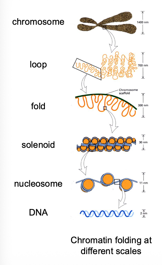
In the mammalian nucleus chromatin folds and loops to form discrete chromosome territories
CTs are polarised so that euchromatin and heterochromatin occupy discrete zones
• Stretches of the genome can be gene-rich or gene-poor
• Different chromatin domains fold into distinct ~1 Mbp DNA foci
• Active and inactive higher-order domains are also separated
Nuclear compartments - function
RNA synthesis only occurs in certain parts of the nucleus
Molecular and cell biology approaches show that sites of mRNA synthesis are clustered within factories that typically contain many genes with 5-10 active RNA pol-II complexes
Transcriptional co-regulation in transcription factories
3D Spatial Clustering:
Upon TNF⍺ stimulation, NF-kappaB target genes loop out from different loci to cluster at a shared transcription factory enriched with RNA Polymerase II.
Co-localizes distant genes on the same chromosome (SAMD4A and TNFAIP2 on Chromosome 14, 50bp apart) and across different chromosomes (SLC6A5 on Chromosome 11).
Hierarchical & Ordered Assembly:
Recruitment follows a set order: 1st SAMD4A → 2nd TNFAIP2 → 3rd SLC6A5.
TALEN Knockout: Deleting SAMD4A prevents downstream genes from co-associating, proving that pioneer locus interaction drives factory formation.
Validation & Significance:
Triple RNA-FISH: Confirms physical co-localization of nascent transcripts at a single sub-nuclear spot.
Takeaway: 3D spatial aggregation in transcription factories enables rapid, synchronized multi-gene responses to extracellular signals.