CVS
 Call Kai
 Learn
Learn Practice Test
 Spaced Repetition
 Match
Match Flashcards
 Knowt Play
1/9

There's no tags or description
Looks like no tags are added yet.
Name | Mastery | Learn | Test | Matching | Spaced | Call with Kai | Chat |
|---|
No analytics yet
Send a link to your students to track their progress
10 Terms
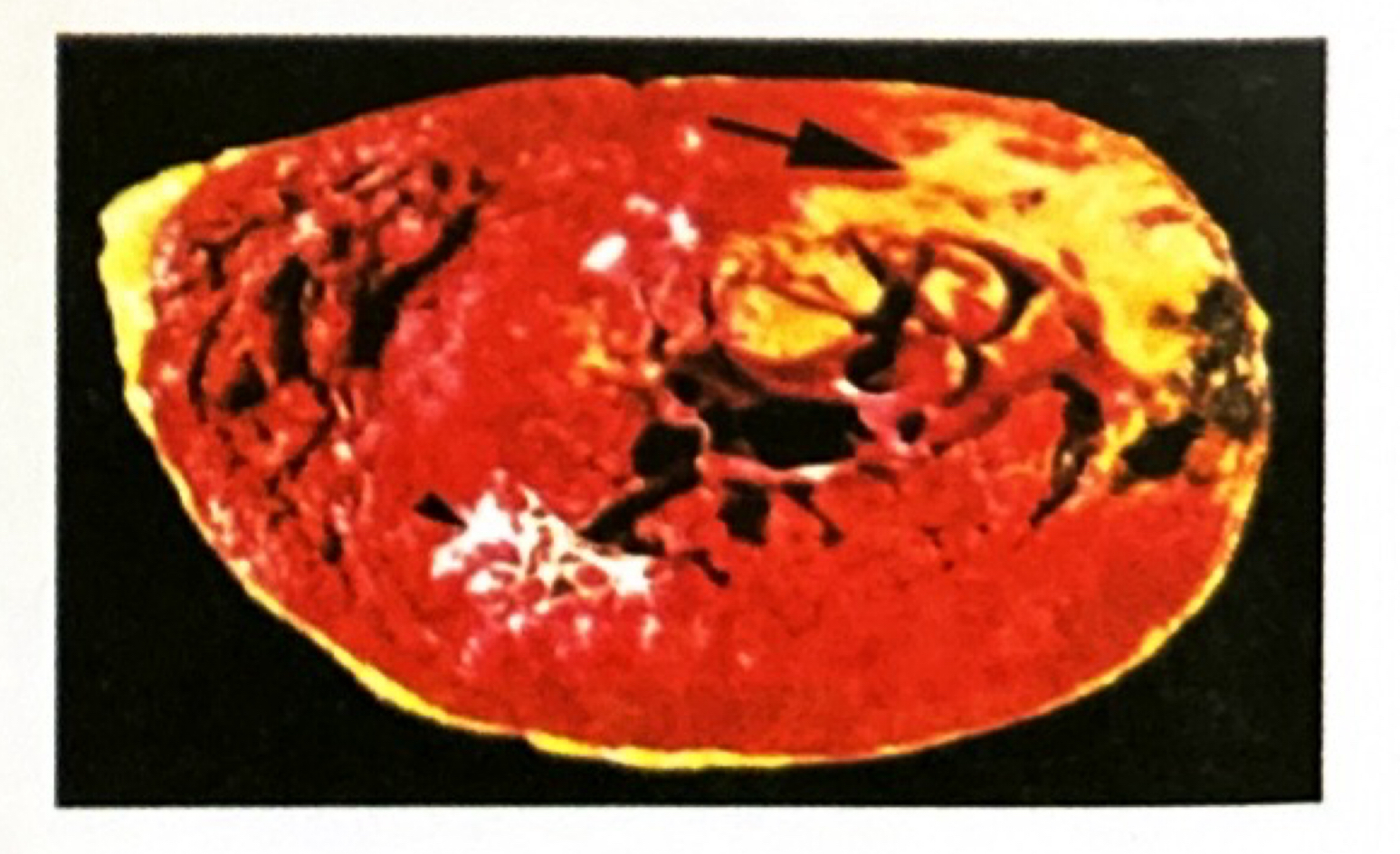
Gross Image Clue: A cardiac ventricular slice from a patient who died 3days after severe substernal chest pain shows a central pale yellow-tan soft region with a hyperemic red border. What is the diagnosis and key gross features according to Ramdas Nayak?
Diagnosis: Acute Myocardial Infarction (2-4days old)
Key Gross Features (Ramdas Nayak):
Color/Appearance: Central yellow-tan softening with a surrounding hyperemic (red) rim of vascular congestion and early inflammation
Location: Most commonly subendocardial or transmural in the distribution of LAD, RCA, or LCX
Diagnostic Hallmark: Yellow-tan center surrounded by a hyperemic hypervascular border

Microscopic Image Clue: Left ventricular myocardium shows loss of cardiac muscle striations and nuclei, hypereosinophilic myofibers, and dense interstitial infiltration by neutrophils. What is the diagnosis and characteristic microscopic features?
Diagnosis: Acute Myocardial Infarction (12-72hours)
Microscopic Features (Ramdas Nayak):
Low-Power: Band of necrotic myocardium with dense acute inflammatory cell infiltration at the periphery
High-Power: Coagulative necrosis of myocytes (loss of nuclei and striations, intense hypereosinophilia)
Infiltrate: Dense neutrophilic infiltrate between necrotic myocytes
Diagnostic Hallmark: Coagulative necrosis of cardiac myocytes with dense neutrophilic infiltrate

Microscopic Image Clue: Myocardial section demonstrates a focal granulomatous lesion containing central fibrinoid necrosis surrounded by lymphocytes, plasma cells, and large macrophages with vesicular nuclei and ribbon-like chromatin. What is the lesion and its pathognomonic cells?
Diagnosis: Aschoff Body in Rheumatic Carditis
Microscopic Features (Ramdas Nayak):
Central Region: Fibrinoid necrosis of collagen
Cellular Infiltrate: T lymphocytes, plasma cells, and pathognomonic histiocytes
Key Pathognomonic Cells:
Anitschkow cells: Large histiocytes with abundant cytoplasm and central caterpillar-like chromatin on longitudinal section ("owl-eye" on cross section)
Aschoff giant cells: Multinucleated giant cells derived from macrophages
Diagnostic Hallmark: Aschoff body containing pathognomonic Anitschkow ("caterpillar") cells


Gross Image Clue: Mitral valve viewed from the left atrium shows severe thickening and fusion of leaflet commissures, thickened and shortened chordae tendineae, and a rigid, narrowed orifice. What is the diagnosis and characteristic gross morphology?
Diagnosis: Chronic Rheumatic Heart Disease (Mitral Stenosis)
Key Gross Features (Ramdas Nayak):
- Valve Leaflets: Diffuse fibrous thickening, calcification, and commissural fusion
- Chordae Tendineae: Thickened, shortened, and fused into a single fibrous column
- Orifice: Narrowed, rigid opening resembling a "fish-mouth" or "button-hole"
- Atrial Changes: Left atrial massive dilatation with MacCallum plaques on the posterior atrial wall
- Diagnostic Hallmark: Commissural fusion with "fish-mouth" or "button-hole" valvular deformity

Morphological Comparison: How do the vegetations of Rheumatic Heart Disease, Infective Endocarditis, Non-Bacterial Thrombotic Endocarditis (NBTE), and Libman-Sacks Endocarditis differ in size, friability, location, and bacterial presence?
1. Rheumatic Heart Disease (RHD):
- Small (1-2mm), firm, warty (verrucous), sterile, along lines of closure (atrial surface of AV valves, ventricular surface of semilunar valves)
2. Infective Endocarditis (IE):
- Large (>5mm), bulky, friable, destructive, non-sterile (contains dense bacterial colonies), can extend onto chordae or valve ring
3. Non-Bacterial Thrombotic Endocarditis (NBTE):
- Small-to-medium (1-5mm), sterile, bland, non-destructive, loosely attached along closure lines in hypercoagulable states
4. Libman-Sacks Endocarditis (SLE):
- Small-to-medium, sterile, granular, located on both surfaces (underside and upper side) of valve leaflets, commissures, and endocardium

Microscopic Image Clue: Heart valve section shows a large, friable mass composed of dense fibrin, platelets, necrotic debris, neutrophilic infiltrate, and large bacterial colonies. What is the diagnosis and key histological components?
Diagnosis: Acute Infective Endocarditis
Microscopic Features (Ramdas Nayak):
- Superficial Zone: Dense layer of fibrin, platelets, and bacterial colonies (microorganisms)
- Base Zone: Intense neutrophilic acute inflammatory reaction and underlying granulation tissue
- Deep Zone: Suppurative necrosis destroying underlying valvular collagen
- Diagnostic Hallmark: Friable vegetation containing dense bacterial colonies mixed with fibrin and neutrophils on destroyed valvular tissue
Gross Image Clue: Abdominal aorta opened posteriorly shows raised, yellow-white focal intimal lesions with ulceration, calcification, and overlying mural thrombi. What is the lesion and its gross structural components?
Diagnosis: Advanced Atherosclerotic Plaque (Atheroma)
Key Gross Features (Ramdas Nayak):
Surface: Raised yellow to white-grey eccentric intimal plaques
Complications: Ulceration, calcification (brittle, eggshell-like), intra-plaque hemorrhage, and thrombosis
Predilection Site: Abdominal aorta > Coronary arteries > Popliteal arteries > Internal carotid arteries
Diagnostic Hallmark: Raised eccentric yellow intimal plaque with central lipid core and fibrous cap

Microscopic Image Clue: Arterial wall section demonstrates a prominent fibrocellular cap covering a necrotic core containing cholesterol clefts, lipid-laden foam cells, necrotic debris, and focal calcification. What is the diagnosis and key histological features?
Diagnosis: Atherosclerotic Plaque
Microscopic Features (Ramdas Nayak):
- Fibrous Cap: Smooth muscle cells, dense collagen, elastic fibers, and scattered lymphocytes
- Necrotic Core: Acellular core containing needle-like cholesterol clefts (empty spaces left after tissue processing), necrotic cell debris, and calcium deposits
- Basal/Peripheral Region: Foam cells (lipid-laden macrophages) and neovascularization (capillaries)
- Diagnostic Hallmark: Needle-shaped cholesterol clefts within a central necrotic core beneath a fibrous cap
Gross Image Clue: Opened pericardial sac demonstrates a dry, roughened, opaque pericardial surface with shaggy, irregular exudate giving a "bread and butter" appearance. What is the diagnosis and key gross features?
Diagnosis: Fibrinous / Serofibrinous Pericarditis
Key Gross Features (Ramdas Nayak):
- Pericardial Surface: Thickened, dull, opaque, and shaggy with dense fibrinous exudate
- Appearance: "Bread and butter" appearance (resembles two buttered slices of bread pulled apart)
- Fluid: Variable serous fluid mixed with fibrin strands
- Etiology Clue: Uremia, acute myocardial infarction (Dressler syndrome/early post-MI), rheumatic fever, or viral infection
- Diagnostic Hallmark: Shaggy, fibrous "bread and butter" surface exudate
Image Clue: Radial artery section from an elderly patient shows ring-like calcifications restricted to the tunica media without narrowing of the arterial lumen or intimal disruption. What is the diagnosis and distinguishing morphological features?
Diagnosis: Mönckeberg Medial Calcific Sclerosis
Morphological Features (Ramdas Nayak):
- Gross: Hard, rigid, pipe-stem-like non-compressible arteries ("pipestem arteries"); intact lumen
- Microscopy (Tunica Media): Basophilic ring-like calcification along the internal elastic lamina and media
- Microscopy (Tunica Intima): Normal intimal layer without occlusion or lipid accumulation
- Clinical/Diagnostic Hallmark: Ring-like medial calcification that does not narrow the vessel lumen