3.1
 Call Kai
 Learn
 Practice Test
 Spaced Repetition
 Match
 Flashcards
 Knowt Play
1/52

There's no tags or description
Looks like no tags are added yet.
53 Terms
Tbx6 expression after gastrulation is the earliest marker for
paraxial mesoderm identity
recall

paraxial mesoderm becomes the
somites (and head)
Tbx6 is expressed in paraxial mesoderm until
it starts to actually make a somite, and then, expression of Tbx6 starts to decline
3 main steps of somite formation
*last two steps occur simultaneously
identify region of rostral SPM (segmental plate mesoderm) that will become the next somite
separate from neighboring cells via MET
cell must acquire R/C axis for things that the cell does later on
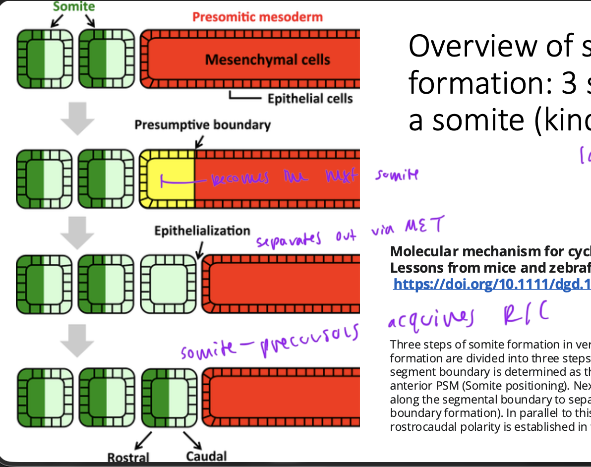
somites are
epithelial (so must undergo MET to get there)
SPM specification for somite lineage occurs early, but
somites don’t actually form until later on
if somites don’t form correctly (and later on) then they won’t interact properly with the neiboring tissues
what causes the embryo to elongate posteriorly
high rate of cell proliferation in tailbud
so new neural tube and SPM formed as the embryo elongates
somite formation must be coordinated on
both sides of the embryo
like vertebrate are fored from half of a somite from one side and half from a somite from the other side
so coordination of somite formation on each side must be coordinated
name for the process of forming somites
somitogenesis
numbering systems for numbering somites

PSM=SPM
pre-somitic mesoderm or segmental plate mesoderm
paraxial mesoderm that is a somite precursor
epithelialization to form somites is
a gradual process (expressing same N-cadherins at incr rate to stick tg)
starts at S-3/-4 and doesn’t finish until S0
so decision to make a somite occurs around S-3/-4

does the MET to form somites occur quickly?
no
fission
physical separation of a somite to form its last wall and be separate from the nearby cells
occurs at caudal edge of s0 and the rostral edge of s1
after fission, the new edge finishes its epithelialization

observations of somitogenesis
somite formation is bilaterally symmetrical
is invariant in a given species but varies across species
there is some clock that determines the periodicity of somite formation and is in the paraxial mesoderm, and is not dependent on external signals
cells don’t change their A/P position within the PSM during somitogenesis (only relative position in the embryo changes)
when somites form, they already know their A/P identity
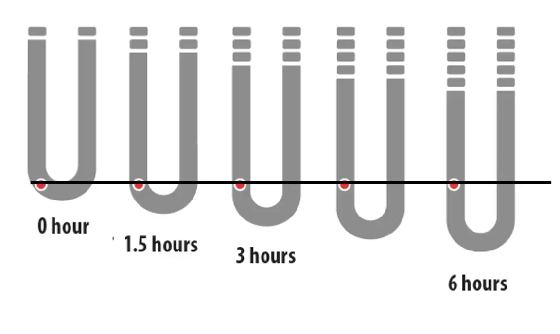
how do we know that the internal clock to dictate somitogenesis is local to the SPM?
because if taken out that tissue and grafted elsewhere, somites form as thet would, and in the correct R/C order, on the same time table
if you graft it rotated, they still form in the OG direction as they should
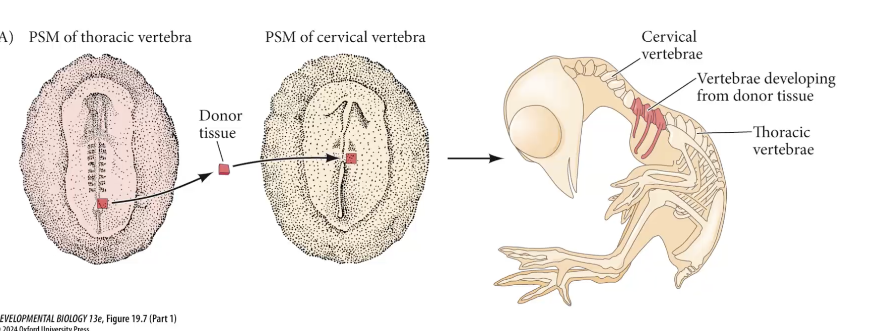
how do we know somite precursors know their AP identity and have an A/P axis identity themselves
if graft older tissue that would make thoracic into younger embryo where cervical should form, you get thoracic in the cervical region
so Hox code of somites is established before somites actually form even
somite A/P polarity
have A/P polarity based on differential gene expression
Tbx18 causes A side and Uncx4.1 causes P
this is important bc the P half of one somite and the A half of another form a vertebrate eventually (called resegmentation)

clock and wavefront model
idea that there are 2 overlapping systems that help form somites correctly
clock: more like a metranome (since keeps relative time, starts ticking once somite precursors are born and leave tailbud area
clock genes regulate timing of the metranome, which causes the precursors to start somitogenesis
moving gradient posterior to suppress somitogenesis too early, as embryo grows posteriorly, the gradient moves as well
anterior has somite activating signals, when metranomes go off, they start forming somites (determination front)
determination front
point around S-3/-4 where cells in PSM escape from the repressive signaling gradient as the tailbud extends posteriorly, and have their clock genes turned on to start the metranome

the inhibitory gradient that suppresses somite formation moves posteriorly over time so that
somites form A—>P
first evidence for the internal clock/metranome
Hes and Hairy are TFs actiavted by Notch signaling
did in-situ hybridization and found Hes and Hairy expr occurring in waves in diff cells
repeating pattern of Hairy and Hes expr, which they found matches the timing of somite formation in a given species
ex: in chx, 90min

if take out PSM cells and dissociate them,
the cells still express Hes and Hairy, but their clocks are no longer synced

clock genes are under control of Notch signaling
Delta is TM protein
bidirectional response (goes to each cell)
part of Notch gets proteolytically cleaved at cytoplasmic domain, is the NICD (Notch intracytoplasmic domain) which has nuclear signal, assembles TF complex to bind to target genes that are expressed in cyclical fashion like Hes, Her, Hairy
Notch must then be replenished, so allows for a waiting time, allowing for this periodic signaling


Reasons why we see oscillations in signaling for clock
Notch must be replenished
also Hes and Her inhibit themselves, and Notch and Delta, so causes big delays, resulting is oscillatory behavior

Why do some species have longer cycles
diff species have diff baseline metabolic rates, which scale linearly with length of signaling pathways
higher baseline metabolic rate, shorter oscillatory period
Tbx6 expressing cells in PSM undergo
oscillatory behavior as well
defines what set of cells will help form the next somite and when
determination front
decides where the next somite will form
gradient of Ff8 and Wnt3A high posterior, low anterior
suppresses somite formation, even when the clock strikes
cells born in tailbud express Fgf8 and Wnt5A
stop transcribing these genes once out of tailbud, but still have some of those mRNA
so gradient of mRNA high tailbud, low rostral, creates signaling gradient that is inhibitory of somite formation, strongest in P side

recall RTK pathway
Ras Raf MEK ERK
phosphorylated ERK shows presence of Fgf8 pathway
so low phosphorylated ERK anterior where Fgf8 levels are lower

Wnt5A and Fgf8 gradients (high P) cause
RA gradient that is high A and low P
since newly made somites express more RALDH than Cyp26, making RA levels high A and low P
SPM makes more Cyp26 than RALDH (so decr RA levels where somites have not yet formed)

location of determination front
sweet spot where enough RA to promote somite formation and not enough Fgf8 + Wnt5A to inhibit it, where somite formation occurs
is at around S-3/-4
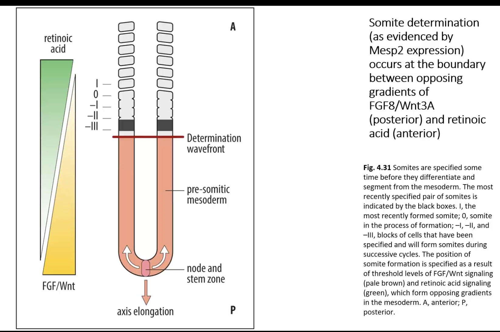
see gradients
cyp26 expression keeps fgf8/wnt5A gradient restricted to posterior PSM

Mesp2 expression
first expressed at S-3/-4
marker for commitment to somite lineage
if delete Msp2, you still have a clock, but no somite formation
adding Fgf8 and Wnt5A inhibits Msp2 expression
taking out RA make weird phenotypes, like differences bilaterally

Tbx6 causes
Notch and Delta expression
Hes and Her activate
Mesp2 expr
closer to tailbud has high
Fgf8 and Wnt5A signaling
so inhibits Msp2 expression even when the clock strikes
S-3/-4 area
is far enough from Fgf8 and Wnt5A inhibitory signals and strong enough in RA signaling to activate Msp2 expression and cause formation of a somite

cyclic gene expression only shown on L side in this image
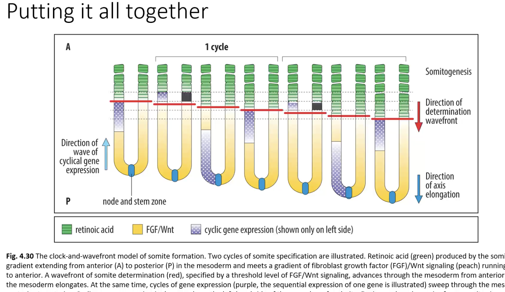
Mesp2 function
activates genes necessary for MET to actually form somites (so causes cadherin upregulation)
sets stage for A/P polarity within each somite
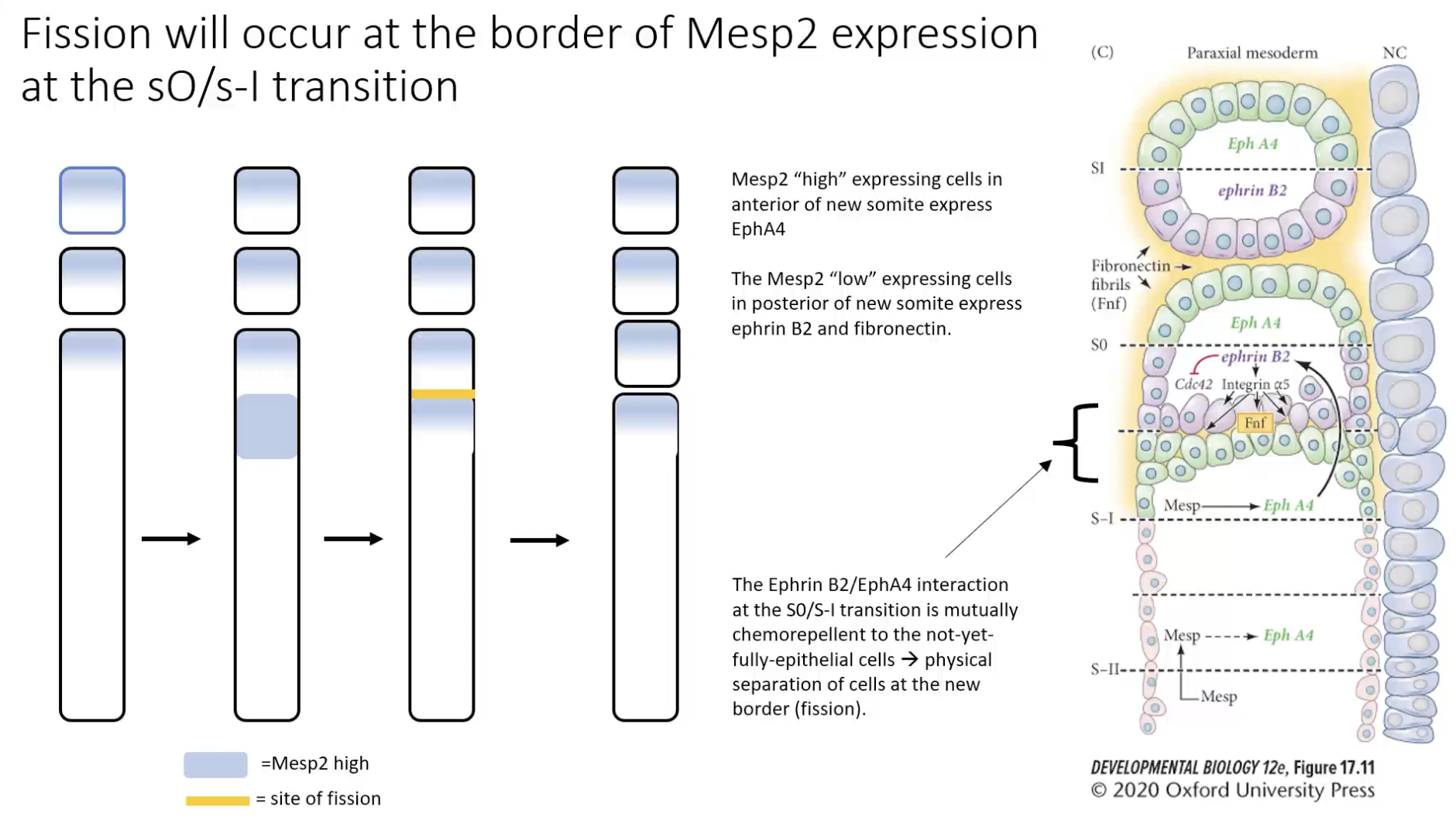
high Mesp2 on A side of each cell and low Mesp2 on P side of each cell
so cells high Mesp2 go A and form A side of a somite and low mesp2 cells form P side of a somite (so sort out)
so when that somite is born, it has intrinsic A/P axis

early A/P variation in a somite dictates
how somites react with their neighbors later on
must know which half of cell is A/P or R/C to know
where its moves S1
A/P in each somite setting stage for fission
difference in Mesp2 expre cause differential Eph receptor or Ephrin (Eph receptor ligand) expr
Ephrin interactions cause bidirectional signaling

high expressing Medsp2 cells express EphA4 (chemorepellent for Ephrinb2 expressing cells)
low expressing Mesp2 cause EphrinB2 expression (chemorepellent for EphA4 expressing cells)
so border between S0 and S1, have mesenchymal cells, Eph4A and EphrinB2 bind each other and move away from each other, creates space to separate S0 from S1
so causes fission
expression of Mesp2 also leads to
downregulation of Tbx6, so stops thinking you’re PSM and start thinking you’re somite

mutations in genes for somitogenesis can lead to
axial skeleton defects (congenital)

recall Hox code within paraxial mesoderm determines the
A/P identity of somites and their derivatives
the Hox code is imparted as cells leave the tailbud region and become part of the PSM
cells born sooner express lower paralog groups, since chromatin around 3’-most region on paralog group opens up first
the chromatin that is open shifts towards the more 5’ end as cells spend more time in the tailbud before joining the PSM (more/longer exposure to Wnt and Fgf8 gradinet)
so the Hox code is already in place when somites start to form

interconnected systems that allow somites to form at the right place at the right time
clock/metranome: Notch/Delta
shifting Wnt and Fgf8 gradient
determination front: determines where somites are specified
Hox gene expression: somite A/P identity based on timing of cells leaving the posterior proliferation zone/decondensation of Hox clusters resulting on differential Hox gene expression

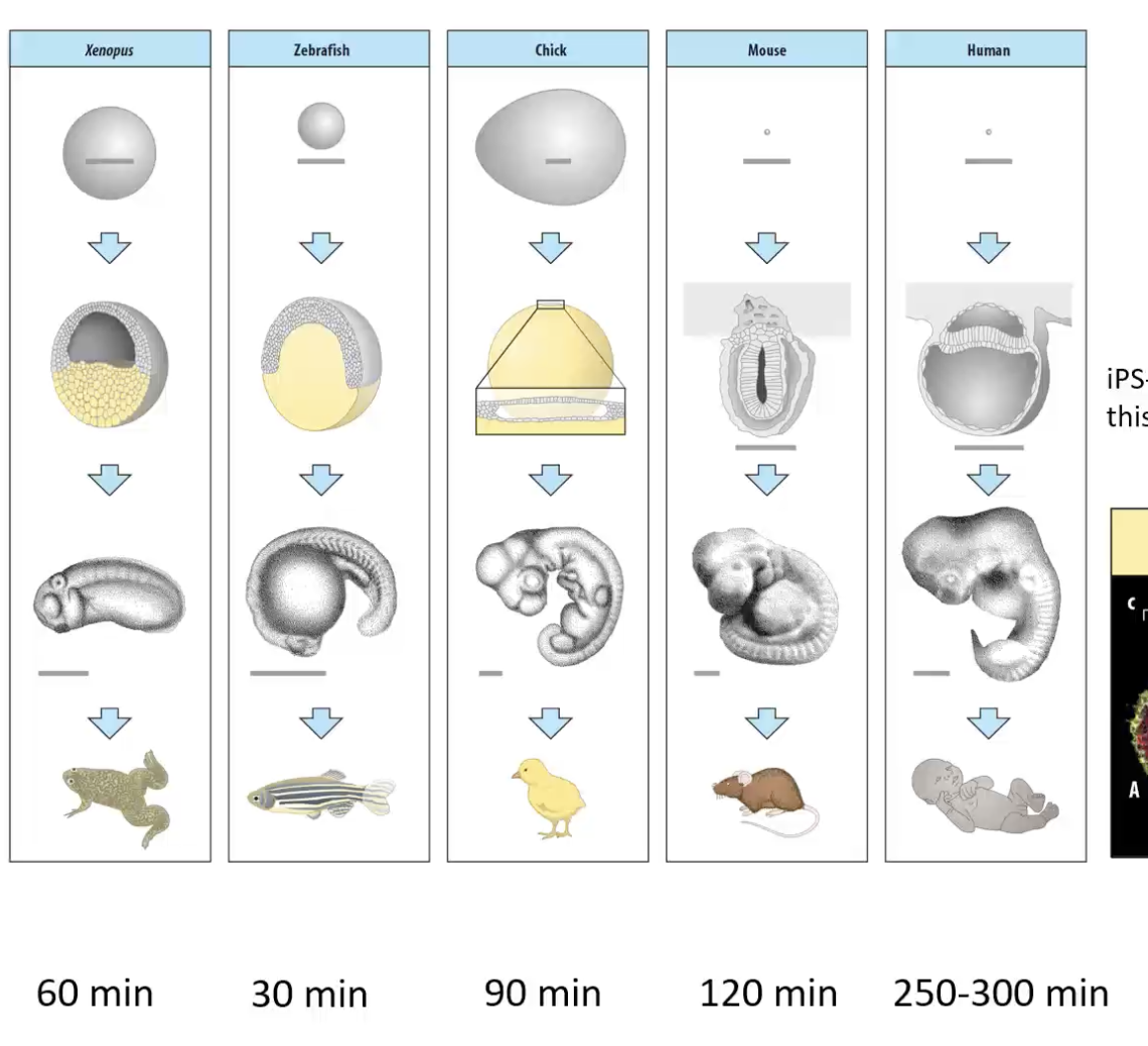
Why is the segmentation clock diff in diff species?
bc diff species have diff baseline metabolic rates
studied by pushing iPS down paraxial mesoderm pathway, culture to initiate Notch signaling to form somites in culture to study the clock in diff species
How does baseline metabolic rate determine segmentation clock timing?
B-catenin ubiquination/acetylation to determine Wnt signaling is dependent on baseline metabolic rates
upstream of B-catrenin, Fgf-signaling dictates glycolysis and metabolism rates, leading to rates of B-catenin signaling

differences in baseline metabolism in diff spcies correlates to
differences in developmental progression and segmentation clock timing
NOT dependent on size of the animal, etc
length of embryogenesis, baseline metabolic rate to form mRNA, DNA, proteins, and segmentation clock timing are all correlated
diff NAD+ to NADH ratios determine diff
rates or protein translation
humans have lower NAD+:NADH ratio than mice (bc cells twice as large) so have lower metabolic rates
slowing down glycolysis rates in mice leads to slower
protein turnover rate, so clock speeds decr
accomplished by ubiquinating Hes