last minute practical bullshitting
 Call Kai
 Learn
 Practice Test
 Spaced Repetition
 Match
 Flashcards
 Knowt Play
1/86

There's no tags or description
Looks like no tags are added yet.
87 Terms

26
right brachiocephalic vein

27
left brachiocephalic vein

29
superior vena cava

34
aorta

31
pulmonary trunk artery

38
brachiocephalic artery

39
left common carotid artery

40
left subclavian artery

32
left and right pulmonary arteries

33
left and right pulmonary veins

15
left atrium

1
right atrium

3
right ventricle

7
pulmonary semilunar valve

8
aortic semilunar valve

5
tricuspid valve

6
bicuspid/mitral valve

4
left ventricle

13
papillary muscles

(not numbered) What are the net-like muscle ridges on the internal wall surface for both ventricles?
trabecular carnae

(not numbered) what connects the papillary muscles to tricuspid and bicuspid/mitral valves?
chordae tendinae

11 (muscle ridges in atria)
pectinae muscles
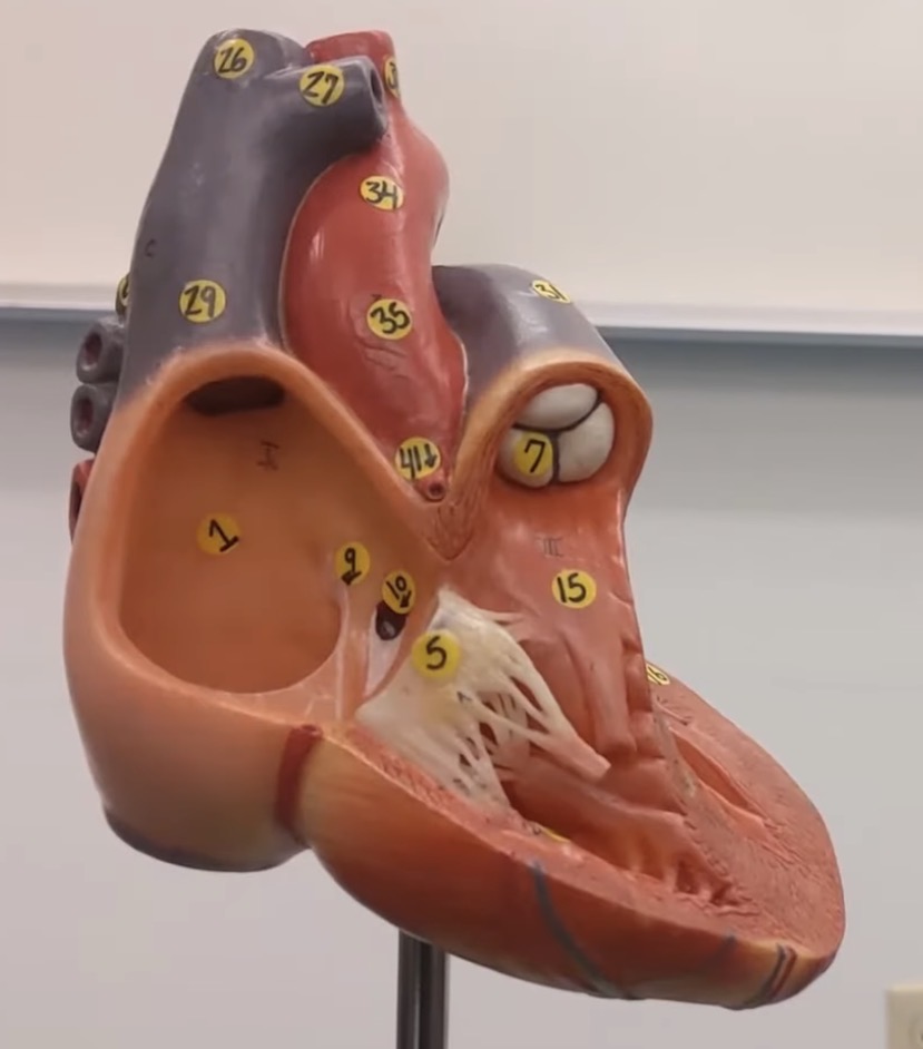
9
fossa ovalis (in adults)

10
opening of coronary sinus

51
coronary sinus

14
interventricular septum

(not numbered) what is the groove being pointed at?
anterior interventricular sulcus

(not numbered) what is the groove being pointed at?
right coronary sulcus

(not numbered) what is the groove being pointed at?
left coronary sulcus
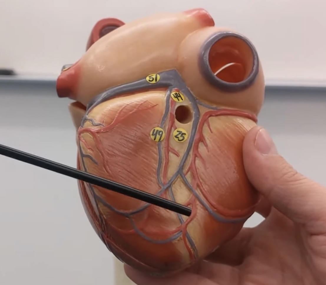
(not numbered) what is the groove being pointed at?
posterior interventricular sulcus

43
anterior interventricular artery

44
posterior interventricular artery

41
right coronary artery
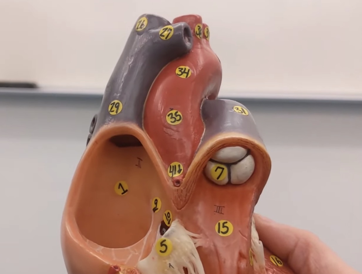
35
ascending aorta

20
apex

36
aortic arch

37
descending aorta

45
right marginal artery

46
left marginal artery

47
circumflex artery

48
great cardiac vein

49
middle cardiac vein

50
small cardiac vein

18
right auricle
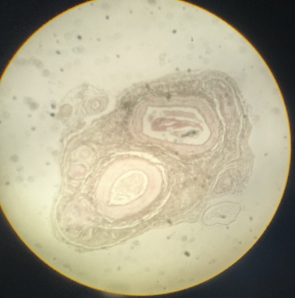
list the 2 major structures from left to right
artery and vein

What kind of tissue is this?
heart muscle

label the WBC A and B respectively
lymphocyte and neutrophil

What kind of tissue is this?
lung

What kind of tissue is this?
mammal lung

What kind of tissue is this?
spinal cord

What kind of tissue is this?
cerebrum

What kind of tissue is this?
cerebellum

What kind of tissue is this?
giant multipolar neuron smear

A
endocardium

B
myocardium

C
pericardial cavity

D
fibrous pericardium

E
parietal layer of serous pericardium

F
epicardium (visceral layer of serous pericardium)
Outer layer that is firmly attached to the fibrous pericardium without space in between, anchors serous membrane
parietal layer of serous pericardium
Innermost layer of serous pericardium that acts as a protective outer barrier for the heart muscle, secretes the lubricating pericardial fluid into the pericardial cavity
epicardium (visceral layer of serous pericardium)
thick muscular layer of heart wall responsible for the heart’s pumping action
myocardium
thin, innermost layer of tissue that lines chambers and valves of heart
endocardium

green line (label structure, if it’s the upper/lower respiratory tract, and if it’s the conducting/respiratory zone)
nasopharynx, upper, conducting

red line (label structure, if it’s the upper/lower respiratory tract, and if it’s the conducting/respiratory zone)
oropharynx, upper, conducting

blue line (label structure, if it’s the upper/lower respiratory tract, and if it’s the conducting/respiratory zone)
laryngopharynx, upper, conducting

a, b, c (label structure, if it’s the upper/lower respiratory tract, and if it’s the conducting/respiratory zone)
nasal conchae, upper, conducting

space in between folds of a, b, c (label structure, if it’s the upper/lower respiratory tract, and if it’s the conducting/respiratory zone)
nasal cavity, conducting, upper

(label structure, if it’s the upper/lower respiratory tract, and if it’s the conducting/respiratory zone)
epiglottis, upper, conducting

(label structure, if it’s the upper/lower respiratory tract, and if it’s the conducting/respiratory zone)
larynx, upper, conducting

(label structure, if it’s the upper/lower respiratory tract, and if it’s the conducting/respiratory zone)
thyroid cartilage, upper, conduting

(label structure, if it’s the upper/lower respiratory tract, and if it’s the conducting/respiratory zone)
cricoid cartilage, upper, conducting

422 (pointed area) (label structure, if it’s the upper/lower respiratory tract, and if it’s the conducting/respiratory zone)
trachea, lower, conducting
The right lung has _ lobes and the left lung has _ lobes. The right/left lung contains the cardiac notch and is larger/smaller.
3, 2, left, smaller

pointed area (label structure, if it’s the upper/lower respiratory tract, and if it’s the conducting/respiratory zone)
bronchi, lower, conducting
the structures that follow the bronchii are _____
bronchioles
the overarching structure that includes the trachea, primary bronchi, secondary bronchi, tertiary bronchi, bronchioles, terminary bronchioles, and respiratory bronchioles
bronchial tree

A (label structure, if it’s the upper/lower respiratory tract, and if it’s the conducting/respiratory zone)
terminal bronchiole, lower, conducting

B (label structure, if it’s the upper/lower respiratory tract, and if it’s the conducting/respiratory zone)
alveolar sac, lower, respiratory

C (label structure, if it’s the upper/lower respiratory tract, and if it’s the conducting/respiratory zone)
alveolar pores, lower, respiratory

D (label structure, if it’s the upper/lower respiratory tract, and if it’s the conducting/respiratory zone)
alveolar duct, lower, respiratory

E (label structure, if it’s the upper/lower respiratory tract, and if it’s the conducting/respiratory zone)
capillaries, lower, respiratory

F (label structure, if it’s the upper/lower respiratory tract, and if it’s the conducting/respiratory zone)
alveolus, lower, respiratory

G (label structure, if it’s the upper/lower respiratory tract, and if it’s the conducting/respiratory zone)
respiratory bronchiole, lower, respiratory
normal inhalation involves what primary muscles?
diaphragm, external intercostals
normal exhalation has which muscles relaxing?
diaphragm, external intercostals
forceful exhalation involves the contraction of what muscles?
internal intercostals, abdominal muscles