Physio E109 Midterm 1: Key Biology Terms & Definitions
 Call Kai
 Learn
 Practice Test
 Spaced Repetition
 Match
 Flashcards
 Knowt Play
1/97

There's no tags or description
Looks like no tags are added yet.
Name | Mastery | Learn | Test | Matching | Spaced | Call with Kai | Chat |
|---|
No analytics yet
Send a link to your students to track their progress
98 Terms
Human Physiology
the science of the mechanical, physcial, and biochemical functions of our bodies
- studied at various levels of organization
cells --> tissues --> organs --> organ systems --> organisms
Homeostasis
ability of bodies to maintain a relatively constant internal conditions
homeostasis is not static, but means body is aiming to return to return our body to a set point
fail: compensation fails and has inability to maintain homeostasis --> illness or disease
succeed: compensation succeeds --> wellness
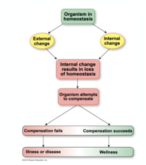
How to maintain homeostasis
in a cellular level: being able to control the state of our cells; occurs through exchange between our cells and what is external through our cells
we have the ability to control inside intracellular state to respond to external changes
exchange between our cells, the ECF, & the external environment that determine the cell's state & how they respond

Fluid Compartments
ICF: inside cell (1/3)
ECF: extracellular spaces (2/3)
Plasma: within circulatory system (non-cellular component of blood which we consider EC)

Maintaining Equilibrium
within our fluids, in order to provide physiological functions; rely on active transport to maintain it
(always present)
Ion concentrations within ICF, ECF, and Plasma all vary with one another

Osmolarity
the number of solutes present in a unit volume of fluid (solute concentration); difference in osmolarity can drive movement of fluids or develop osmotic pressure
separated by selectively permeable membrane
Hyperosmotic: higher [solute]; lower [free H2O]
Hypoosmotic: lower [solute]; higher [free H2O]
# of solutes = solute concentration x volume
fluid move from low --> high concentration of solutes (water follows solute) solute would be high to low
Tonicity
feature of a fluid within which a cell is held & a way of describing if fluid is able to move into or out the cell
mvt of water depends on osmolarity
Hypotonic solution: Less solutes in soln → H2O moves into cell → LYSED
Isotonic solution: Equal amount of solutes in cell and solution → NORMAL
Hypertonic solution: More solutes in soln → H2O moves out cell → SHRIVELED
Calculating Osmolarity
S/V=C
S = #solutes (mOsm)
V = Volume (L)
C = solute conc. (mOsm/L)
S = C x V

Diffusion
mvt of molecules from an area of high conc to low conc; diffusion across cell membrane depend on size of molecule and membrane permeability
vibration = hit each other, repel, and move further apart (Brownian motion) & disperse molecules away form itself
Rate of mvt depends on size of molecules (bigger size = higher inertia = slower @ lower frequency)
Fick's Law of Diffusion
the variables & parameters that are likely to determine the rate at which things are going to diffuse across a membrane
Faster diffusion across membrane:
- Increased membrane permeability
- Smaller, lipid soluble molecules (larger lipid solubility)
- Stronger/greater concentration gradient
- Larger surface area
Slower diffusion across membrane:
- Decreased membrane permeability
- Larger, non lipid soluble molecules
- Weaker/smaller concentration gradient
- Smaller surface area
Fick's Law: Calculation
plug & chug with correct units

Membrane Proteins
2 types of functions:
1. Communicating/Signaling: structural, enzymes, membrane receptor proteins; critical for maintaining cellular function & responding to external to internal changes to return cells to homeostasis
2. Transport: channel proteins, carrier proteins
Membrane Transport Proteins
Channel Proteins: creates a water-filled pore; increases the membrane permeability to molecules that are lipophilic
- gates: opens and closes from signals
- open channels: only open (pores)
Carrier Proteins: never forms an open channel between the two sides of the membrane; open channel rely on conformation changes that allow specific molecule to transport across membrane
- uniport carriers transport only one kind of substrate
- symport carriers move two or more substrates in the same direction across the membrane
- antiport carriers move 2 substrate in opposites directions
Facilitated Diffusion (Membrane Transport)
mechanism that allows us to maintain the mvt of molecules into the cell by altering intracellular concentration through metabolic modification (uniport transport)
high conc --> low conc
no ATP required (concentration gradient~ PASSIVE diffusion); need a either a channel or carrier protein to help molecules get across
ex: GLUT1- Na+/glucose symport

Co-transport: SGLT (Membrane Transport)
mvt of 2 solutes into the cell at the same time
Na+ and glucose move into cell together via active transport
Basic Cascade (Membrane Communication/Signaling)
1. EC signal molecule or ligand binds to cell membrane receptor
2. Intracellular signal activated
3. Binding triggers
4. Response cellular responses modifying state of the cell
Four Categories of Membrane Receptors
1. Receptor channel membrane receptors- channel with extracellular binding site that can open/close channel
2. Receptor enzyme complex- ligands binding to a receptor-enzyme activates an intracellular enzyme
3. GPCR (similar to receptor enzyme complex)- extracellular binding site and G protein that can open ion channels or change enzyme activity; Ligand binds and activates G protein; G protein activates adenylyl cyclase (converts ATP → cAMP); cAMP activates PKA (phosphorylates other proteins→ cellular response)
4. Integrin receptors- ligand binding to integrin receptors (extracellular binding site) can change cytoskeleton (motility)

Transduction Pathway
Signal molecule binds to membrane receptor protein and activated the intracellular signal molecules which alters target proteins and creates a response

GCPR Signal Transduction and Amplification
1. signal molecule binds to GPCR, which activated G-protein
2. G proteins turns on adenylyl cyclase, an amplifier enzyme; take signaling molecules, induce a robust change by amplifying a signal
3. adenylyl cyclase converts ATP to cyclic AMP
4. cAMP activates protein kinase A; secondary messenger that interacts with protein kinase
5. protein kinase A phosphorylates other proteins, leading ultimately to a cellular response

4 Ways for Membrane Transport
Passive
Simple diffusion: Passive diffusion without the use of any membrane proteins. Small, uncharged molecules can diffuse down their concentration gradients (Ex. O2)
Facilitated diffusion:Passive diffusion with use of membrane proteins; need either a channel or carrier protein to help molecules get across (Ex. GLUT1- Na+/glucose symport)
Osmosis: A specific type of facilitated diffusion for water; uses aquaporins (channel proteins) to flow down concentration gradient
Active
Active Transport: Requires ATP and use of either channel or carrier proteins; molecules to move against their concentration gradients; low conc → high conc (Ex. Na+/K+ pump)
Feedback Control Pathway
1. Stimulus - deviation from a set point of a physiological parameter to which we're trying to return
2. Sensor - stimulus has to be picked dup by a sensor
3. Integrating Center - processing to formulate response w/ sensor; aspects of CNS taking in info & integrate appropriate response to that deviation
4. Target Cell - responsible to provide accurate response to that stimulus
5. Response (can cascade through negative feedback)
Realistic Negative Feedback Pathway (endocrine)
1. Stimulus = deviation from homeostatic set point
2. Hypothalamus = releases tropic hormone 1
3. Pituitary Gland = releases tropic hormone 2
4. Endocrine Gland = Releases hormone
5. Target Tissue
6. Responses
If the target endocrine gland secretes a hormone, those hormones move to the target tissue to initiate a response.
If the target endocrine gland does not secrete a hormone, it is considered the target tissue/organ and responds to the signal accordingly

Feedback Loops
Positive: response/output increases reaction pathway activity (less common)
Ex. Depolarization
As Na+ enters the cell and depolarization occurs, more channels open for further depolarization
Negative: response/output decreases/stops reaction pathway activity (more common)
Ex. Body temperature
Cold → shiver → heat
Stop shivering when you are warm enough
Local Communication
Direct
1. Gap junctions: direct interchange of signals between adjacent cells that can directly communicate w/ one another
- Cytoplasm connected
2. Contact-dependent signals: cells interacting through receptor & signal in order to produce change in neighboring cells
Nearby (most common)
1. Autocrine: Cells secret signal into extracellular fluid --> not going to enter blood stream but w/ diffuse w/ neighboring cells w/ appropriate reception
- cells may send signal into EC fluid & react w/ its own receptors (autocrine control)
- Signal acts on itself
2. Paracrine: signaling molecule can diffuse across extracellular fluid & interact w/ receptors or neighboring cells --> inducing a local change in the condiiton od those cells
- Signals acts on neighboring cells

Long-Distance Communication
Endocrine System (hormones)
- Chemical signals
- Released into the bloodstream and travel long distances → takes much longer and is less targeted (slower)
- Cells with the correct corresponding receptor will interact with the signal to induce a response while other cells will remain unaffected
- long-lasting response
- production of proteins
- gene expression changes
Nervous System (nerves)
- A local electrical signal is converted into a chemical signal
- Very rapid and targeted to specific cells
- Action potential depolarizes synapse, Ca2+ enters the cell, neurotransmitters released and move across synaptic cleft to interact with receptors and induce a response
- short-lived

Hormones
Produced by glands, specialized cells, or neurons; dispersed throughout the body
1. alter gene expression and protein synthesis; slower process (alter membrane permeability)
2. alter existing proteins by phosphorylation of proteins (change rates of enzymatic reactions)
Peptide (Hormone)
series of amino acids
- not lipid soluble
- exocytosis
- dissolve in plasma (hydrophobic)
- can be premade and stored in vesicles
- Very short half life (fast) and short, rapid responses
- receptors on cell membrane
- can modify existing proteins

Peptide Hormone Synthesis, Packaging, & Release
1. mRNA on ribosome binds to preprohormone (longer version of active hormone). --> goes to ER
2. Enzyme in ER chops off signal sequence making inactive prohormone --> goes to Golgi
3. Secretory vesicles bud off and chopped into active state → releases hormones by exocytosis into ECS (blood)
4. Hormone moves into the circulation for transport to its target

Peptide Hormone Action
- activate membrane receptors and signal transduction
- rapid cellular response because hormones act on existing proteins
Steroid Hormone
has fatty acid chain
- lipid soluble
- simple diffusion
- Made on demand- bound by transport proteins (making it slow)
- Long half life; sustained response (long-lasting)
- Alters gene expression
- initiated by hormones
Steroid Synthesis
- Made in endocrine glands & Derived from cholesterol
1. Once released, it moves into the cell and binds to cytoplasmic/nuclear receptors
2. generates ATP --> cAMP
3. pKA becomes active and allows protein to phosphorylate
4. interacts with lipid droplet (LDL)
5. remove fatty acid to make free cholestrol
6. moves to mitochondria and smooth ER (shuttling)
7. sent to bloodstream
*alters gene expression*
Steroid Hormone Action
1. bound to plasma protein carriers. unbound gets diffused into target cells
2. steroid hormone receptors in the cytoplasm or nucleus
3. steroid binds to membran receptors
4. receptor hormone complex binds to DNA and activates/repressed genes
5. activated genes = mRNA and goes back to cytoplasm
6. translation produces new proteins in cell
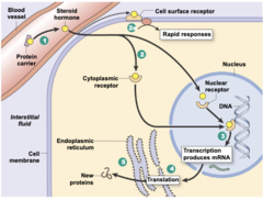
Hormone Interactions
increase or decrease level of response for endocrine signal
- combines effects of multiple hormone
Permissiveness (hormone interaction)
once hormone enhances cellular response to another hormone
ex. 2 hormone on same cell; the presence of 1 hormone enhances the response to another

Synergism (hormone interaction)
the combined effect of multiple hormones exceeds the sum of their individual effects
ex. 2 hormones present = stronger response not changing sensitivity and work better together

Antagonism (hormone interaction)
One hormone decreases the cellular response to another hormone
ex. one hormone can down regulate another hormone
Hormone Regulation
Trophic Hormones: endocrine regulation of hormonal release

Hypothalamus-Anterior Pituitary Axis
Main drivers of endocrine control & regulates release of growth hormones
1. neurons synthesizing trophic hormones release them into capillaries of portal system
2. portal vessels from hypothalamus carry the trophic hormones directly to the anterior pituitary
3. endocrine cells in anterior pituitary release their hormone into the second set of capillaries for distribution to the rest of the body

Endocrine Pathology: Hypersecretion
The appropriate amount of hormone is not being produced and secreted; can be due to problems with endocrine cells
hyposecretion - not enough hormone production/secretion
hypersecretion - too much hormone production/secretion
Endocrine Pathology: Target Cell Pathologies
Target cell receptors are important for allowing cells to receive signals
- Receptors on target cell not working properly
- Downregulation/loss of target cell receptors
- transduction of signals in target cells: signal pathway is not being initiated properly
Organization of the Nervous System
CNS = brain and spinal cord
Sensory neurons (afferents) = signals towards CNS
Efferent neurons = info from CNS to rest of body
1. autonomic neurons
- sympathetic: controls "fight or flight"
- parasympathetic: controls "rest or digest"
- controls everything but skeletal muscles
2. somatic motor neurons
- controls skeletal muscle

Glial Cells (NS)
Astrocytes: maintenance of extracellular spaces surrounding neurons; ions outside of neurons are important, so astrocytes clean up the area for neurons
Oligodendrocytes: lay down myelin, but limited to CNS
Schwann cells: generate myelin sheaths, but associate w/ PNS
Stem Cells: develop into any cells in NS
Neurons
- in chains with NT moving across; where info is processed & transmitted from one region to another
dendrites: regions where neuron interacts with pre-synpatic neurons; where signal from previous neuron determines if it will propagate or initiate AP (input signal)
cell body: integrating centers with propagation of info in NS, decision to initiate AP happens here (integration)
axon: extension of cell body
axon hillock: regions where a certain threshold of info needs to be met in order for AP can be propagated; signal need to pass here in order to go to next neuron! (output signal)
myelin sheath: surround axon; increase rate at which neuron propagates AP
Chemical Disequilibrium
what causes a membrane potential (like potential energy)
Nerst Equation uses ionic disequilibrium ~ find membrane potential with ion concentrations in/outside cell (K+, Na+, Cl-) and membrane permeability
Resting membrane potential ~ -70 mV
Phases of Action Potential
1 - resting state (-70 mV bc our membrane is more permeable to K+ and its channel is open to maintain it)
2 - depolarization phase (Na+ channels open into cell; positive feedback loop)
3 - overshoot (peak; Na+ channel inactivate)
4 - repolarization phase (K+ channels open now bc they are slower; K+ comes in)
5 - hyperpolarization (dip; brief period where K+ channels remain open until inactivated)
6 - return to resting (recovery; K+ channels close)

Refractory Period
Absolute Refractory Period: when Na+ channels are inactivated; another AP cannot be initiated since Na+ have to be open in order for it to happen
Relative Refractory Period: channels are offline & determines upperlimit of frequency of AP that can be sent out
- inactive = current cannot trigger to open; closed = current can

Action Potential Propagation
Local Current Flow: local charge that moves & interacts with neighboring sites
Graded Potential: signal that can have a negative or positive sign & any magnitude ~ small current within cell (what opens gated channels & triggers AP)
Depolarized section of the axon: creates a local current that moves in both directions & has the capacity to initiate another AP to the neighboring cell
THEREFORE, voltage gated channels are looking for a local deparlization event & flow of a local current as a trigger, changing confirmation of ion channels and initiating AP to start
AP moves in one direction!
The RATE of AP depends on the axon to current loss; increased resistance to current loss = maintain signal at longer distance

Propagation Rates
Speed of AP in neuron is influenced by:
- SIZE (diameter of axon; larger axon = faster since there's less resistance within the axon)
- MYELINATION (resistance of axon membrane to ion leakage out of the cell; myelinated axons = faster since it provide insulation to the axon and allows GP & local current to move at a rapid rate)
Saltatory Conduction (in myelinated axons)
- "Jumping" across the nodes of ranvier
- Decreased current leakage!
Myelin Degeneration
if myelin is compromised in any way = no regions of Na+/K+ voltage gated channels AND no adequate resistance to long, strong GP
- results in LOSS OF FUNCTION (disconnection of 2 ends of the axon due to action potential not being able to be propagated adequately)
Action Potentials
- All or none
- larger/longer graded potential = increased frequency of action potential
- Impacted by Na+ and K+
- Generated by voltage gated channels
- Depolarizing
- Refractory period

Graded Potentials
- No threshold
- Triggers action potential if it is large enough to meet the threshold
- Can have any sign, duration, size, etc.
- Can be depolarizing/hyperpolarizing (MP can be more neg or pos)
- impacted by any ions: Na+, K+, Ca2+, Cl-
- Generated by voltage, ligand, mechanically gated channels
- No refractory period; signals can sum
- tend to decrease in strength as they move down the cell body (so if input signal isn't strong enough, AP won't fire)

Graded Potential: Summation
Inhibitory: Inhibitory graded potentials work to prevent the signal from being initiated
- signals depending on their size, may cancel one another out and prevent it from reaching threshold
Excitatory: If there are multiple excitatory signals, they can add up to reach threshold and initiate an action potential
Types of summation (Integration)
Spatial summation: Signals from different areas sum together; if they add up to reach threshold → AP is initiated
Temporal summation: Graded potentials do not experience refractory periods. Therefore, multiple signals can be sent quickly to one another another and add up; if they add up to reach threshold → AP is initiated

Electrical Synpases
- Less common
- found primarily in the CNS, smooth muscle, and cardiac muscle
- Connected through gap junction like structures (connexins) ~ direct physical contact with GP moving through them
- depolarization at axon terminal → moves through gap junction as graded potential → initiates depolarization in postsynaptic neuron

Chemical Synpases
- more common
- found at the ends of neurons where electrical signals → chemical signals (outside terminal and across synaptic cleft)
- rich in mitochondria (used in the making of NT and metabolic activity)
- has synaptic vesicles that hold NT ; AP induces exocytosis and allows the vesicles to bind to the membrane to release NT
2 types of effects that occur:
Ionotropic (fast)
NT binds to receptors to open ion channels (ex. with Na+ channels at post-synaptic and allow changes in MP)
Metabotropic (slow)
NT binds to receptors to open/close ion channels and modify proteins/protein synthesis (ex. GPCR, G-protein transduction pathway with effector)
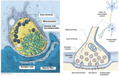
Events at the Synapse: Process
1. Action potential reaches axon terminal
2. Ca2+ channels open
3. Ca2+ enters the cell
4. Ca2+ initiates the release of neurotransmitters (triggers exocytosis)
5. Vesicle with neurotransmitters moves to membrane
6. Neurotransmitters released via exocytosis
7. Neurotransmitters diffuses across the synaptic cleft and bind to receptors on post-synaptic cell
8. Na+ channels open (depolarization) & AP propagated

Synthesis and Recycling of Neurotransmitters
Synthesis: acetyl CoA + choline = acetylcholine (Ach)
Breakdown & Recycling (synaptic cleft):
- Once it has been in the synaptic cleft for a while interacting with receptor → broken down by acetylcholinesterase
- Choline is transported back into axon terminal with Na+; Acetate diffuses into surrounding medium
- Recycled and packaged into vesicles to make Ach (vesicles available for depolarization)

Synaptic Transmission
Many neurotoxins target specific parts of a synapse and disrupt synaptic transmission; manipulation of ns can shut it down and block signals
- botulinum toxin (botox - bacteria): disrupts vesicle docking and no release of axon terminal
- bungarotoxin (sea snakes): binds to and block nitinic Ach receptors
Calcicludine (green mamba): blocks voltage gated Ca 2+ channels
Disruption of synpactic transmission is a symptom of many neurological disorders
CNS Architecture
Spinal Nerve Regions: serve as pathways through which info is interacting w/ CNS
-cervical, thoracic, lumbar, and sacral
Nerve Projections: the nerves connecting the body to the CNS
- info generated outside of NS coming in is utilizing the pathways
- info generating by CNS that we want to send out to the rest of the body we are aiming to control

Brain Anatomy
- Frontal lobe- cognitive function and voluntary movement
- Parietal lobe- sensory information processing
- Occipital lobe- vision
- Temporal lobe- processes emotions and connects them to sensory experiences
- Hypothalamus- maintain homeostasis
- Thalamus- relays sensory info to the cerebral cortex
- Pituitary- secretes hormones
- Pons- breathing
- Cerebellum- coordinated movement
- Medulla oblongata- involuntary function
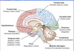
Brain Matter
White & gray matter indicate where there are regions with a lot of myelination & regions in CNS w/cell bodies or unmyelinated neurons
White Matter: myelinated axons; contains very few cell bodies; info moving quickly through region
Gray Matter: Unmyelinated axons, many cell bodies, dendrites, axon terminals

Hypothalamus
- activates sympathetic NS (fight or flight)
- maintains body temp (thermal regulation)
- controls body osmolarity (solute concentration)
- controls reproductive functions
- controls food intake (feeling hungry or full)
- influences cardiovascular control center in medulla oblongata
- regulates release of hormone form the pituitary gland
Spinal Cord Anatomy (gray matter)
Visceral Sensory Nuclei: sensory info from our viscera is comign down the dorsal root ganglion & synapsing with cells in the regions
Dorsal Root Ganglian (affarent): getting sensory info from periphery and sending it toward spinal cord --> nerve projections --> dorsal horn (region that get afferent info)
Ventral Root (efferent): sending info out to rest of body from ventral horn
Somatic Motor Nuclei: where efferent signals are sent to somatic systems (muscles & skin), the cell bodies of those neurons are housed in here or ventral horn
Autonomic efferent nuclei: housing cell bodies of neurons that will send efferent info to body (lateral horn)
EFFERENT: coming out of ventral or lateral sides
AFFERENT: coming into dorsal side
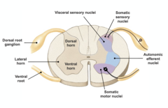
Spinal Cord Anatomy (white matter)
responsible for communication within the CNS, specifically between the brain and the spinal cord
descending tract: info from brain --> spinal cord
ascending tract: info through spinal cord --> brain
(sensory info --> dorsal root ganglia --> integrated in gray matter --> to brain using ascending tract)
Rapid Reflex Pathway
1. stimulus
2. sensory input
3. dorsal horn
4. intervention (interneuron: small neurons that links dorsal horn with efferent side of things)
5. visceral horn (lateral)
6. response (where info is sent to target)
(Since it is so fast:
Little to no integration,
No feedback,
Lots of myelination,
limit number of synapses!!
Sensory info sent to the brain after to process what happened)
ex. knee tap
1. stimulus
2. sensory input
3. CNS
4. autonomic/somatic neurons
5. target cell/muscle
6. response

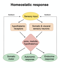
Homeostatic Response
- Slower
- Lots of feedback regulation to maintain within a range of normal values
- Constantly being monitored
- Antagonistic branches
- Mechanisms that work in opposition to one another
(Ex. parasympathetic vs. sympathetic)
Use of same neurotransmitters, segregate use of PNS or CNS through receptors
Ex. muscarinic receptor (PNS), alpha and beta1 receptors (CNS)
Withdrawal Reflex
1. pain from heat
2. info goes to spinal cord via afferent sensory neuron
3. dorsal root ganglia
4. dorsal horn
5. interneuron (not gray matter, but is myelinated and a connection between the different horns -- slowest part of the process)
5. efferent side (ventral horn and out the ventral root)
6. efferent somatic motor neuron (tells muscles to fire quickly)
7. rapid reflex response!

Homeostasis Responses
- Slower
- Lots of feedback loops to constantly maintain within a range of normal values
- Must constantly be monitored
- Antagonistic branches: Multiple mechanisms that work in opposition to each other
(Ex. parasympathetic/ sympathetic)
- Same neurotransmitters used, but different receptors for different responses

Autonomic NS
- spatially segregated
Effectors: controls smooth & cardiac muscle, exocrine & endocrine glands, lymphoid & adipose tissue
Parasympathetic
- Acetylcholine (Ach)
Sympathetic
- Norepinephrin (NE)
Adrenal Sympathetic Pathway
- Epinephrin
the 2 branches of the ANS affect the same target cells but produce antagonistic effects by interacting with different receptors on the same cell

Types of Muscles
Skeletal
- one of the biggest in the body
- attached to skeleton
- striated (banding pattern tells how motor proteins and other structural components are put together)
- multinucleated (multiple nuclei in each cell since it's a bigger cell; more nucleus to regulate the cell = achieves diffusion, keeping nuclear domain reasonable)
- fastest
- somatic motor
Cardiac
Found in the heart, striated, sarcomeres, uninucleated, intermediate contraction, controlled autonomically
Smooth
Found in internal organs/vessels, smooth in texture, contains oblique bundles, nucleated cells, slowest contraction, controlled autonomically, SLOWEST
Fundamental Function
For muscle contraactions to work, muscles must convert chemical energy --> mechanical energy
- heat is the biproduct
Antagonist Pairs of Muscle
as they act as a joint, one moves the joint in one direction and the other moves the join in the opposite direction
(organized in antagonistic pairs)
ex. tricep relaxes when bicep contracts
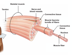
Organization of Skeletal Muscle
- The smallest component of skeletal muscles (besides the cell themselves) are the myofibrils
- Many myofibrils come together to form muscle fibers
- Many muscle fibers (muscle cell) then come together to form a muscle fascicle
- Many muscle fascicles then come together to form the skeletal muscle itself
- muscle fascicle: group of muscle cells incased in their own connective tissue wrapping
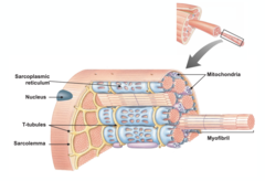
Muscle Fiber Structure
Sarcoplasmic reticulum: stores and releases the calcium required for contraction
Nucleus: houses genetic material
T-tubules: invaginations that allow for faster signal conduction; extensions of the cell membrane to send signal quickly bc cell is too big
Mitochondria: produces ATP that the cell requires to convert chemical to mechanical energy
Myofibril: performs the actual contractions for the muscle; where contractile proteins are stored (more myofibril = more force generated; ATP --> force)
Sarcolemma: muscle cell membrane/sheath around muscle fibers interacts with extracellular environment
Ultrastructure of Muscle
Sarcomeres: fundamental unit of contraction
Titin: stabilizes thick filament; springy structural cytoskeleton; regulates the position of the thin and thick filaments; provides structural integrity and provides tension when muscle is not active
Z disk: boundaries of sarcomere and attachment sites for thin filaments; forces generated by our motor proteins need to go through the Z disc to be transmitted out to the rest of the cell
M line: center of sarcomere
Thin filament: composed of actin, troponin, and tropomyosin
- have actin chain
- tropomyosin: wraps around actin and prevents myosin from grabbing it (when Ca binds to it, it allows myosin to interact with actin); blocking binding sites of g-protein
- troponin: regulatory proteins
Thick Filament: composed of myosin and myosin heads; works with actin to contract muscle
-myosin head: site where they interact with actin & serves regulatory function int he sue of hydrolization of ATP during contraction

Summary of Muscle Contraction
Events at neuromuscular junction (input of neural signal from NS & interact w/skeletal muscle cells; synapse between motor neurons & skeletal muscle) --> excitation contraction coupling (descriptor of how across a synapse, electrical signal is generating within the skeletal muscle cells a Ca2+ signal) --> Ca2+ signal --> Contraction-relaxation Cycle (ATP to ADP and remove calcium) --> muscle twitch or sliding filament theory

Neuromuscular Junction
Place where somatic neuron is coming in & creating a synapse w/ skeletal muscle cell or fiber
1. Action Potential Arrival: AP depolarizes axon terminal
2. Calcium Ion Influx: Depolarization causes calcium to enter axon; vesicle then dock and attack to cell membrane
3. Ach Release: ACh is released into synapse through exocytosis
4. Ach & post-synaptic cell: ACh binds to closed channels --> open and influx in Na+ and an efflux in K+
(more Na+ comes in than K+ comes out)
5. depolarization occurs, provides GP and propagate to both directions --> initiate AP
nervous system can regulate the force output of skeletal muscles by varying the frequency of action potentials sent to the neuromuscular junction.

Excitation Contraction Coupling
1. action potential
2. release of Ach
3. ion channels open (NA+ comes in)
4. generate depolarization
5. AP is initiated in both directions away from the neuromuscular junction
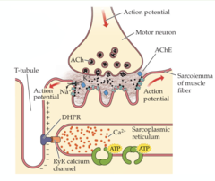
The Calcium Signal
Activation
1. depolarization
2. RyR calcium channel separates from DHPR, spilling out Ca2+ from sarcoplasmic reticulum
3. Ca2+ moves down concentration gradient (passive)
4. interacts with motor protein
Relaxation
1. no more depolarization signal
2. RyR channel closes
3. triggers calcium pumps to bring Ca2+ back into SR from cytoplasm (active)
Calcium plays an important role in muscle contraction as it binds to binding sites on troponin and causes a shift in tropomyosin.
Shifting the tropomyosin now exposes the myosin binding sites and now myosin can interact with troponin (troponin loses affinity for actin) and cause muscle contraction.

Contraction Cycle
1. When ATP is hydrolyzed (dephosphorylates), myosin undergoes a conformational change and this conformational change allows it to weakly bind actin (cross bridge forms = connection between myosin and actin)
2. When the inorganic phosphate leaves the myosin head, it causes another conformational change in myosin and this conformation change allows myosin to strengthen its bond to actin and ultimately cause the power stroke
3. When ADP is lost, the cross-bridge enters the transient rigor state (brief).
4. During the rigor state, myosin remains bound to actin
5. Upon ATP binding, the myosin head will undergo another conformational change that allows it to release from actin. (detachment of cross-bridges and Ca2+ uses it to move Ca back to SR)

Regulation (muscle contraction)
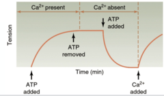
- ATP & Ca2+ regulate muscle contractions in different ways (required for contraction)
- both are required for a muscle to initiate the process of cross-bridge cycling
- when Ca2+ is removed & ATP is present --> force decays and tension drops
- when ATP is removed after cross-bridge cycling is initiated, tension does not drop (cross-bridge can't go back to relaxed state)
Ca + ATP = increase tension
Ca - ATP = remain the same
ATP - Ca = tension drops
Sliding Filament Theory
theory that actin filaments slide toward each other during muscle contraction, while the myosin filaments are still (Z-disc move to center toward M-line)
determines at what lengths of sarcomeres are we capable of generating high forces & what lengths are we compromised the ability to form cross-bridges
A band (entire length of thick filament- overlaps with thin filaments) does not change length!
H zone and I band shorten as Z lines move inward!
H zone → smaller section of A band; only contains thick filaments
I band → only contains thin filaments

Length-Tension Relationship
highest force = optimal overlap (sarcomere length is important)
If the sarcomere is too long, there is not enough overlap between thick and thin filaments
- As a result, the maximum amount of cross bridges cannot form and this prevents the sarcomere from functioning at its maximum potential --> leads lower overall tension in the muscle
If the sarcomere is too short, there is too much overlap between thick and thin filaments
- Too much overlap means the sarcomere is already mostly contracted
- lower probability of cross-bridges generated
- the sarcomere cannot contract much further which also ultimately lowers tension in the muscle
Shorter → some negative interaction; actin competing for space; z disks run into myosin filament
Longer → less overlap between actin and myosin; limiting number of cross bridges
**Isometric → generating force, but not changing length
intrinsic property: geometry of sarcomere determines the conditions in generating high forces vs low forces

Force Velocity Relationship
- inverse relationship between the amount of force a muscle can contract at and the speed at which that muscle can contract at (as speed/velocity increases, force of muscle or load at which is contracts decreases)
- At the end of the day, there is a trade-off
This trade-off is mainly driven by the time required for cross-bridge detachment
- slow contraction = more cross-bridges; take configuration that counteracts force rather than contributing to it
- fast contraction = actin filament moves towards center, cross-bridges don't have time to attach or detach and pulls direction opposing forces by other cross-bridges (opposing force production)

Muscle Twitch
when the muscle develops tension in response to stimulus once (smallest contractions)
Slight delay between action potential in motor neuron and action potential in muscle cell (sarcolemma)
Process
Action potential motor neuron
Action potential in muscle cell
1. Latent period: Action potential travels down t-tubules, release Ca2+, etc;
- Takes some time - no force generated yet
2. Contraction phase (Powerstroke)
3. Relaxation phase: Ca2+ sequestered back into SR; Length of relaxation phase dependent on how fast Ca2+ can be removed from the cytoplasm
**pattern is very stereotyped; always looks the same!**

Twitch Summation
when multiple muscle twitches occur in rapid succession and the tensions of these muscle twitches combine with one another
- mechanism NS can regulate muscle force; adding AP closer together allows forces to sum
Unfused Tetanus
rapid succession fast enough to maintain muscle tension above a certain level
However, there is still enough time between twitches for the muscle to relax a little (this explains the small drops in tensions)
Fused Tetanus
rapid succession fast enough to maintain muscle tension at maximum tension
For this type of tetanus, muscle twitches occur so rapidly that the muscle does not get to relax a little between each twitch
Muscle twitches occur faster for fused tetanus and slightly slower for unfused tetanus
- NS can regulate the amount of tension regenerated in our skeletal muscles through change in the frequency of AP that are sent
(regulating force ~ fine-tuning force)
- more force = more cells recruited

Muscle Force Regulation
1) through changes in AP frequency
- higher frequency = higher force
2) through changes in the number of muscle cells (motor units) activated
- more motor units = higher force
Motor Unit Recruitment
a motor unit consists of one motor neuron and all the fibers it innervates
(only a certain set of fibers)
Not all motor units have the same number/size of muscle cells; they contribute different amounts of force! (small MU = small force)
Start small → intermediate → large when recruiting motor units as needed
(More energetically efficient)

Fiber Types
Slow
- Found in small motor units
- Can achieve intermediate tension in a long period of time
- Slow oxidative (more efficient)
- lower Vmax
- Takes longer to develop tension/release tension
- slow myosin ATPase
- a lot of mitochondria
- has myoglobin (red)
Intermediate
- Found in intermediate motor units
- Can achieve somewhat high tension in a short amount of time
- Fast oxidative (but somewhat inefficient)
- intermediate Vmax
- has myoglobin (red)
Fast
- can develop force more rapidly
- develops and relaxes very quickly
- faster myosin ATPase = attach and hydrolyze ATP quicker
- fast glycolytic
- large fibers
- greater Vmax
- found in large motor units
- Can achieve high tension in a short amount of time
- no myoglobin (white)
- easily fatigued
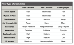
Contractile Speed Comparison
Slowest to Fastest
Smooth muscle --> cardiac muscle --> skeletal muscle
Smooth Muscle Characteristics
internal organs
smooth
oblique bundles
uni-nucleated
slowest (only forces needed to forms cells themselves)
autonomic
line outer edges of hollow organs (serves as sheet and changes shape of themselves)
long contractile speed (not forceful bc no big loads; slow)
Layers of Smooth Muscle
Organized into layers that are antagonistic
Oblique: Diagonal
Longitudinal: Up and down along organ's vertical axis (along length)
Circular: Side to side (circumference of the organ)
longitudinal and circular are antagonistic
**Tip: think of a water filled balloon and what happens when you squeeze different parts! Other parts of the balloon change shape to compensate!**
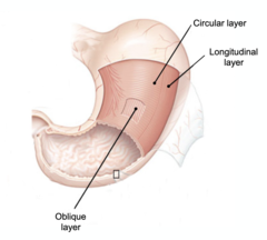
Anatomy of Smooth Muscle
- Consists of dense bodies (like z-discs), filament bundles of actin and myosin
- single nucleus
- Muscle can contract in multiple directions as opposed to skeletal muscle
- Similar to skeletal muscles, thick myosin filaments interact with thin actin filament to cause contraction
- no need for many contractile proteins since smooth muscle only changes its own shape
- obliquely orientated filaments
- Cross bridges oriented in opposite directions and move in opposite directions to pull actin and contract the muscle
- less myofilaments compared to skeletal muscle = less force

Varicosities in Smooth Muscle
the autonomic NS sends signal down the postganglionic autonomic neuron until it reaches varicosities
varicosities: expansions of neurons (operate as a replacement for axon terminals)
- attached directly on and spreads throughout muscle
- holds NT and release them to regulate the smooth muscle
- rather than relying on synapse for single cell, the ANS induces an excitatory signal causing the varicosities to release NT out into EC space and wash over smooth muscle layer
- function is to hold neurotransmitters and release them to regulate the smooth muscle
Types of Smooth Muscle: Single Unit
- Muscle fibers joined by gap junctions so that the muscle contracts as a single unit
- Found in the walls of all visceral organs except the heart
- Also found in small intestine
- coordinated process = moves as 1 unit
1. NT from varicosity reaches receptor on muscle cell
2. receptor causes depolarization & induces GP
3. GP moves through gap junction
4. induces contraction and travels cell to cell
5. slow function, but well coordinated

Types of Smooth Muscle: Multiple Unit
- Rarely possess gap junctions so they are not electrically coupled
- Contraction does not spread from one cell to the next, but is instead confined to the cell that was originally stimulated
- Found in large blood vessels and the eye
- neuron has to extend and intertwine with all the cell
- cell is looking for EC signaling in order to contract
- recruited in multi-unit pathway: recruits some but not all sheets of smooth muscle
excitatory signal --> coordinated response the entire tissue, thus being more selective
Calcium Signal in Smooth Muscle
smooth muscles can be tonic: refers to a muscle that always has some baseline level of tension
- sometimes can be closed at all times and excitatory signal opens it
- SO muscles are always generating tension
Process
Signal (Ex. Neurotransmitter, hormone, stretch or depolarization, etc.)
Receptor: Increased IP3 (Inositol triphosphate- important for smooth muscle contraction)
IP3-R: R = receptor; IP3 binds to receptor on SR and causes the release of Ca2+ (Similar function to Ryr receptor in skeletal muscle)
Sarcoplasmic reticulum (SR)
Ca2+ released
Muscle contraction
Alternative pathway of bringing Ca2+ into the cell
Signal
Membrane channel (Extracellular Ca2+ enters cells)
Increased Ca2+ entry
Muscle contraction
Pathway: Ca2+ stores low in SR
- Low concentration of Ca2+ in SR detected
- Store operated Ca2+ channels in the membrane open and enter the cell
- Ca2+ is brought to SR and used in muscle contraction
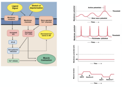
Smooth Muscle Contraction
1. Increase in Calcium
- Intracellular Ca2+ concentrations increase when Ca2+ enters cell and is released from SR
2. Calmodulin
- The Ca2+ that enters the cells finds its way to calmodulin, binds to it, and activates calmodulin (acts as troponin)
3. Calmodlin and MLCK
- The activated calcium bound calmodulin then activates myosin light chain kinase (MLCK)
4. MLCK Function
- After MLCK is activated, it phosphorylates light chains in myosin heads and increases myosin ATPase activity
5. Muscle Tension Created
- active myosin cross-bridges slide along actin and create muscle tension

Smooth Muscle Relaxation
1. Decrease in Calcium
- The free calcium in the cytosol decreases when the calcium is pumped out of cell or into the sarcoplasmic reticulum
2. Calmodulin Unbinding
- As calcium concentration decreases, it also unbinds from calmodulin and this ultimately leads to the deactivation of MLCK
3. Myosin Phosphatase
- myosin phosphatase comes along and removes phosphate from myosin, which results the decrease in myosin ATPase activity
4. Muscle Tension Drops
- Since there is less ATPase activity, the muscle tension also decreases because cross bridges no longer form or are active
