Ventricular Dysrhythmias

0.0(0)
Studied by 0 people
call kaiCall Kai
learnLearn
examPractice Test
spaced repetitionSpaced Repetition
heart puzzleMatch
flashcardsFlashcards
GameKnowt Play
Card Sorting

1/19

encourage image

There's no tags or description

Looks like no tags are added yet.

Last updated 2:35 AM on 4/16/25
Name
Mastery
Learn
Test
Matching
Spaced
Call with Kai
Chat

No analytics yet

Send a link to your students to track their progress

20 Terms
1
New cards
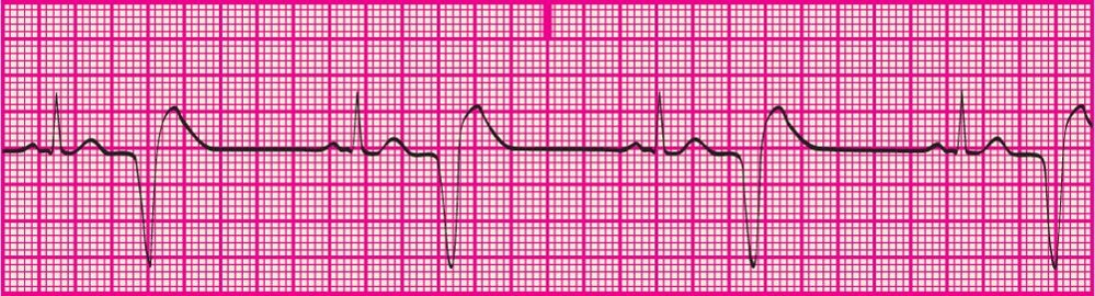
<p>A pattern of PVC in which every other complex is a PVC</p>

A pattern of PVC in which every other complex is a PVC

Bigeminy
2
New cards
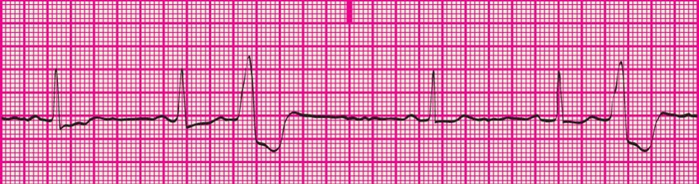
<p>a pattern of PVCs in which every 3rd complex is a PVC</p>

a pattern of PVCs in which every 3rd complex is a PVC

Trigeminy
3
New cards
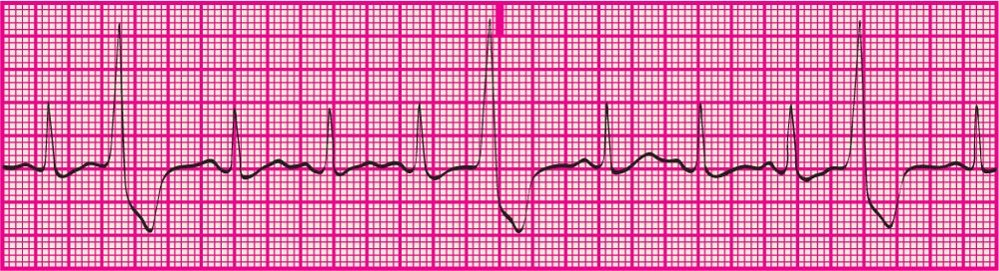
<p>every 4th complex is a PVC</p>

every 4th complex is a PVC

Quadgeminy
4
New cards
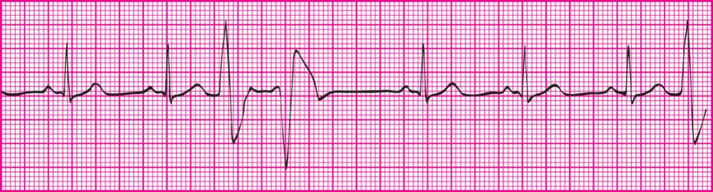
<p>Two PVCs that occur back to back</p>

Two PVCs that occur back to back

Coupling
5
New cards
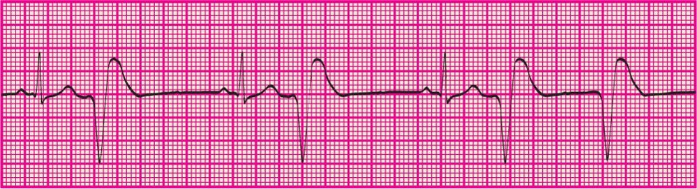
<p>Six or more PVCs per minute</p>

Six or more PVCs per minute

Frequent PVCs
6
New cards
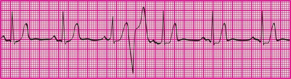
<p>PVC that occurs during the normal R-R Interval without interrupting the underlying rhythm</p>

PVC that occurs during the normal R-R Interval without interrupting the underlying rhythm

Interpolated PVC
7
New cards
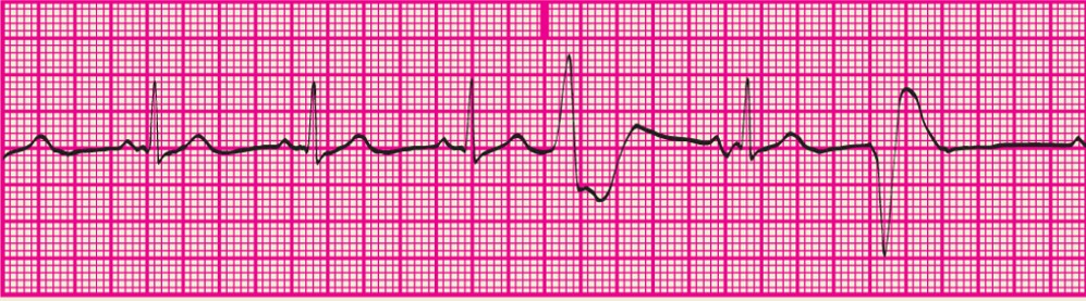
<p>Varied shapes and forms of the PVCs, suggesting more than one irritable focus</p>

Varied shapes and forms of the PVCs, suggesting more than one irritable focus

Multifocal PVC
8
New cards
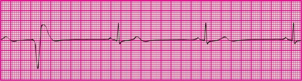
<p> Less than 6 PVCs per minute</p>

Less than 6 PVCs per minute

Occasional PVC
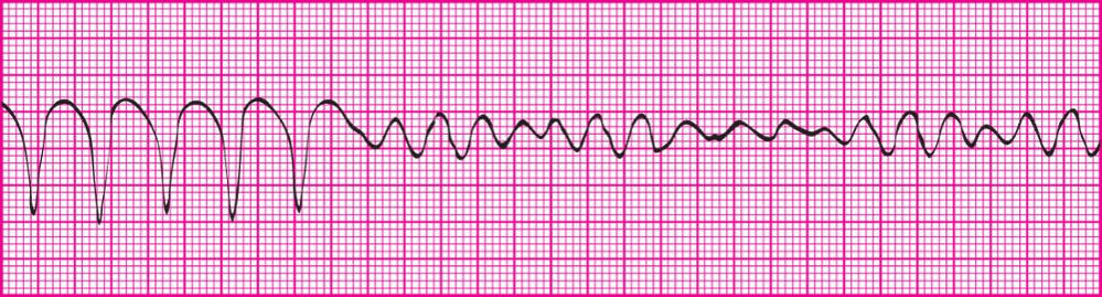
9
New cards
<p>A witnessed change in any cardiac rhythm, including starting, stopping or both; for ex- sinus rhythm to SVT or Ventricular Tach to Ventricular fibrillation</p>

A witnessed change in any cardiac rhythm, including starting, stopping or both; for ex- sinus rhythm to SVT or Ventricular Tach to Ventricular fibrillation

Paroxysmal Event

10
New cards

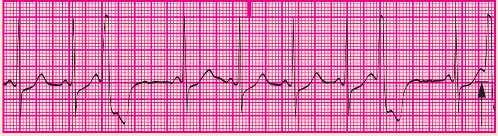
An ectopic impulse originating in either ventricle that occurs too early in the cycle


Premature Ventricular Complex (PVC)
11
New cards
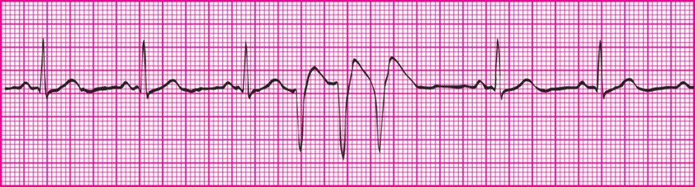
<p>PVC that occurs on the downslope of the T wave or the vulnerable period of the relative ventricular refractory period</p>

PVC that occurs on the downslope of the T wave or the vulnerable period of the relative ventricular refractory period


R on T PVC
12
New cards
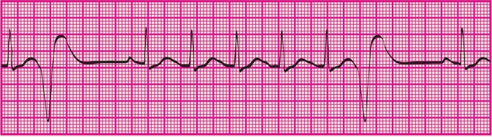
<p></p><p>Three or more PVCs occuring in a row at a rate greater than 100bpm</p>


Three or more PVCs occuring in a row at a rate greater than 100bpm

Run of Ventricular Tachycardia or Salvo or Triplet PVC
13
New cards
<p>Early Complexes that have a similar shape, suggesting only one irritable focus present</p>

Early Complexes that have a similar shape, suggesting only one irritable focus present

Unifocal PVC
14
New cards
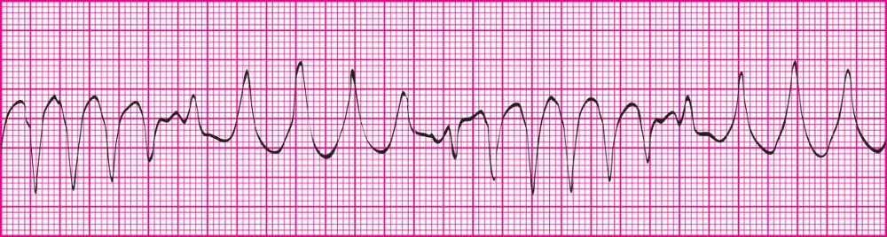
<p>Depolarization impulses move from one ventricle to the other;  occurs due to electrolyte deficiencies; treatment includes magnesium sulfate</p><p></p><p>Rhythm: P-P interval usually not identifiable; R-R interval usually regular, can be slightly irregular at times.</p><p>Rate: Ventricular rate- 100‒200 beats per minute; Atrial rate- Cannot be determined</p><p>P wave: Absent</p><p>PR interval: Cannot be determined</p><p>QRS: Duration greater than 0.12 second; Wide, bizarre appearance; T wave in opposite direction (usually down) from QRS complex</p><p></p><p>Lost atrial kick and decreased ventricular filling time result in decreased cardiac output.</p><p>Approximately 50% of patients become unconscious immediately.</p><p>Notify licensed practitioner immediately.</p><p>Save rhythm strips in medical record to document rhythm changes.</p>

Depolarization impulses move from one ventricle to the other; occurs due to electrolyte deficiencies; treatment includes magnesium sulfate


Rhythm: P-P interval usually not identifiable; R-R interval usually regular, can be slightly irregular at times.

Rate: Ventricular rate- 100‒200 beats per minute; Atrial rate- Cannot be determined

P wave: Absent

PR interval: Cannot be determined

QRS: Duration greater than 0.12 second; Wide, bizarre appearance; T wave in opposite direction (usually down) from QRS complex


Lost atrial kick and decreased ventricular filling time result in decreased cardiac output.

Approximately 50% of patients become unconscious immediately.

Notify licensed practitioner immediately.

Save rhythm strips in medical record to document rhythm changes.

Torsades De Pointes (SVT or VT)
15
New cards
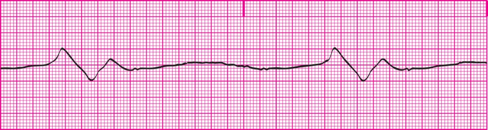
<p>Occurs when all the pacemakers of the heart have failed; the heart is dying; ventricular rate is less than 20bpm</p><p></p><p></p><p>Rhythm: There is no P-P interval; R-R may or may not be regular</p><p>Rate: Ventricular- Less than 20bpm; Atrial- cannot be determined</p><p>P wave: No P wave is present</p><p>QRS: 0.12 secs or greater; wide, bizarre appearance</p><p>Profound loss of cardiac output.</p><p>Patient will be unconscious.</p><p>Notify health care practitioner immediately.</p><p>This is a medical emergency; BLS and ACLS interventions will be initiated.</p><p>ECG strips must be saved and put in medical record.</p><p><em>**Medical Emergency</em>**</p>

Occurs when all the pacemakers of the heart have failed; the heart is dying; ventricular rate is less than 20bpm


Rhythm: There is no P-P interval; R-R may or may not be regular

Rate: Ventricular- Less than 20bpm; Atrial- cannot be determined

P wave: No P wave is present

QRS: 0.12 secs or greater; wide, bizarre appearance

Profound loss of cardiac output.

Patient will be unconscious.

Notify health care practitioner immediately.

This is a medical emergency; BLS and ACLS interventions will be initiated.

ECG strips must be saved and put in medical record.

**Medical Emergency**

Agonal Rhythm
16
New cards
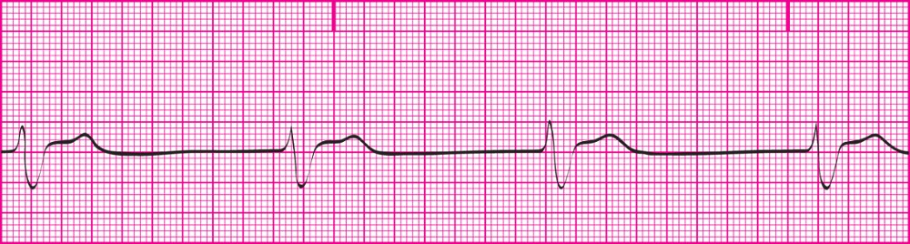
<p> Impulse created by the ventricular pacemaker; SA node &amp; junctional Pacemaker failed to initiate impulse; presents with classic QRS, slow ventricular rate; &amp; no P waves; 20-40bpm</p><p></p><p>Rhythm: R-R interval is regular; P-P interval cannot be determined</p><p>Rate: Ventricular- 20-40bpm; Atrial- Cannot be determined</p><p>P wave morphology: No P wave is present.</p><p>PR interval: Cannot be measured</p><p>QRS duration and morphology: 0.12 seconds or greater; Wide, bizarre appearance</p><p></p><p><strong>**This is a medical Emergency**</strong></p><p>Profound loss of cardiac output</p><p>The patient will likely be unconscious.</p><p>Notify health care practitioner immediately.</p><p>This is a medical emergency.</p><p>Likely to require medication and/or pacing.</p><p>ECG strips must be saved and put in medical record.</p>

Impulse created by the ventricular pacemaker; SA node & junctional Pacemaker failed to initiate impulse; presents with classic QRS, slow ventricular rate; & no P waves; 20-40bpm


Rhythm: R-R interval is regular; P-P interval cannot be determined

Rate: Ventricular- 20-40bpm; Atrial- Cannot be determined

P wave morphology: No P wave is present.

PR interval: Cannot be measured

QRS duration and morphology: 0.12 seconds or greater; Wide, bizarre appearance


**This is a medical Emergency**

Profound loss of cardiac output

The patient will likely be unconscious.

Notify health care practitioner immediately.

This is a medical emergency.

Likely to require medication and/or pacing.

ECG strips must be saved and put in medical record.

Idioventricular Rhythm
17
New cards
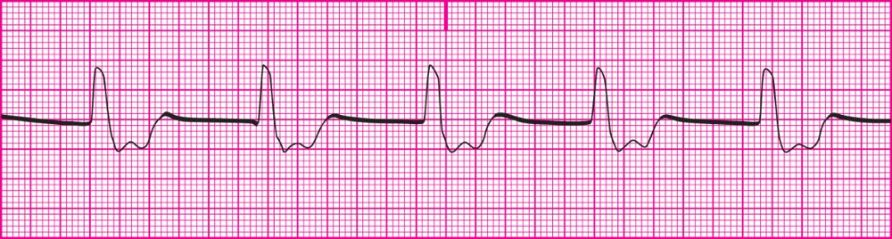
<p>impulse created by the ventricular pacemaker due to SA and AV node failure; QRS is wide &amp; bizarre; P waves are absent; 40-100bpm</p><p></p><p>Rhythm: R-R interval is regular; P-P interval cannot be determined</p><p>Rate: Ventricular- 40‒100 bpm; Atrial- Cannot be determined</p><p>P wave: No P wave is present</p><p>PR Interval: Cannot be identified</p><p>QRS: 0.12 second or greater; Wide, bizarre appearance</p><p></p><p>Decrease in cardiac output due to slow ventricular rate and loss of atrial kick.</p><p>Patient may be unconscious.</p><p>Notify health care practitioner immediately.</p><p>May require medication and/or pacing.</p><p>ECG strips must be saved and put in medical record.</p>

impulse created by the ventricular pacemaker due to SA and AV node failure; QRS is wide & bizarre; P waves are absent; 40-100bpm


Rhythm: R-R interval is regular; P-P interval cannot be determined

Rate: Ventricular- 40‒100 bpm; Atrial- Cannot be determined

P wave: No P wave is present

PR Interval: Cannot be identified

QRS: 0.12 second or greater; Wide, bizarre appearance


Decrease in cardiac output due to slow ventricular rate and loss of atrial kick.

Patient may be unconscious.

Notify health care practitioner immediately.

May require medication and/or pacing.

ECG strips must be saved and put in medical record.

Accelerated Idioventricular Rhythm
18
New cards
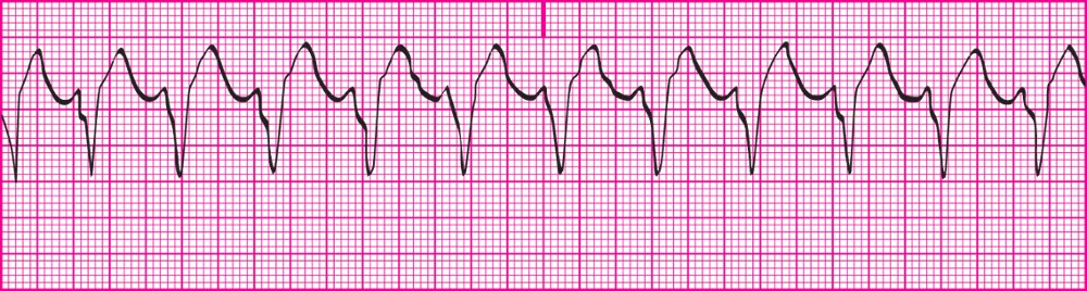
<p>Occurs when there is 3 or more PVCs in a row &amp; the HR is over 100bpm; Ventricles are continously contracting &amp; relaxing with no period of repolarization.</p><p></p><p>Rhythm: P-P interval usually not identifiable; R-R interval usually regular, can be slightly irregular at times.</p><p>Rate: Ventricular rate- 100‒200 beats per minute; Atrial rate- Cannot be determined</p><p>P wave: Absent</p><p>PR interval: Cannot be determined</p><p>QRS: Duration greater than 0.12 second; Wide, bizarre appearance; T wave in opposite direction (usually down) from QRS complex</p><p></p><p>Lost atrial kick and decreased ventricular filling time result in decreased cardiac output.</p><p>Approximately 50% of patients become unconscious immediately.</p><p>Notify licensed practitioner immediately.</p><p>Save rhythm strips in medical record to document rhythm changes.</p>

Occurs when there is 3 or more PVCs in a row & the HR is over 100bpm; Ventricles are continously contracting & relaxing with no period of repolarization.


Rhythm: P-P interval usually not identifiable; R-R interval usually regular, can be slightly irregular at times.

Rate: Ventricular rate- 100‒200 beats per minute; Atrial rate- Cannot be determined

P wave: Absent

PR interval: Cannot be determined

QRS: Duration greater than 0.12 second; Wide, bizarre appearance; T wave in opposite direction (usually down) from QRS complex


Lost atrial kick and decreased ventricular filling time result in decreased cardiac output.

Approximately 50% of patients become unconscious immediately.

Notify licensed practitioner immediately.

Save rhythm strips in medical record to document rhythm changes.

ventricular Tachycardia
19
New cards
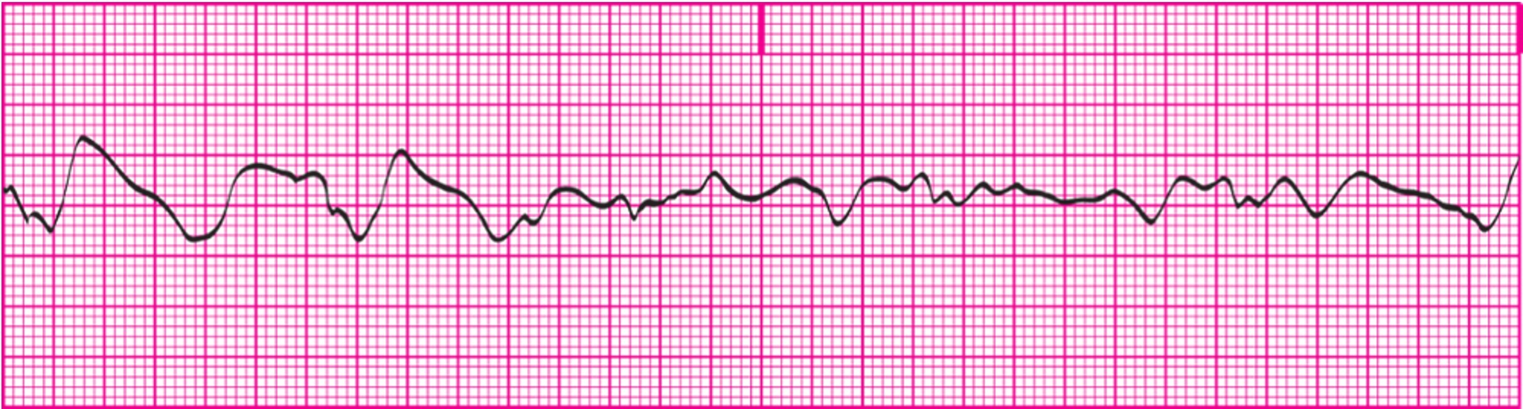
<p>chaotic, asynchronous electrical activity within ventricular tissue; Ventricle walls quiver, preventing ejection of blood; No cardiac output</p><p>Rhythm: P-P and R-R intervals cannot be determined because neither P waves nor R waves are present.</p><p>Rate: Neither the atrial nor the ventricular rate can be determined.</p><p>P wave morphology: No P waves are present.</p><p>PR interval: There is no PR interval.</p><p>QRS duration and morphology: Cannot be determined.</p><p>Always check the patient first.</p><p>In true *<em>Rhythm name</em>* : ***Medical Emergency***</p><p>Patient is unconscious, apneic, and pulseless</p><p>True emergency situation</p><p>Initiate Code Blue and begin CPR/ACLS procedures immediately.</p>

chaotic, asynchronous electrical activity within ventricular tissue; Ventricle walls quiver, preventing ejection of blood; No cardiac output

Rhythm: P-P and R-R intervals cannot be determined because neither P waves nor R waves are present.

Rate: Neither the atrial nor the ventricular rate can be determined.

P wave morphology: No P waves are present.

PR interval: There is no PR interval.

QRS duration and morphology: Cannot be determined.

Always check the patient first.

In true *Rhythm name* : ***Medical Emergency***

Patient is unconscious, apneic, and pulseless

True emergency situation

Initiate Code Blue and begin CPR/ACLS procedures immediately.

Ventricular Fibrillation

20
New cards
<p>Absence of ventricular activity and depolarization; no electrical activity is present in the myocardium; straight line/ flatline; Always confirm waveform in at least two different leads to rule out “fine” ventricular fibrillation.</p><p></p><p>Rhythm: No waveforms are present.</p><p>Rate: No atrial or ventricular rates are present.</p><p>P wave morphology: No P waves are present.</p><p>PR interval: Cannot be measured</p><p>QRS duration and morphology: Not measurable</p><p></p><p>Situation is life-threatening.</p><p>Patient will be unconsciousness and apneic.</p><p>Patient is in cardiac arrest; initiate emergency procedures.</p><p>Always a Code Blue situation.</p>

Absence of ventricular activity and depolarization; no electrical activity is present in the myocardium; straight line/ flatline; Always confirm waveform in at least two different leads to rule out “fine” ventricular fibrillation.


Rhythm: No waveforms are present.

Rate: No atrial or ventricular rates are present.

P wave morphology: No P waves are present.

PR interval: Cannot be measured

QRS duration and morphology: Not measurable


Situation is life-threatening.

Patient will be unconsciousness and apneic.

Patient is in cardiac arrest; initiate emergency procedures.

Always a Code Blue situation.

Asystole



