(MMI) Lab Module 3: Plates and Plating Techniques
 Call Kai
 Learn
Learn Practice Test
 Spaced Repetition
 Match
Match Flashcards
 Knowt Play
1/32

Earn XP
Description and Tags
Students will apply aseptic, pure culture, and selective isolation techniques to cultivate, enrich, and isolate microorganisms from mixed cultures
Name | Mastery | Learn | Test | Matching | Spaced | Call with Kai | Chat |
|---|
No analytics yet
Send a link to your students to track their progress
33 Terms
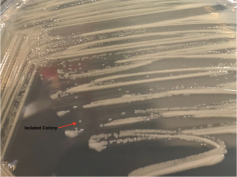
Isolation
•refers to a single, pure culture (colony/CFU) of an organism obtained from a mixed culture
Colony Forming Unit (CFU)
•a group of cells originating from a single cell or small group of cells

Four-Quadrant Streak (GOLD STANDARD FOR ISOLATION)
•used for high cell density or mixed samples (a loop is used)
•is “diluted” quadrant by quadrant until isolates are formed
•Qualitative Streak Method

Zig-Zag Streak
•used for low cell density or pure samples (a swab is used)
•sample is not diluted
•Quantitative Streak Method
Colony
•a visible mass of cells
•Isolated colonies in either the 3rd or 4th quadrants or both

•Good techniques for plating should include:
•Good use of plate space
•No unnecessary intersections or overlapping of quadrants
•No cuts or gouges into the agar
•Decreasing bacterial density with each subsequent quadrant

Factors Influencing Colony Morphology
•Temperature
•Incubation Time
•Nutrient Conditions
(Morphology is read from the 3rd or 4th quadrant)
COLONY MORPHOLOGY CATEGORIES
S S P O T M E
Size
Shape/Whole Colony
Pigment
Optical
Texture
Margin
Elevation
Size
: the physical size of an individual colony
punctiform (<1mm), small, medium, large, etc.
Shape/Whole Colony
: the physical shape of an individual colony
round, irregular, filamentous, rhizoid, etc.
Pigment
: color produced by the bacterium
yellow, red, green, etc.
Optical
Opacity of the colony
transparent, translucent, opaque
Texture
The physical “feel” of a colony
dry, wet, mucoid, etc.

Margin
the physical appearance of the edge of a colony
smooth/entire, irregular, lobate, etc.

Elevation
how tall/short the colony is in relation to the agar level
convex, umbonate, flat, raise, etc.

What is a great instrument to use for magnifying colonies up close (morphology) ?
Stereoscope

Morphology Example (SSPOTME)
Size: punctiform
Shape: round
Pigment: yellow
Optical: opaque
Texture: moist
Margin: smooth/entire
Elevation: convex
Differential Medium (Experiment 3-3)
to tell the difference between, or differentiate, different organisms
•Differentiation based on hemolysis
Hemolysis
•Lysing, or breaking down, of red blood cells
Either complete or partial lysing of cells
Carried out by an enzyme known as hemolysin (an exotoxin)
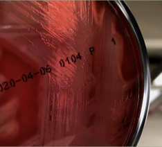
Make an observation, what type of hemolysis is this?
No change around growth | Organism does not hemolyze RBC’s | Gamma or γ - hemolysis |

Make an observation, what type of hemolysis is this?
Greenish discoloration around growth | Organism partially hemolyzes RBC’s | Alpha or α - hemolysis |

Make an observation, what type of hemolysis is this?
Clearing around growth | Organism hemolyzes RBC’s completely | Beta or β - hemolysis |
Significance of Hemolysis in Microbiology
•Indicates pathogenicity of an organism
Pathogenicity: ability of an organism to cause disease
Many organisms are beta-hemolytic
Streptococcus spp. are…
are frequent producers of hemolysins. There are known as Streptolysins, toxic proteins produced by Group A Streptococcus that destroy red blood cells
Streptolysin O (Oxygen Sensitive)
AEROBIC ENVIRONMENT (O2) | ANAEROBIC ENVIRONMENT (NO O2) |
No activity | Maximum activity |
Streptolysin S (Oxygen Stable)
AEROBIC ENVIRONMENT (O2) | ANAEROBIC ENVIRONMENT (NO O2) |
Minimum Activity | Maximum activity |
Streptolysins have better activity in _________ _________.
anaerobic environments
This is why we stab the blood agar plat
electrical tape

What kind of hemolysis and streptolysin is being demonstrated?
1. We can see in this image that the streptolysin appears to work the same at the surface of the agar as it does inside the stab. This would be an example of Streptolysin S

What kind of hemolysis and streptolysin is being demonstrated?
2. Here we can see that the alpha-hemolysis is more apparent in the stab than it is on the surface of the plate. This tells us that this is Streptolysin O

What kind of hemolysis and streptolysin is being demonstrated?
3. And here we can see that there is no hemolysis which means that no streptolysin is present.
Capable organisms (Beta Hemolysis (Complete Lysis))
Streptococcus pyogenes (Group A Strep / GAS)
Streptococcus agalactiae (Group B Strep / GBS)
Staphylococcus aureus
Capable organisms (Alpha Hemolysis (Incomplete/partial Lysis))
Streptococcus pneumoniae
Viridans Group Streptococci
Organisms that are NOT capable of Hemolysis (Gamma Hemolysis)
Enterococcus faecalis & Enterococcus faecium
2. Staphylococcus epidermidis
Staphylococcus saprophyticus