Exam 2 - CAD, Systolic Function, Stress Echo, Diastolic Function, Heart Pressures, Right Heart
 Call Kai
 Learn
 Practice Test
 Spaced Repetition
 Match
 Flashcards
 Knowt Play
1/206

There's no tags or description
Looks like no tags are added yet.
207 Terms
Coronary Arteries
RCA
LAD
Cx
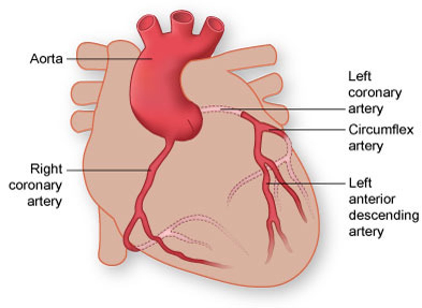
Coronary Artery Dominance
90% - RCA supplies inferior portions
10% - Cx supplies inferior portions
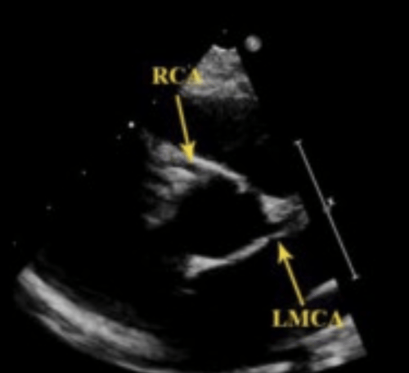
Causes of CAD
Atherosclerosis*
Congenital anomalies
Coronary artery spasm
Embolus
Injury to the chest or thorax
Uncontrollable Risk Factors of CAD
Age
Male gender
Family history
Type 1 Diabetes
Controllable Risk Factors of CAD
Smoking
Hypertension
High cholesterol
Obesity
Stress
Alcoholism
Symptoms of CAD
Angina Pectoris - ischemic chest pain
Tightness or burning in chest
Pain in arm/shoulder
Shortness of breath
EKG changes
CAD Treatments
Drug therapy
Stents
CABG
Kawasaki Disease
Dilation of the coronary arteries
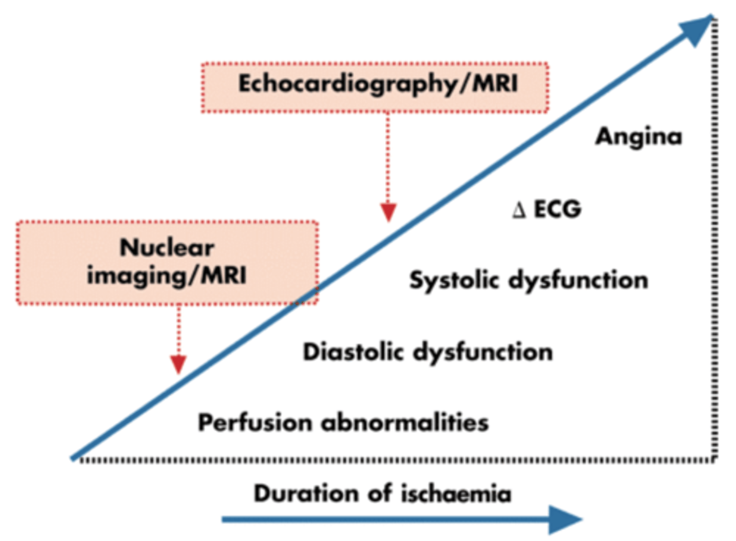
Ischemic Cascade
Perfusion defects
Diastolic function abnormality
Strain abnormalities
Systolic wall motion abnormality
EKG changes
Chest pain
Myocardial Ischemia
Reversible
Inability to meet oxygen demands during exercise
Symptoms when blockage reaches ≥ 70% area stenosis while exercising
Wall motion abnormality during exercise - not at rest
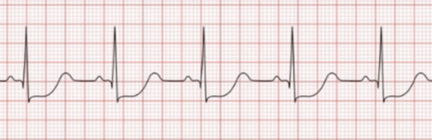
Ischemia EKG Changes
Depressed S-T segment
Stunned Myocardium
Wall motion abnormalities last days to weeks
Re-perfused ischemic muscle
Hibernating Myocardium
Wall motion abnormalities last weeks to months
Oxygen is chronically depressed
Myocardial Infarction
Irreversible wall motion abnormalities
Symptoms with lesions ≥ 90% area stenosis even at rest
Acute Myocardial Infarction
Wall motion is reduced or absent
Normal wall thickness in diastole
Reduced or absent wall thickness in systole
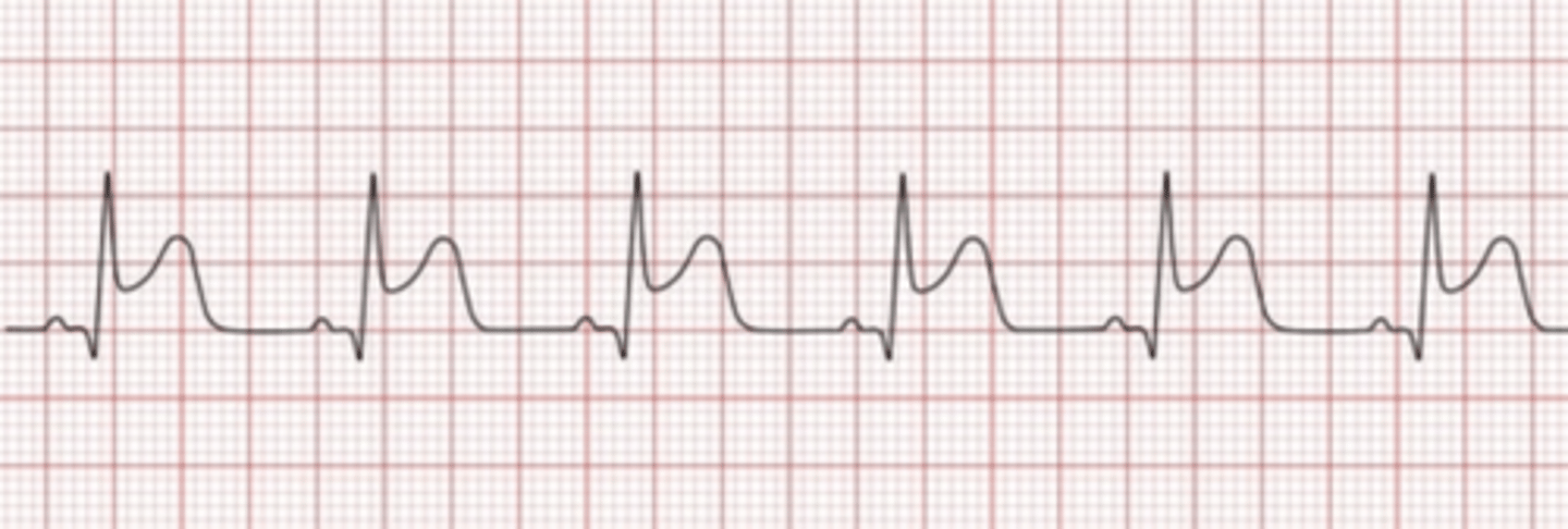
Acute MI EKG Changes
Elevated S-T segment
Chronic Myocardial Infarction
Abnormal wall motion
Thin wall thickness in diastole
Absent wall thickening in systole
Increased wall echogenicity
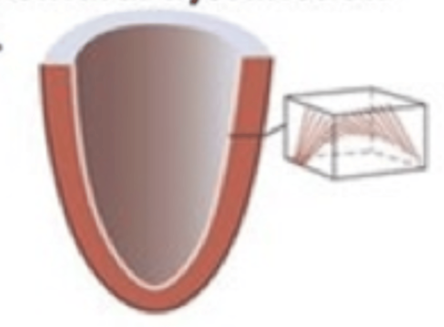
Transmural Infarction
Includes entire wall thickness
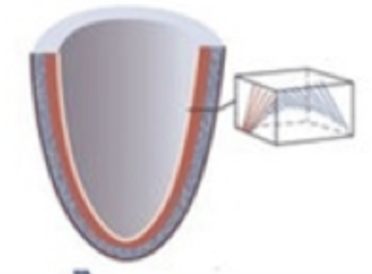
Subendocardial Infarction
Does not penetrate entire wall thickness
Myocardial Remodeling
Seen with myocardial infarctions
~ 6 weeks to complete
Tissue becomes necrotic
Necrotic area replaced with fibrous scar tissue
New wall is thinner & denser - more prone to aneurysms, akenesis and dyskenesis
Complications of a Myocardial Infarction
Myocardial remolding
Pericardial effusion
Papillary muscle dysfunction or rupture
LV aneurysm
Ventricular septal or free wall rupture
Ventricular thrombus
LV pseudoaneurysm
CHF
Arrhythmias
Sudden cardiac death
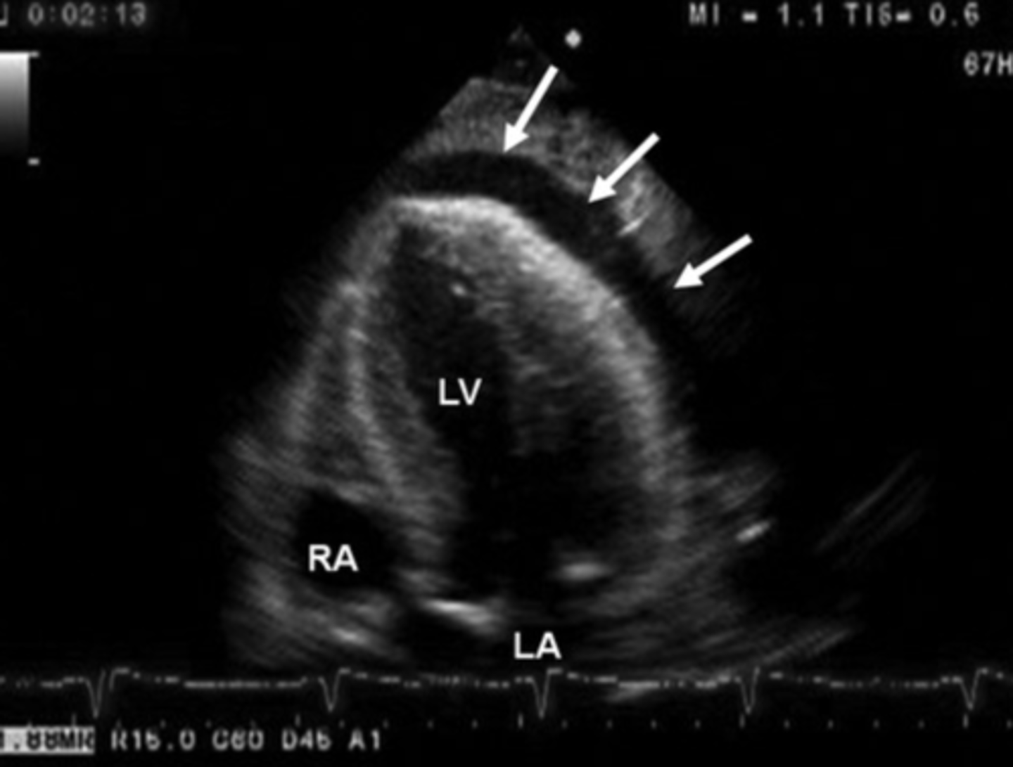
Pericardial Effusion
Small circumferential fluid space
Can be life threatening
Papillary Muscle Dysfunction or Rupture
Damaged myocardium in the area of a papillary muscle
Leads to tear in myocardium or chordae
Causes valvular dysfunction
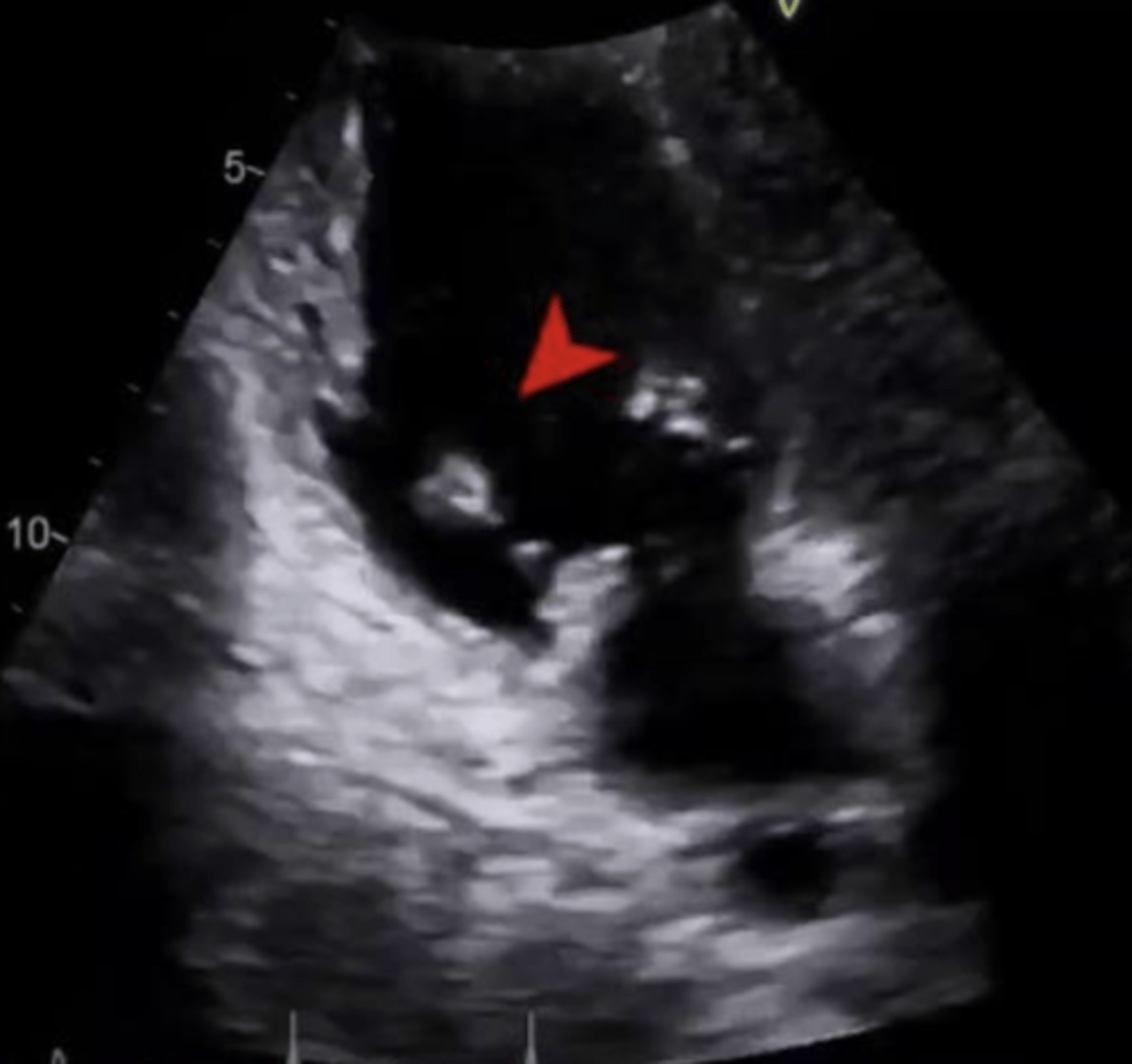
Ventricular Thrombus
Thrombus in region of akinetic myocardium
RV thrombus - lungs
LV thrombus - MI, stroke, peripheral blockage
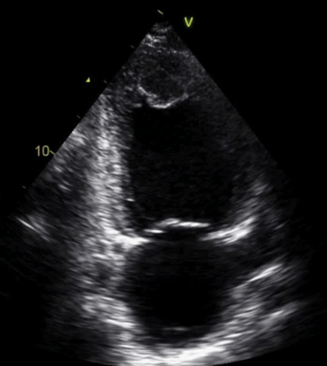
LV Aneurysm
Weakening of muscle causing region to bulge or rupture
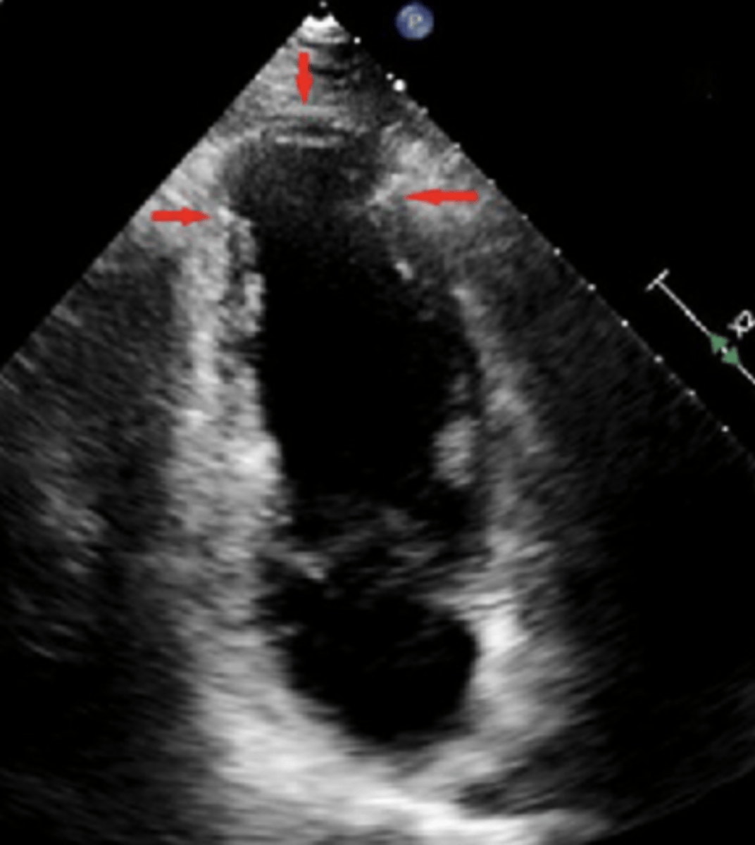
Ventricular Septal Wall Rupture
Leads to VSD
Causes systolic murmur
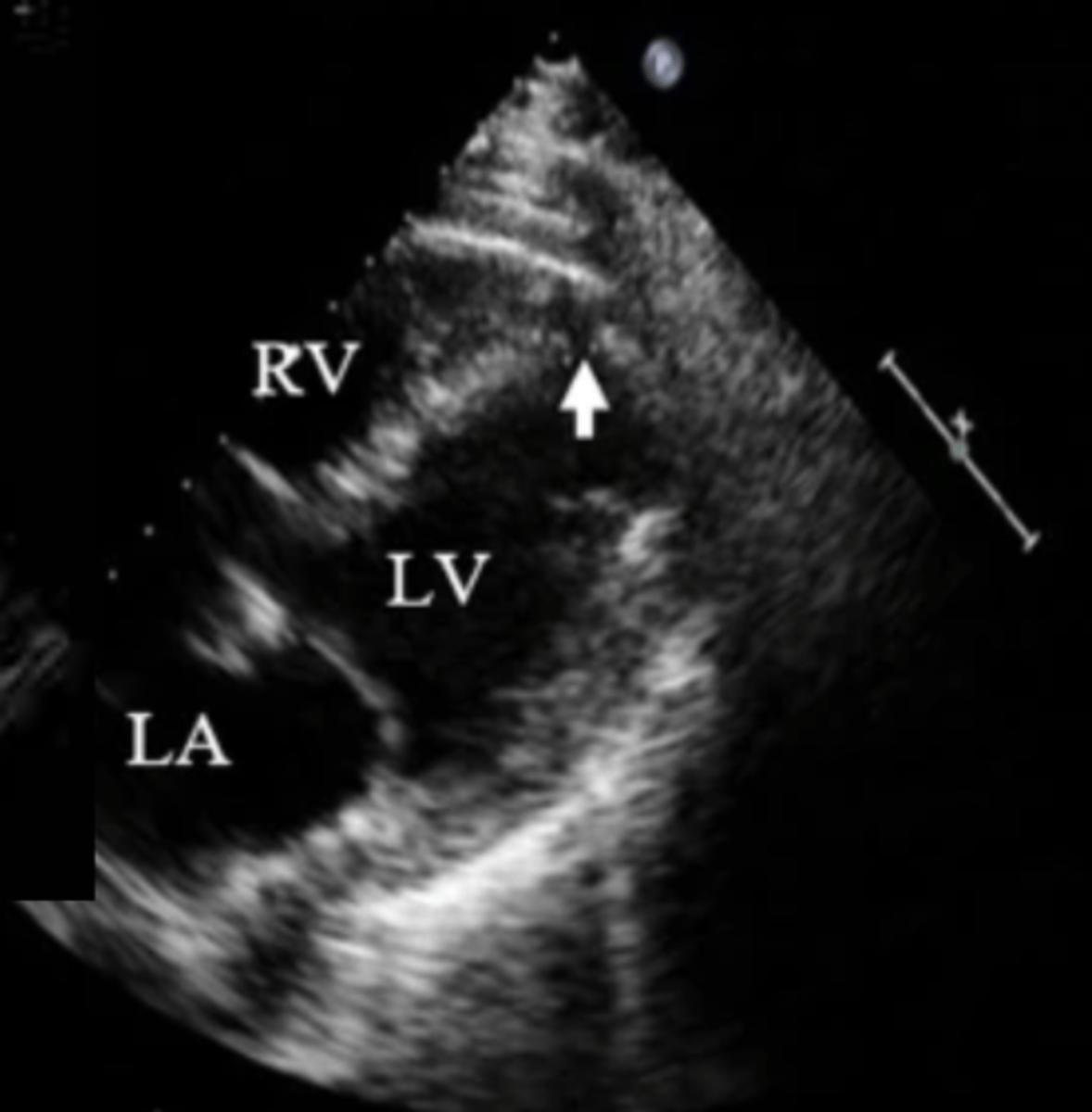
Ventricular Free Wall Rupture
Sudden hemodynamic changes due to tamponade
Rarely survived
Causes systolic murmur
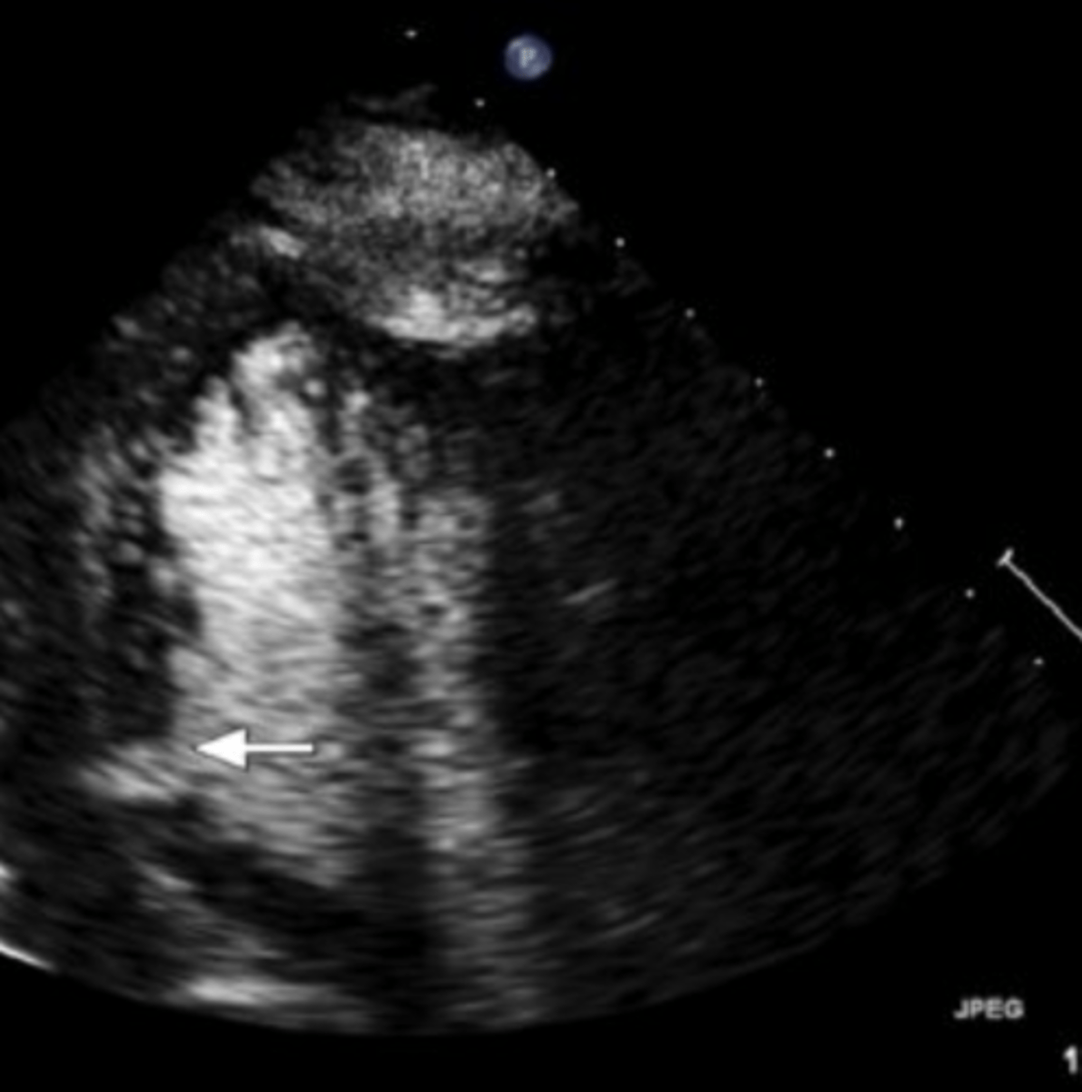
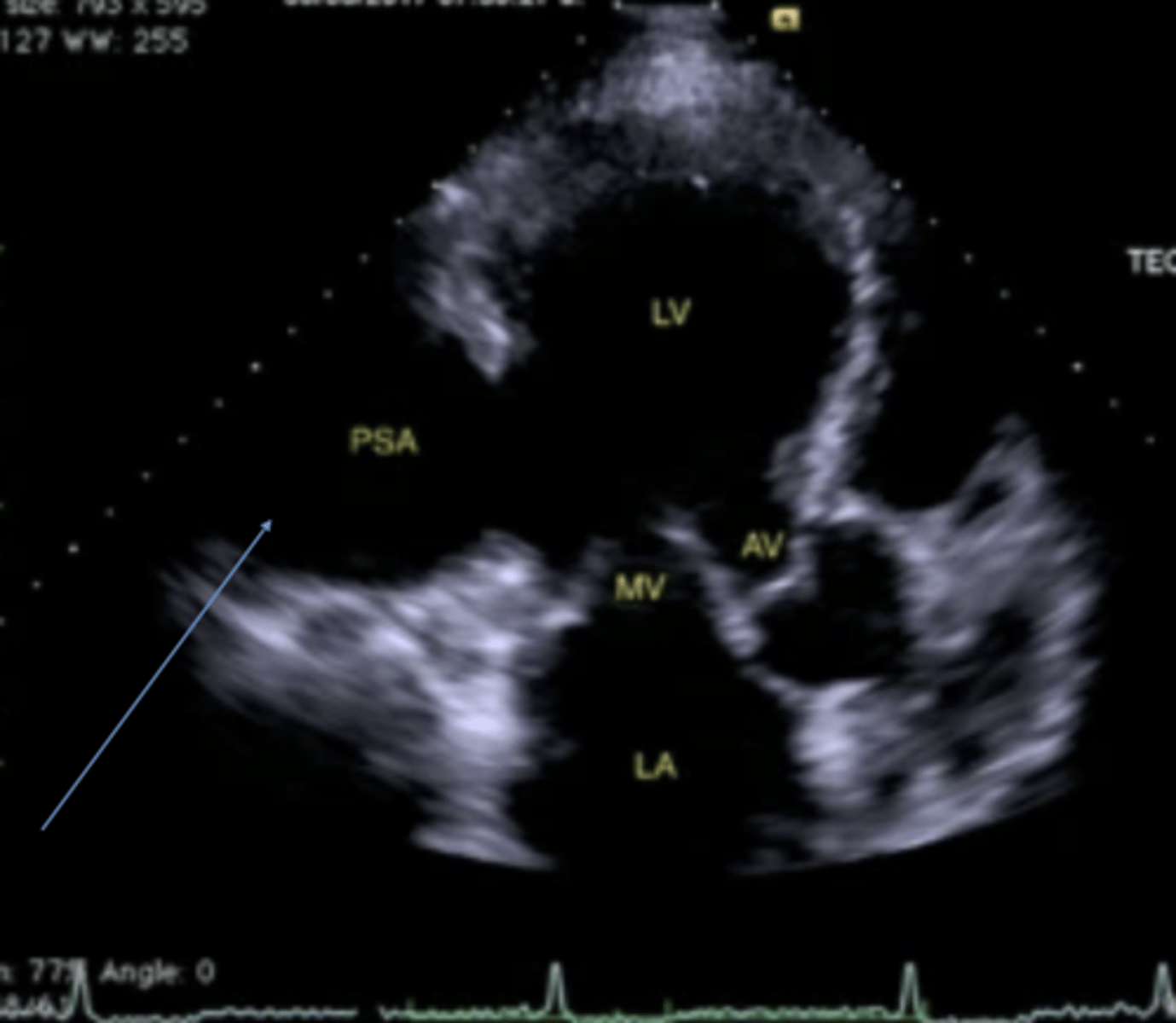
LV Pseudoaneurysm
Contained rupture of free wall
Usually due to thrombus
Look for neck connection
To and fro flow
Congestive Heart Failure (CHF)
Weakened myocardium overtime due to ischemia
Thin walls & dilated chambers
Causes of CHF
CAD
HTN
Cardiomyopathy
Diastolic dysfunction
Chronic ischemia/infarction
Arrhythmias
Infarction in SA or AV node
AFIB
Rt heart block
Lt heart block
Complete heart block
Sudden Cardiac Death
Occurs from tachy-arrhythmias
Preload
Filling & stretching of muscle fibers during diastole
Length of muscle fiber stretch is related to the strength of the next contraction
Frank-Starlings Law -> more diastolic filling = more stretch = more contraction/SV
Factors Affecting Preload
Venous return - total blood volume & blood distribution
Filling time - atrial contraction
Afterload
Resistance the blood encounters during systole - pressure needed to pump blood into the body
Factors Affecting Afterload
Aortic pressure
Peripheral resistance
Distendability/compliance of vessels
Increased Afterload
Leads to decreased stroke volume
Load Dependent Measurements
SV
Myocardial performance index (Tei)
TDI
GLS
Load Independent Measurements
dP/dt
LVOTacc
Wall Thickness
More specific finding than wall movement alone
Should thicken during systole
Wall Motion Scoring
0 = Not visualized
1 = normal or hyperdynamic
2 = hypokinetic
3 = akinetic
4 = dyskinetic
16 total segments (17 w/ perfusion)
assess each wall segment twice
Hyperdynamic
Increased contractility
Compensatory wall motion - making up for damaged wall segment
Due to LVH or dehydration
Hypokinetic
Slow moving/sluggish
Graded as mild, moderate, or severe
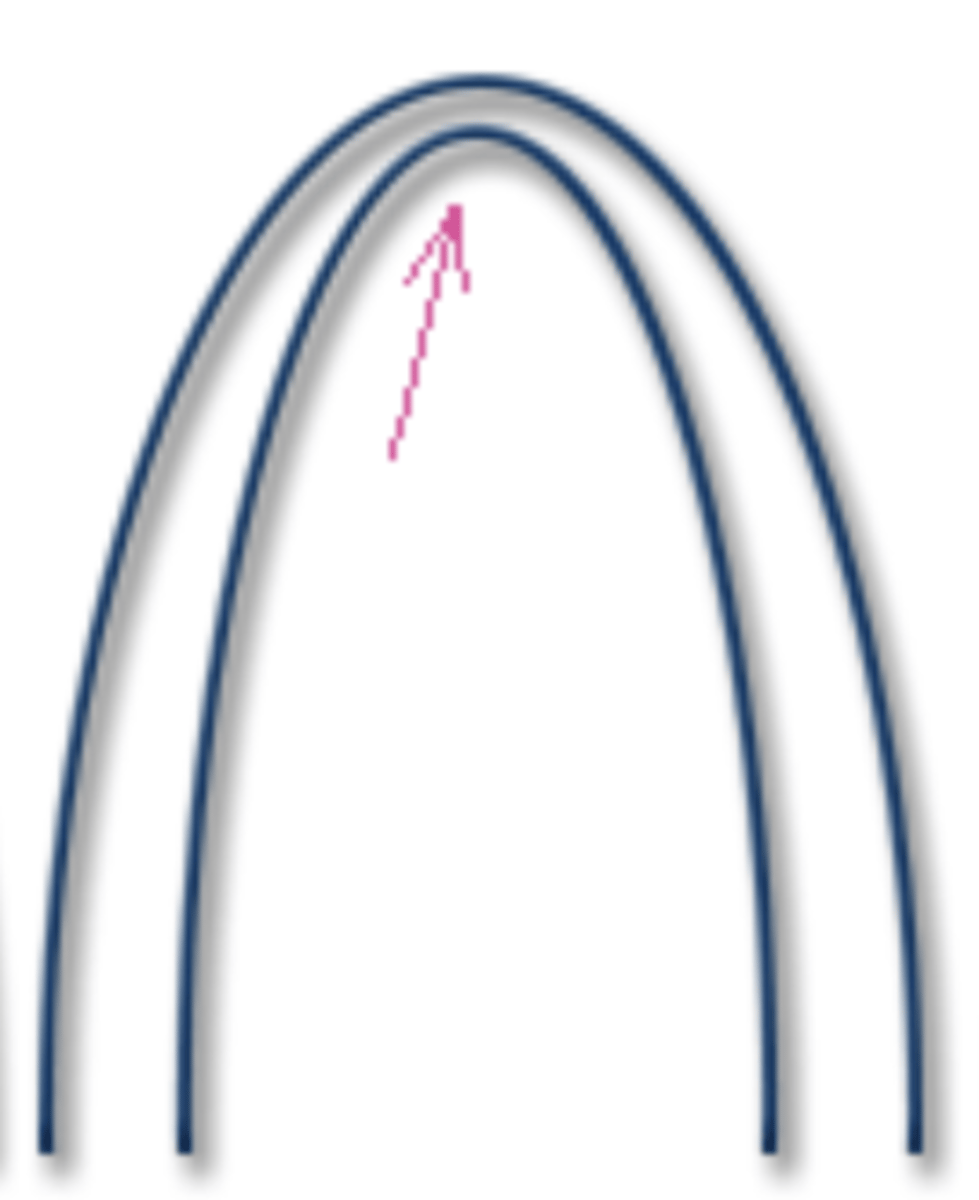
Akinetic
No squeeze
Myocardial thinning during systole
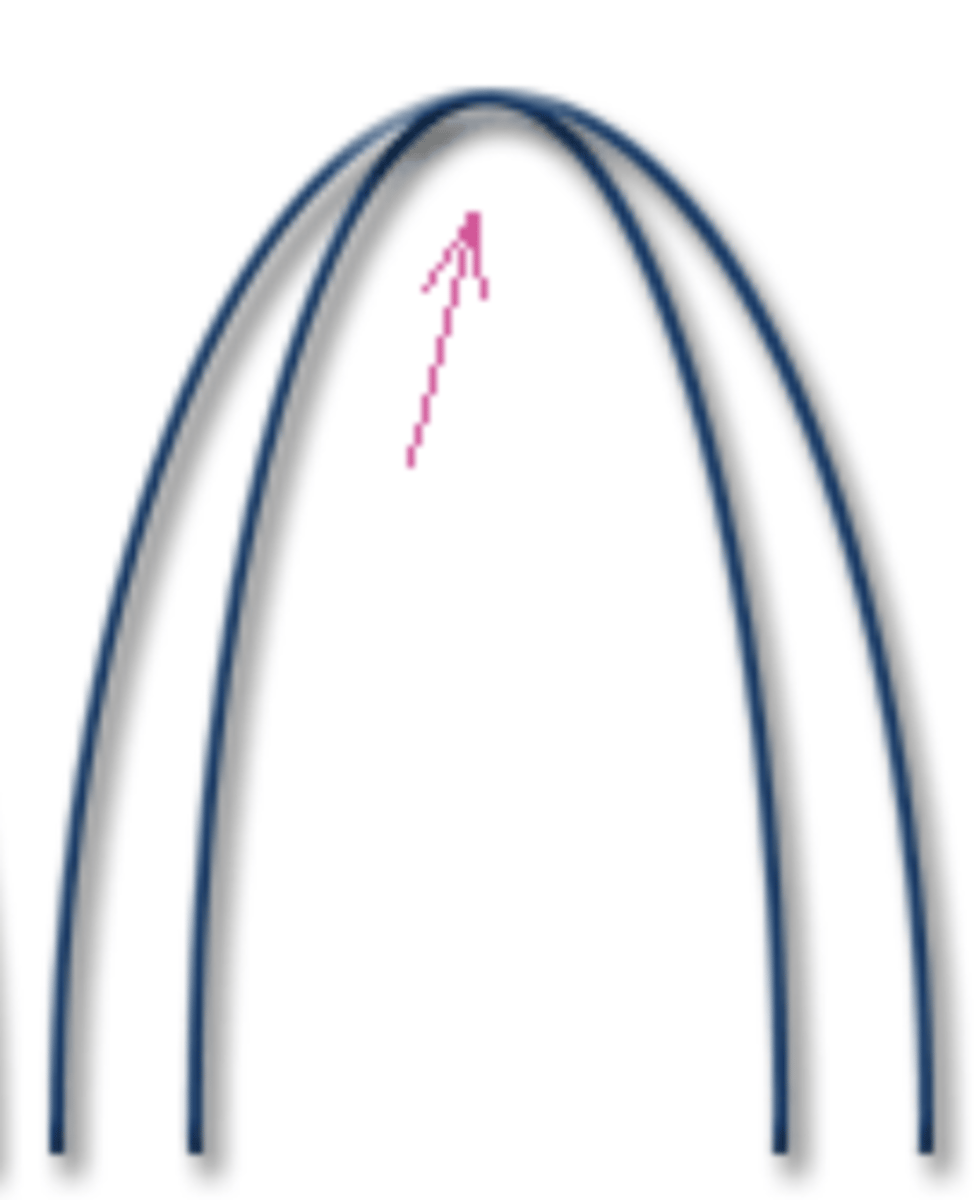
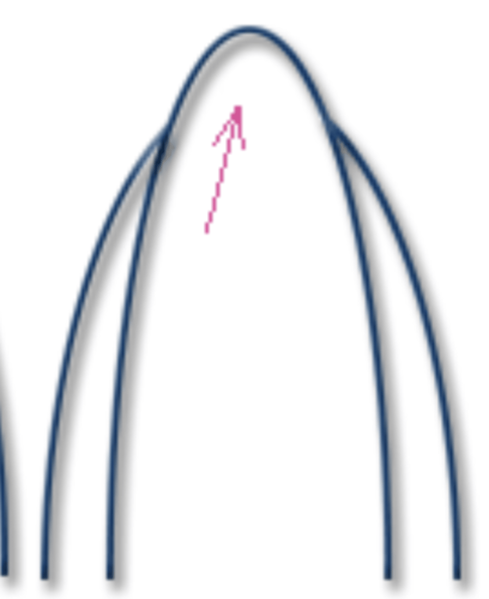
Dyskinetic
Wall bulges/protrudes out during systole
Aneurysmal motion
Paradoxic
Compensatory motion of septum
Wall moves in opposite direction
Rocking motion
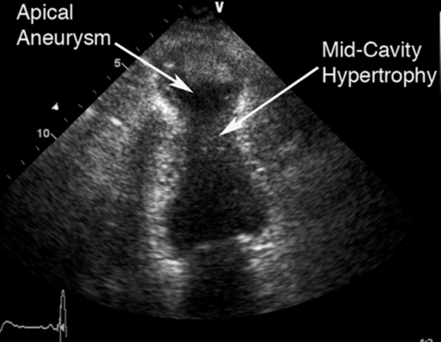
True Aneurysm
2-4 weeks to develop
Common after anterior MI - affects apex & septum
Bulges in systole & diastole
Increases risk of embolism and arrhythmia
Limitations to 2D Wall Motion Assessment
Movement influenced by adjacent segments
Adjacent normal tissue appearing as hypokinetic
Adjacent abnormal tissue pulled by hyperkinetic tissue
Measurements that Assess Global LV Function
SV
dP/dT
LVOTacc
Myocardial performance index (Tei)
TDI
GLS
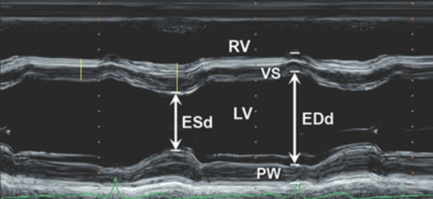
2D Linear Measurements
-> SV, CO, EF, FS
*only recommended to find FS
Preferred over M-mode -> volumes are better
PLAX at MV leaflet tips, inner edge to inner edge
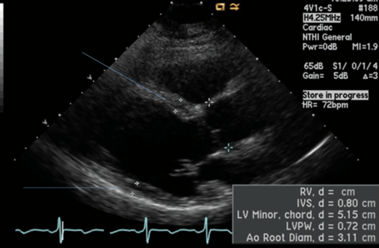
End-Systolic 2D Linear Measurement
Smallest LV or AV closure
End-Diastolic 2D Linear Measurement
Largest LV or MV closure
Fractional Shortening (FS)
% of shortening of LV during systole
NL = ≥25%
Proportional to systolic function
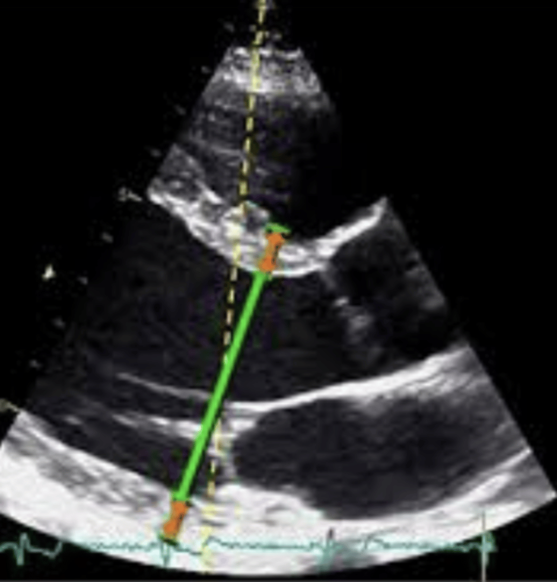
Fractional Shortening (FS) Formula
((LVIDd - LVIDs) ÷ LVIDd) x 100
2D Volume Measurements
-> SV, CO, CI, EF
Preferred over linear methods
Should be indexed with BSA
Simpson's Biplane
Preferred choice of measuring LV function
Trace *only* endocardial boarders of LV in systole and diastole of the same cycle
Use enhancement when endocardial boarders are not well visualized
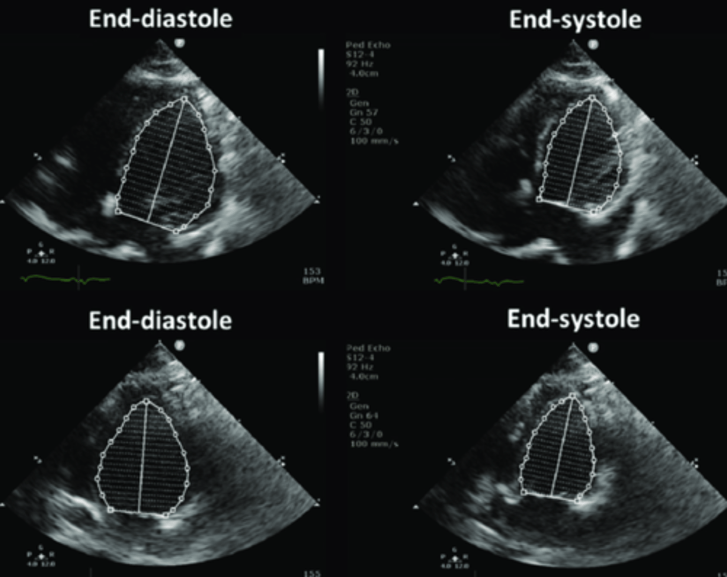
End-Systolic Simpson's Biplane
Frame preceding initial early diastolic MV opening
End-Diastolic Simpson's Biplane
At initial coaptation of the MV leaflets
Stroke Volume (SV)
Volume of blood ejected with each heart beat
NL = 70-100 mL/beat
2D Stroke Volume (SV) Formula
EDV - ESV
Cardiac Output (CO)
Volume of blood ejected each minute
NL = 4-8 L/min
Cardiac Output (CO) Formula
(SV x HR) ÷ 100
Cardiac Index (CI)
Cardiac output adjusted for BSA
Cardiac Index (CI) Formula
CO ÷ BSA
Ejection Fraction (EF)
% of blood volume ejected during systole
2D/3D Ejection Fraction (EF) Formula
((EDV - ESV) ÷ EDV) x 100
Doppler Ejection Fraction (EF) Formula
(SV ÷ EDV) x 100
3D Volume
-> SV, CO, CI, EF
Preferred over 2D volume when available
Does not assume bullet shape
Displayed as 3D rendering, graph of volume, and regional wall motion images
Limited by 2D image quality & temporal resolution
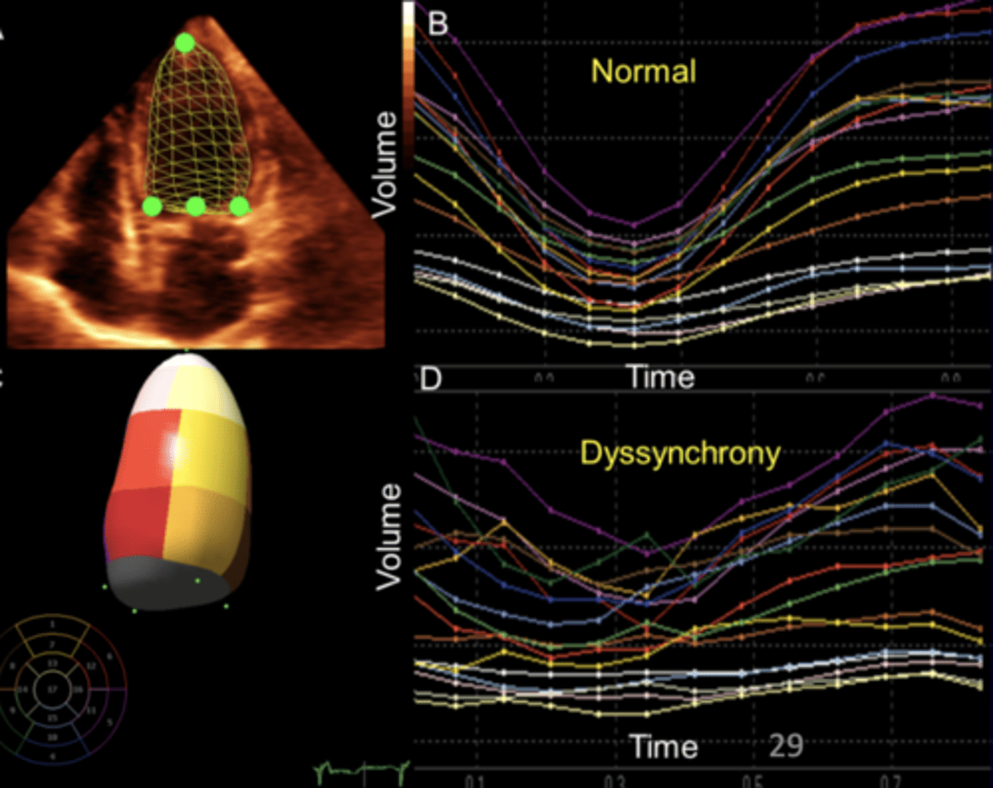
3D Volume Technique
Optimize 2D image
Guide 3D acquisition & obtain volume sets
Machine renders shape
Use contrast to find endocardial boarders
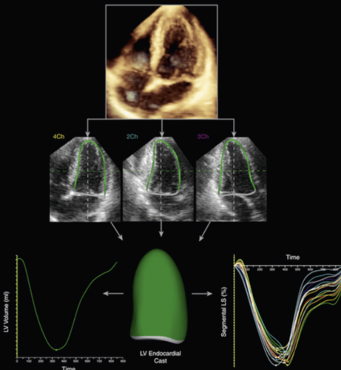
2D LV Mass Measurements
Predictor of cardiovascular events
Trace both interior & exterior sides of the wall at end-diastole
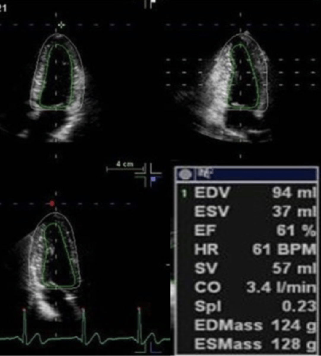
Limitations to 2D Measurements
LV foreshortening - has to be at apex
Assumption of a bulled-shaped LV
Poor endocardial wall visualization
Stroke Volume (SV) using Doppler
-> CO, CI, EF
Global assessment
Load dependent
Inaccurate with moderate-severe AI
2mm error in LVOT diameter = 20% error
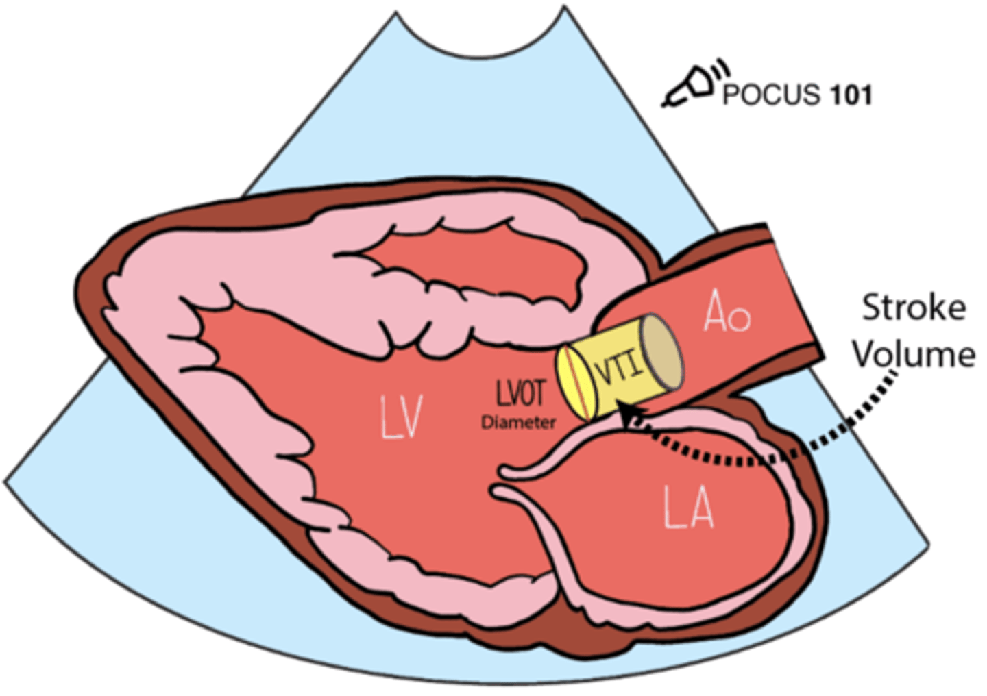
Doppler Stroke Volume (SV) Formula
CSA x VTI
dP/dt
Change in pressure (dP) over time (dt)
Global assessment
Load independent
Measured during isovolumic contraction when MR is present at 1m/s & 3m/s velocity
Increased time or MR severity = decreased dP/dt = decreased LV function
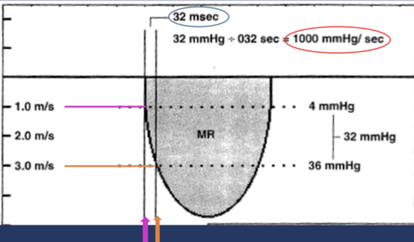
Limitations of dP/dt
Must be measured in CW
Doppler cursor must be parallel to flow - underestimated if not
Not accurate with severe MR
Significant AS or HTN may show NL value
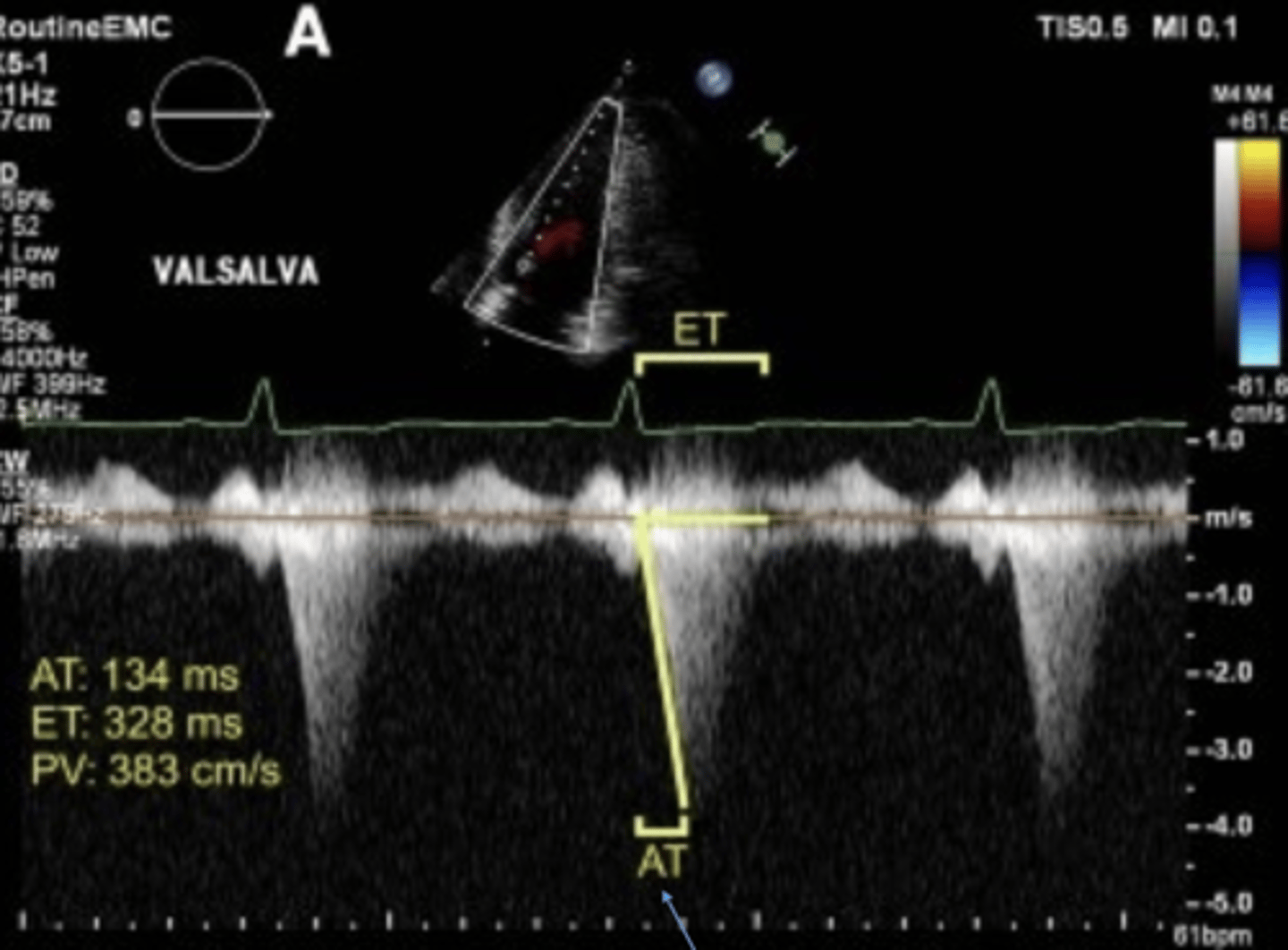
LVOT Ejection Acceleration Time (LVOTacc)
Time to peak LVOT velocity
Global assessment
Load independent
Correlates with LV contractility
Myocardial Performance Index (Tei)
Assesses dilated cardiomyopathy, amyloidosis pulmonary HTN, RV dysplasia
Global assessment
Load dependent
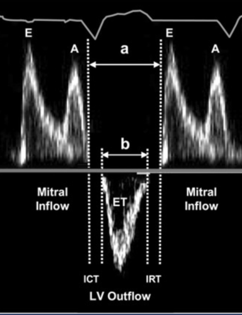
Myocardial Performance Index (Tei) Formula
(IVCT + IVRT) ÷ ET
Tissue Doppler (TDI)
Global assessment
Load dependent
Accurate even when 2D is suboptimal
Evaluate lateral MV annulus
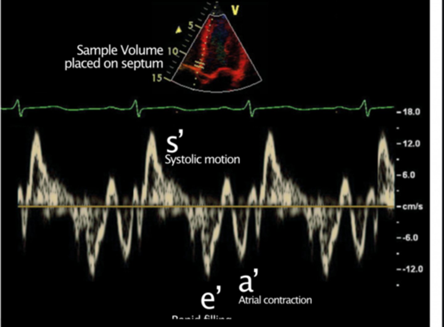
Tissue Doppler (TDI) Velocities
Higher on free wall than septal
Decrease as you move apically
Decrease with age
S' > 5.4 cm/s
Predicts LV EF > 50%
Reduced S'
Correlates with poor LV function
Limitations of Tissue Doppler (TDI)
Detects passive LV movements - tethering
Angle dependent
Global Longitudinal Strain (GLS)/Speckle Tracking
Difference between systolic and diastolic length
Global assessment
Load dependent
Shoes abnormal wall motion earlier than EF
Average of all wall segments & all apical views
Expressed as a negative number - indicates shortening
NL = > - 20%
Lower absolute value = more abnormal (-10 is more abnormal)
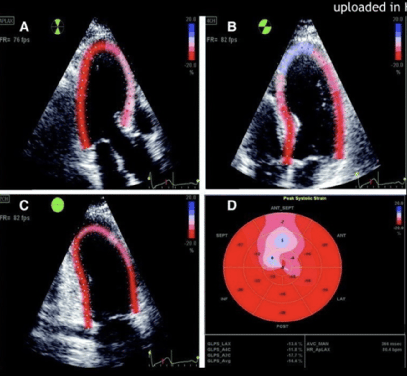
Global Longitudinal Strain (GLS) Formula
(Systolic myocardial length - diastolic myocardial length) ÷ diastolic myocardial length
Limitations of Global Longitudinal Strain (GLS)
Cannot use if tracking is suboptimal in 2 or more adjacent segments
Suboptimal 2D image
Foreshortening
Incorrect measuring/including papillary or trabeculations
Vendor variations
Negative number - confusing
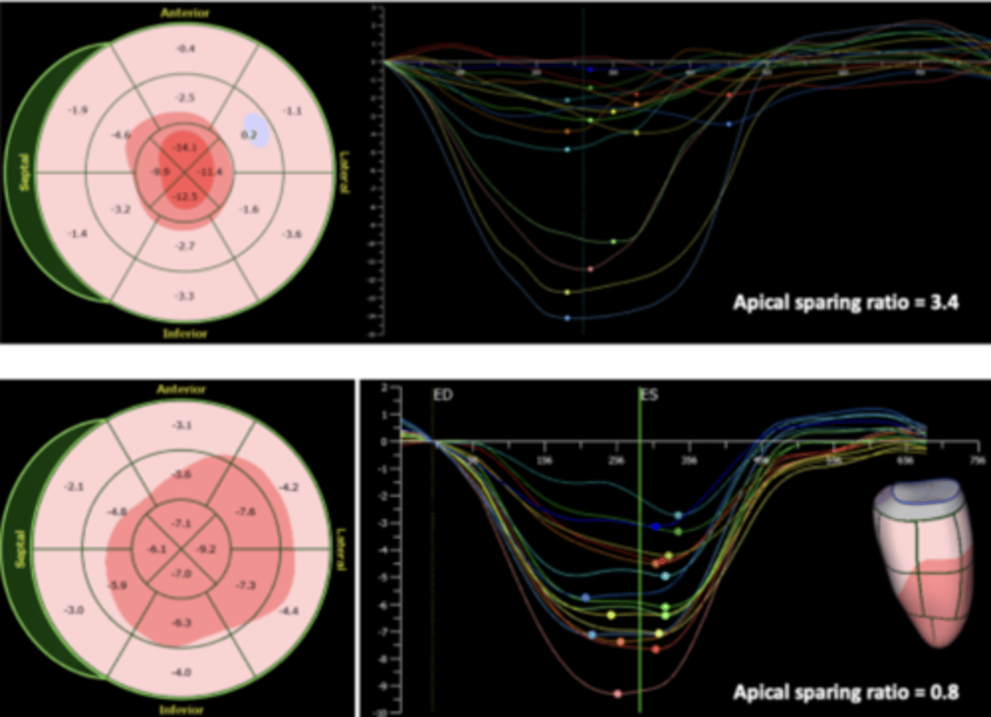
Strain Curves
Assesses all wall segments
Compares movement patterns
Shape of curve is more important than peak
Other modalities that measure LV volume, CO, and EF
Angiography - gold standard
Radionuclide ventriculography/PET - nuclear medicine
Cardiac CT/MRI
Limitations of Doppler Measurements
HR & loading conditions
Angle > 0º
Doppler waveform definition & trace
Movement from respiration
< 3-5 beats recorded
Ultrasound Enhancing Agents (UAE's)
Used when 2 or more adjacent wall segments are not visualized
Used for LV function, wall motion, and myocardial perfusion
Ex. Definity, Lumason, Optison
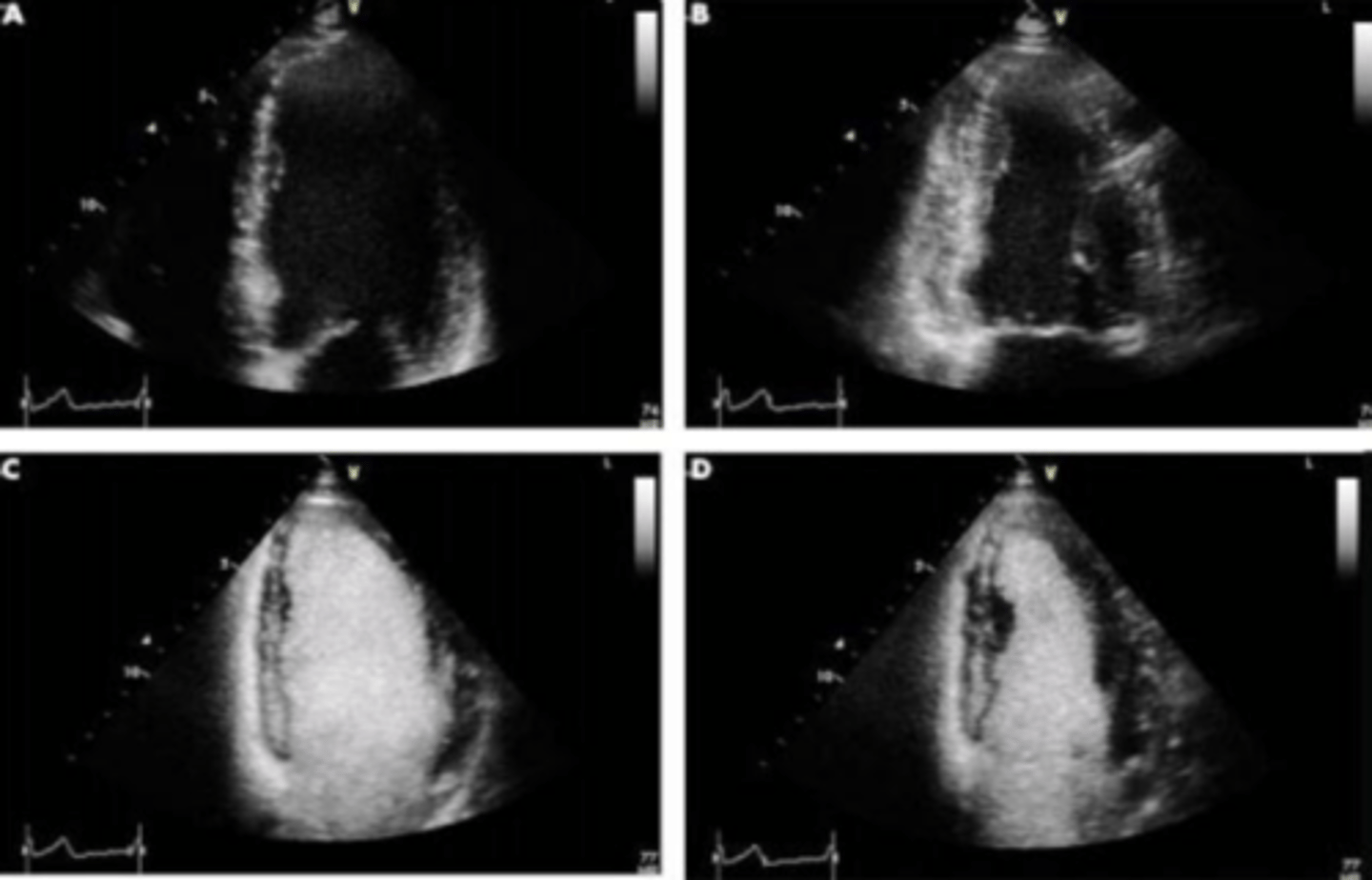
Equipment Setting for Ultrasound Enhancing Agents (UAE's)
Very low multipulse setting
<0.2 MI
Detects fundamental waves
Easier to see basal segment
Techniques for Ultrasound Enhancing Agents (UAE's)
Place focus at annulus level
Clip 4-5 beats
7 Parameters for Baseline Resting Stress Echo Assessment
Regional wall motion
Global function
E/e' ratio
MR and other valve assessments
Systolic Pulmonary Artery Pressure - right heart pressure
RV function
Contraindications
Dobutamine Stress Echo
Lower doses increase contractility
Dobutamine Stress Echo Protocol
Resting images
Increased dosages of dobutamine every 3 minutes
Low dose images every 10 micrograms
Images at calculated peak heart rate (~85%)
Post images when heart rate is below 100pbm or returns to baseline
Continually monitor patients O2 levels, BP, and EKG
Treadmill Exercise Stress Echo
3 minute stages until system starts or changes in ST segment, rhythm, or blood pressure
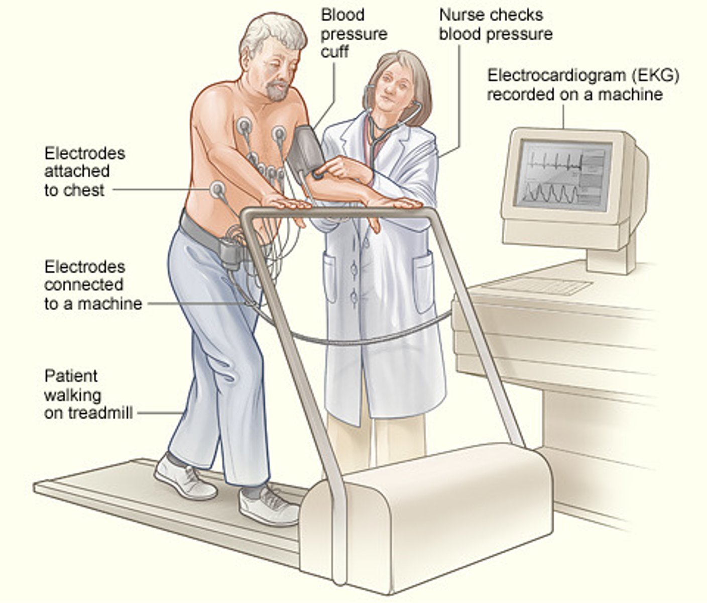
Treadmill Exercise Stress Echo Protocol
Resting
Peak post-exercise within 1 minute
Early recovery
Multistage Bicycle Stress Echo
2 minute stages
Pedaling at 60 revolutions
Can assess intermediate stages
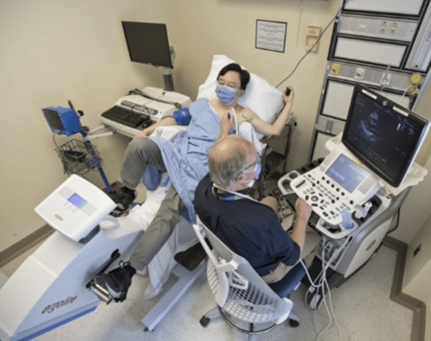
Multistage Bicycle Stress Echo Protocol
Resting
Low stress
Intermediate
Peak stress
Early recovery
Positive Stress Test
New wall motion abnormality
Decrease/no change in global EF
Increase LV systolic volume in PSAX view