Cardiac US Physiology (Part 2)
 Call Kai
 Learn
 Practice Test
 Spaced Repetition
 Match
 Flashcards
 Knowt Play
1/51

Earn XP
Description and Tags
Cardiac Sono
52 Terms
Cardiac output (CO)
is the volume of blood pumped from the LV (and circulated in the body) each minute.
CO Formula
SV x HR
Normal CO
4-8 liters/min (l/min).
Cardiac index (CI)
is the CO corrected for the body surface area (BSA).
Normal Cardiac Index
3-4 liters/min/m²
Body surface area (BSA)
is the calculated surface area of the human body.
Normal BSA
1.73m²
BSA Formula
Height x Weight
Ejection fraction (EF)
is related to stroke volume and is the percentage of preload (volume) that is ejected with every beat of the heart.
EF Formula
EDV-ESV X 100%
Normal EF
50 - 70%
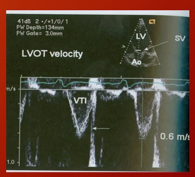
Velocity time integral (VTI)
represents how far the blood travels in centimeters with each ejection.
Preload
the volume (load) in the heart at end diastole
The degree of fiber stretch related to the quantity of blood in the chamber prior to contraction.

Frank Starling Law (length-tension relationship):
This law concludes that there is a direct relationship between the degree of myocardial muscle fiber stretch and the force generated by the succeeding contraction.
The more blood that enters the ventricle during diastole (preload) → the greater the force of the contraction (systole) required to eject the blood
Increased preload
any state of fluid overload (a leak)
Valvular regurgitation
abnormal backwards flow through a closed valve – doesn’t close properly.
MR, AR, TR, PR
Ventricular septal defect (VSD)
defect in the IVS that allows abnormal flow between the LV and RV.
Atrial septal defect (ASD)
defect in the IAS that allows abnormal flow between the LA and RA.
Patent Ductus Arteriosus (PDA)
occurs when the ductus arteriosus (fetal opening between the aorta and pulmonary artery) does not close properly at birth.
Increased Preload (Volume) results in what?
Dilation
Afterload
is the resistance that the heart must pump against (pressure overload)
Hypertrophy
Increased Muscles Mass
*Increased afterload results in
hypertrophy (thickening) of the myocardium.
Afterload is increased by any state of pressure overload:
Stenosis: AS, PS
Hypertension:
Systemic HTN
systolic BP above 140mmHg and diastolic BP above 90mmHg.
Pulmonic HTN
systolic pulmonary artery pressure above 30 mmHg
As and Ms in an Increase or Decrease in Afterload and Pressure?
Increase
Preload (Leaks)
Increased Volume
-Dilation/ Enlargement
-Shunts
ASD, USD, PDA
LAE correlates to
Atria (MS / TS) + Increased Afterload & Pressure
MR correlates to
Increased Preload
Increased Volume
Dilation
What happens before MR?
LAE

PS - Increase Afterload, but what happens 1st?
RVH

AS - Increased Afterload, but what happens 1st?
LVH
Abnormal volume in any cardiac chamber is accompanied by an eventual rise is
Pressure
Abnormal pressure in any cardiac chamber is accompanied by an eventual increase in
Volume
What is the physiologic advantage to dilation in the face of MR or AI?
Dilation allows the receiving chamber to accommodate a larger volume of blood, without experiencing a large increase in pressure, which could lead to pulmonary edema.
Pulmonary Edema
is the accumulation of fluid in the lung tissues and aveoli, usually resulting from increases Left sided pressure (i.e. MS, MR…)
Interval-strength relationship:
The longer the interval between heartbeats → the stronger the contraction required to eject the blood.

post extra systolic potentiation.
The PVC is weak and creates a long interval before the next beat which allow extra time for increased filling (preload). The increased preload requires a stronger contraction in order to eject the blood.
Force velocity relationship –
the greater the force required to eject the blood during systole – the slower the velocity of the muscle fiber shortening. (you pick up heavy weights slower).
Excitation-contraction coupling
the link between the heart’s propagated action potential and the mechanical events of muscular contraction.
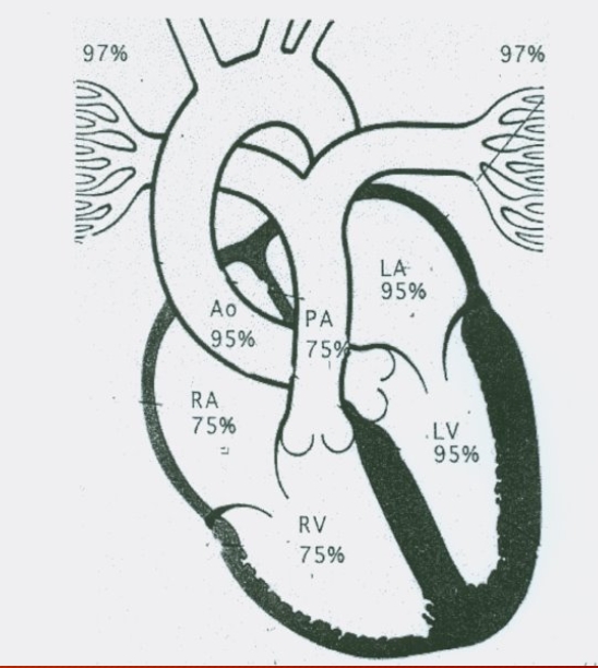
Right side of the heart is the what Circuit?
Pulmonary
Venous (deoxygenated blood) return to the heart (O2 sat – 75%) – coronary sinus lowest O2 sat in the body (60%).
Left side of the heart is the what Circuit?
Systemic
Oxygenated blood return from the lungs (O2 sat 95%).
Sends blood to the body to perfuse the organs and tissues
Pulmonary Components:
Pulmonary Artery
Arteries
Capillaries
Veins
Systemic Components:
Aorta
Arteries
Arterioles
Capillaries
Venules
Veins
Vena Cava
Which side of the heart pumps more blood?
It Should be Equal
The Similarity between Pulmonary and Systemic
They pump about the same amount of blood
Normal metabolic needs of the body are met when the cardiac index is between
2.5 – 4.5
If the cardiac index falls below the patient can go into severe shock
2.0 liters/min/m²
Perfusion of any organ in the body is a product of
Arterial pressure and Vascular resistance.
Local control
determined by the rate of tissue metabolism (vasodilation/constriction).
Central nervous system control (CNS)
(nerves located in the lower pons and medulla oblongata) – vasomotor nerve fibers regulate vessel diameter and function by the release of norepinephrine or epinephrine.