Bones of Skull & Pharynx
 Call Kai
 Learn
 Practice Test
 Spaced Repetition
 Match
 Flashcards
 Knowt Play
1/66

There's no tags or description
Looks like no tags are added yet.
67 Terms
Neurocranium (cranium skeleton)
(cranial skeleton) – forms a vault for the brain
Viscerocranium
– forms the facial skeleton
Neurocranium has this many bones.
8
Sutures are
fibrous joints between bones

Frontal Bone
•Paired bone that ossifies in adulthood to form a single bone
•Forms bony forehead, anterior brain case and the roof of the orbital cavity

Frontal bone

Parietal Bones
•Paired, large bones
•Form the greater portion of the superior and lateral surfaces of the cranium

Parietal bone
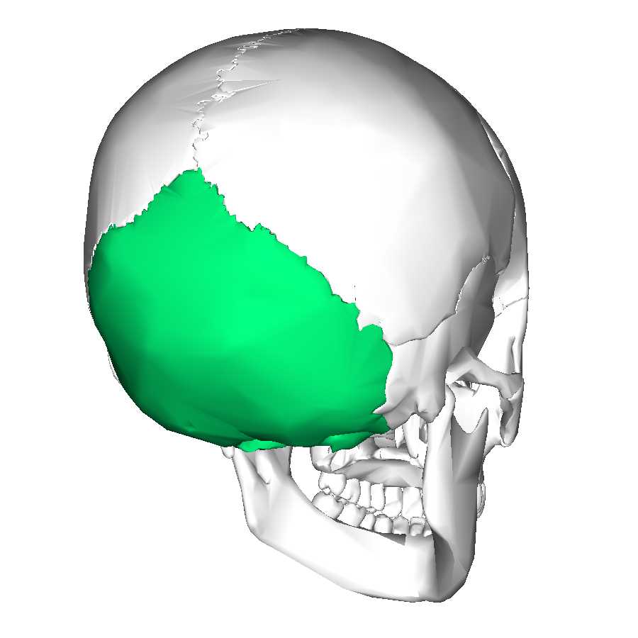
Occipital Bone
•Forms the posterior and inferior surfaces of the brain case (on the back of your head)
• Important features:
•Foramen Magnum
•Occipital condyles

Occipital bone
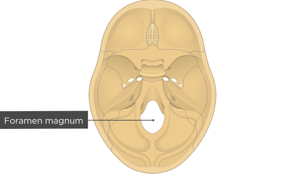
Foramen magnum
The hole in the occipital bone, that allows the brain to transition from brain to spinal cord

Temporal Bones
•Paired bone that forms the lateral base and sides of the brain case.
•Important features:
- zygomatic process
- mandibular fossa
- mastoid process (lumpy rounded protrusion behind ear)
- styloid process (thin projection just anterior to the mastoid process)
- external auditory meatus/ear canal (ears sit right over top of that)

Temporal bone

Ethmoid Bone
•Complex, delicate structure at core of cranium
•Forms part of the walls of the cranial, nasal and orbital spaces
•Important features:
oCrista galli
oPerpendicular plate
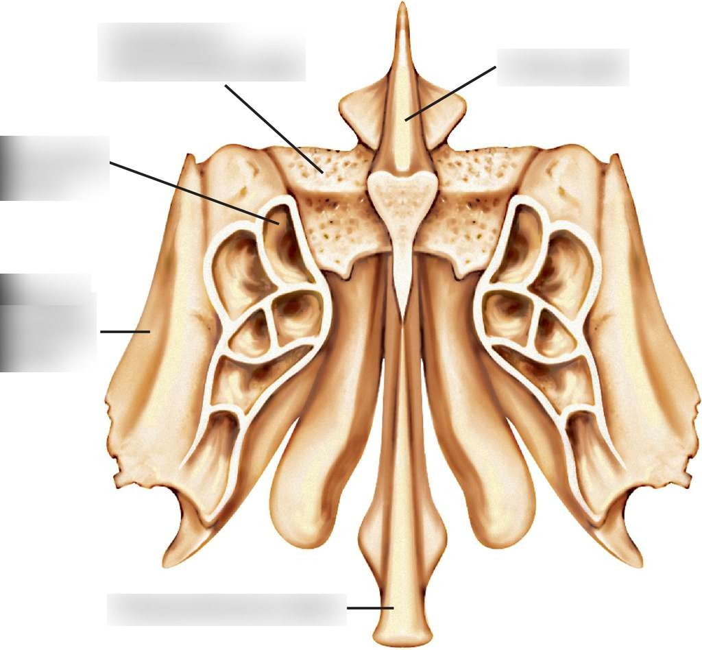
Ethmoid Bone

Sphenoid Bone
•Very complex bone located at the base of the skull; significant contributor to cranial structure
•Important features:
body
Lesser wings – superior to body
Greater wings
Medial & Lateral Pterygoid plates – downward projections

sphenoid bone
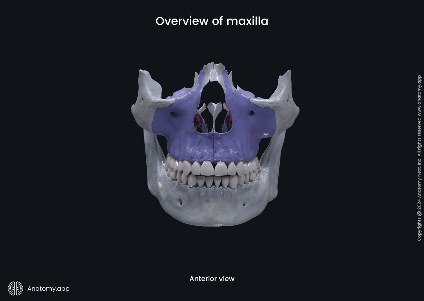
Maxillae
•Paired bones, contribute to roof of mouth, nose, upper dental arch
•Each bone has a body and 4 processes
Zygomatic process
Frontal process
Alveolar process
Palatine processes

Maxillae

Palatine Bones
•Paired; contributes to formation of 3 cavities: nasal, orbital and oral
Feature:
Horizontal process
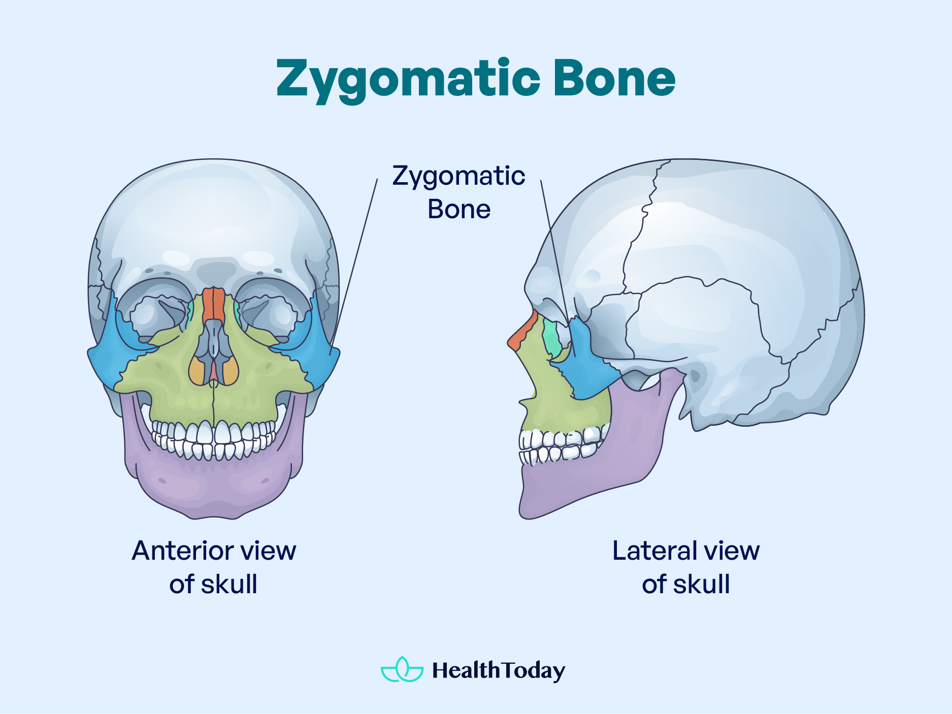
Zygomatic Bones
•Paired; articulates with maxilla, frontal, temporal and sphenoid bones
•quadrilateral-shaped body

Zygomatic Bones
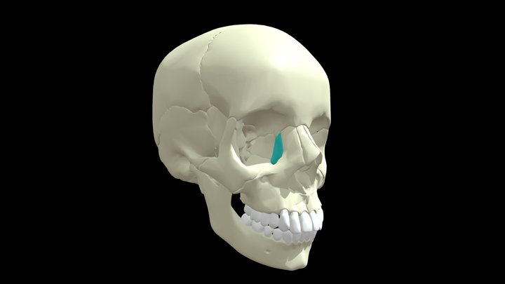
Lacrimal Bones
•Paired; smallest facial bones
•Forms part of the medial wall of the orbital cavity and lateral wall of nasal cavity
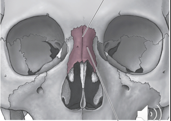
Nasal Bones
Paired
•forms the bony bridge of the nose

Nasal bones

Lacrimal bones

Vomer
•Forms inferior and posterior portion of the nasal septum

Vomer

Mandible
•Single bone by ~age 2; horseshoe-shaped bone
Important features:
•Body
•2 vertical rami
•Condylar process and coronoid process, separated by mandibular notch
•Head of condylar process articulates with cranium at the mandibular fossa of the temporal bone; forms temporomandibular (TMJ) joint

Mandible

Pharynx Functions
•Provides a common pathway for food intake and breathing
•Contracts during swallowing, clearing food from the pharynx into the esophagus
•Acts as a resonating chamber for speech

Pharynx

Muscles of the Pharynx
•3 ‘constrictor’ muscles
surround the pharynx and contract during swallowing
all are open anteriorly, connect to posterior median pharyngeal raphe. Except lower fibers of the inferior constrictor muscle which form a complete ring
organized like stacked cups

Superior constrictor (“1st cup”)
Description: weakest constrictor
Origin: medial pterygoid plate
Insertion: posterior median pharyngeal raphe, occipital bone
Action: constricts pharynx (in swallowing and velopharyngeal closure)
Innervates: Accessory (XI)

A?
Superior constrictor

•Middle constrictor (“2nd cup”)
Description: fan-shaped
Origin: stylohyoid, hyoid bone
Insertion: posterior median pharyngeal raphe
Action: constricts pharynx
Innervates: Accessory (XI)

Middle constrictor

•Inferior constrictor (“3rd cup”)
a.Thyropharyngeus – thickest, strongest
•O: oblique line of the thyroid cartilage
•I: posterior median pharyngeal raphe
•A: constricts pharynx
•Inn: CN XI
b.Cricopharyngeus –narrow band
•O: cricoid cartilage
•I: other side of cricoid cartilage
•A: keeps esophagus closed at UES, at rest
•Inn: pharyngeal branch, eSLN and RLN of X

C?
Inferior constrictor
3 other muscles, pharyngeal elevators, insert into pharyngeal wall (not constrictors) they
elevate pharynx during swallowing

•Salpingopharyngeus muscle
Description: long, thin; paired
O: Eustachian tube
I: lateral wall of pharynx
A: elevates pharynx; shortens pharynx (keeps eustachian tube closed at rest)
Inn: CN XI

Salpingopharyngeus muscle

Stylopharyngeus
Description: long, thin; paired
O: styloid process
I: lateral wall of pharynx
A: elevates pharynx; shortens pharynx
Inn: CN IX (glossopharyngeal)

Stylopharyngeus

Palatopharyngeus muscle
Description: forms the palatopharyngeal arch
O: palatine raphe
I: thyroid cartilage, inferior portion of pharynx
A: lifts the pharynx and larynx, directs bolus into pharynx during swallowing
Inn: CN XI

Palatopharyngeus muscle

Esophagus
•Collapsible, muscular tube of approximately 25 cm in length
•Posterior to trachea, anterior to vertebral column
•From the laryngopharynx, it passes inferiorly, and pierces the diaphragm through the esophageal hiatus

Esophagus Upper Border
Upper Esophageal Sphincter (UES)
UES is comprised of the cricopharyngeus muscle
Normally closed - contraction of CP muscle
UES opens by: relaxation of the CP muscle

Esophagus

Upper Esophageal Sphincter (UES)
Primary peristalsis
muscle contraction triggered by arrival of food at UES; contraction wave continues the length of the esophagus
Secondary peristalsis
if residue remains in esophagus, second peristaltic wave is triggered
Mandibular System
Mandible
•Dentition
•Muscles of Mastication
•Temporomandibular Joint (TMJ)
Teeth
are vital components of the speech mechanism
oProvide articulatory surface for speech sounds
oIntegrity of dentition is important – minor speech deficits may arise as a result of dental abnormalities
provide the mechanism for mastication
oOrientation of them key to effective mastication
•are important contributors to an individual’s characteristic facial structure
Muscles of Mastication
•Jaw moves in 3 dimensions:
–Superior/inferior (up/down)
–Anterior/posterior (forward/backward)
–Laterally (side-to-side)
•Therefore, muscles need to:
–Depress the jaw (mouth openers)
–Elevate the jaw (mouth closers)
–Retract the jaw (draw jaw backward)
–Protrude the jaw
–Swing the jaw laterally

These three muscles do what to mandible
depress it/move it downwards
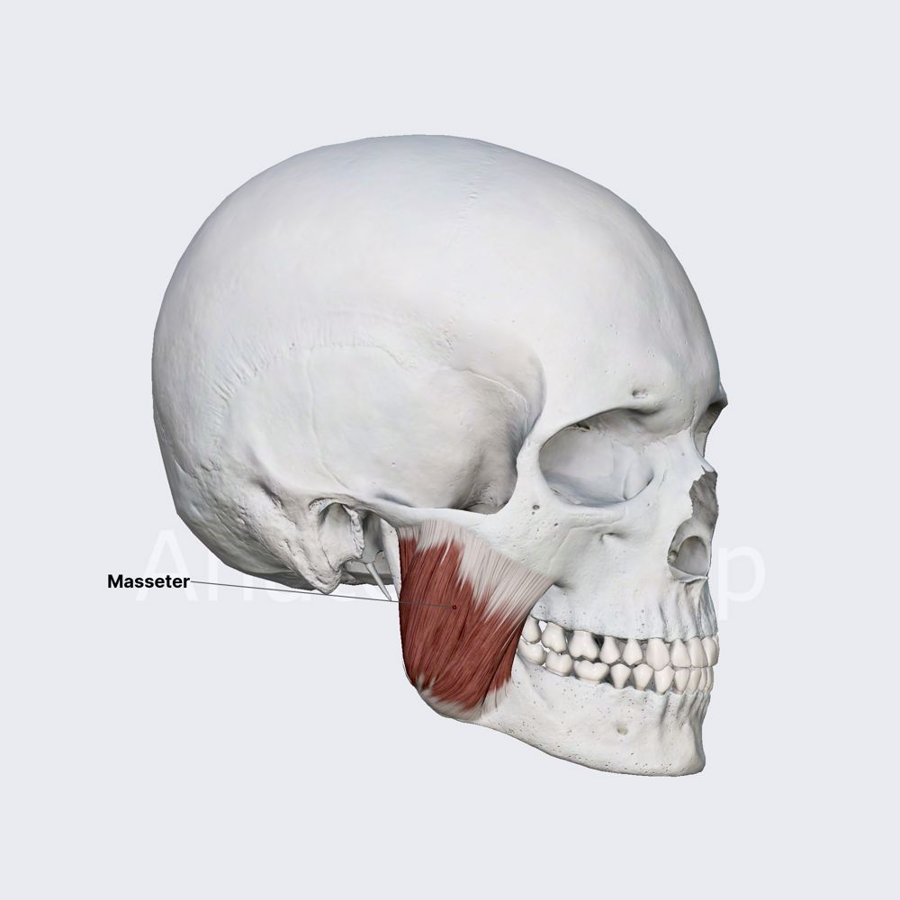
Masseter
•Description: thick, powerful; most superficial muscle of mastication “big chewer”
O: zygomatic process of maxilla, zygomatic process of temporal bone
I: angle and ramus of mandible
A: elevates mandible (major role in chewing)
Inn: mandibular branch of Trigeminal (V)
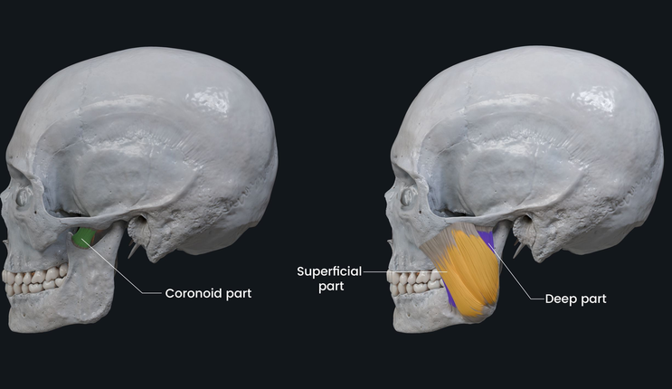
Masseter
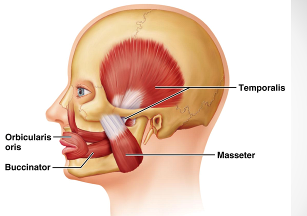
Temporalis
•Description: large, fan-shaped muscle, deep to masseter
O: sphenoid, frontal, parietal, temporal bones (pterion)
I: coronoid process of mandible via a tendon that runs deep to the zygomatic process
A: elevate and retract the jaw
Inn: mandibular branch of Trigeminal (V)

temporalis muscle

Lateral Pterygoid
•Description: deep to masseter
−O: greater wing and lateral pterygoid plate of sphenoid
−I: condyle of mandible, temporomandibular joint (TMJ)
−A: bilateral contraction protrudes jaw; unilateral contraction moves jaw laterally
−Inn: mandibular branch of Trigeminal (V)

Lateral Pterygoid
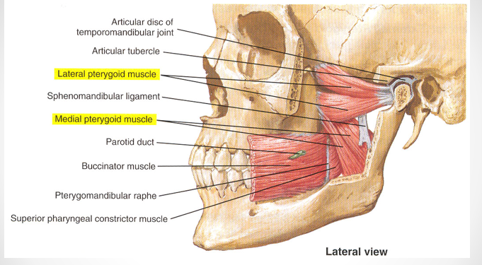
Medial Pterygoid
•Description: deep to lateral pterygoid
O: lateral pterygoid plate
I: angle and ramus of mandible
A: bilateral contraction elevates jaw (jaw-closing) and protrudes jaw; unilateral contraction moves jaw laterally
Inn: mandibular branch of Trigeminal (V)

Medial Pterygoid
Central Pattern Generator
•network of neurons
it sets the basic “chewing rhythm” (once you put food in your mouth you don’t have to keep thinking about chewing.)

Temporomandibular Joint (TMJ)
•integrity of TMJ important for speech
•major movements occur during chewing and yawning
•TMJ disorders occur in 5% of population, associated with teeth grinding
•mandible articulates with the mandibular fossa in temporal bone
-shallow socket, thus TMJ is easily dislocated
•2 kinds of movement:
-hinge-like jaw opening/closing
-condylar process may glide anteriorly when mouth is opened widely
Role of Mandible in Speech
•mandible provides a moving platform for the tongue
•if the tongue is weak, the mandible may compensate with exaggerated movement