echo exam 3 (combined)
 Call Kai
 Learn
 Practice Test
 Spaced Repetition
 Match
 Flashcards
 Knowt Play
1/224

There's no tags or description
Looks like no tags are added yet.
225 Terms
what are the layers of the aortic wall? (3) which one is the most important and composes most of the wall?
tunica intima
tunica adventitia
tunica media (most important, composes 80%)
what does the aortic wall do? (2)
damping - stores force generated by ventricle in systole
expulsion - expels stored energy in systole
(basically first dilates the aorta to allow blood in, then snaps back to push flow forward)
what are the aortic regions? (3) what does the aorta become after passing the diaphragm?
ascending aorta
aortic arch
descending aorta
becomes the abdominal aorta
what are the key segments of the ascending aorta? (4)
annulus
sinuses of valsalva
sinotubular junction
ascending aorta
what branches arise from the aortic arch? (3)
innominate (brachiocephalic)
left common carotid
left subclavian
what is the aortic isthmus? what is it a common site for?
part of aorta distal to left subclavian artery and descending aorta
common site of coarctation
common site of traumatic injury (bc aorta is fixed to thorax here)
where does the abd ao start? where does it run in comparison to the ivc? what is the average diameter?
starts at diaphragm
runs to the left of the ivc
average diameter : 2 cm (1-3 cm is normal range)
what is supravalvular as?
definition : congenital narrowing of the ascending aorta, just distal to coronary arteries
how common is obstructive lesion supravalvular as?
least common site for congenital as… very rare
what features usually accompany supravalvular as? (2)
dilation of the coronary arteries, sometimes ostial obstruction (complete blockage of a vessel)
thickening of the AoV cusps, with some AI
what are obstructive lesions?
general term for any abnormality that blocks or restricts blood flow
what are the types of supravalvular as? (3)
hour-glass shaped
discrete membrane
hypoplastic asc ao
what are the characteristics of hour-glass type svas? (5)
fibromuscular thickening, resulting in hourglass narrowing
just above aortic sinuses
most common type
cusps may be thickened
some degree of associated PA stenosis… (bc PA and Ao develop at same time) can be supravalvular or peripheral
what syndrome is hour-glass type svas associated with? what are the features of that syndrome? (4)
williams syndrome
features:
supravalvular as
elfin facies
mental retardation
peripheral pulmonic stenosis
what are the characteristics of fibrous membrane svas? (2)
seen in normal sized aorta
located near sinotubular junction
what arteries does hypoplastic svas often also effect? what is it also known as? what syndrome is it associated with?
often involves the origins of the brachiocephalic arteries
aka : strand stenosis
associated with hypoplastic left heart syndrome
what is the defining characteristic of each type of svas?
hour-glass : aorta pinches right above valve
discrete membrane : normal sized aorta with small membranous shelf
hypoplastic : small aorta for an extended segment
where does coarctation occur?
AORTIC ISTHMUS
in descending aorta
distal to left subclavian artery
near ductus arteriosis
what is coarctation?
localized narrowing of the descending ao
what other congenital heart defects are usually associated with coarctation? (2)
bicuspid AoV
MV malformations
what is the ‘classic figure 3’ when talking about CXR?
it’s a sign of aortic coarctation.
curved part = area of stenosis
straight part = dilated subclavian artery
double = post-stenotic dilitation
what are the signs of coarctation on CXR? (2)
classic figure 3
rib notching (from dilated vessels)
what are the types of subaortic valvular obstruction? (2)
discrete membrane
tunnel type
what are the characteristics of discrete membrane subaortic obstruction? where does it extend from and to? what is the echo appearance
thin, membranous, crescent-shaped shelf formed right under AoV or in LVOT
membrane usually extends from ANTE septum to AMVL
echo appearance : thin linear echo in LVOT, perpendicular to IVS
what does the m-mode look like for a pt who has discrete membrane subaortic obstruction?
coarse systolic flutter of Aov leaflets (fr turbulent and accelerated flow)
midsystolic partial closure (fr LVOT obstruction)
what is aortic dilation? how common is it?
increased diameter of aorta
most common aortic abnormality
what are the causes for aortic dilation in asc ao? (5)
htn
atherosclerosis
cystic medial necrosis
post-stenotic dilation
collagen vascular and inflammatory disorders
marfans
rheumatoid arthritis
SLE
reiter syndrome
when dilation becomes severe, it is then considered?
aneurysmic
what are aneurysms? what % increase in dilation would be considered anuerysmal?
localized areas of abnormal dilation of a vessel
usually more than 50% increase in a vessel = aneurysmic
what is criteria to call an aortic aneurysmal?
over 5 cm in asc ao
over 4 cm in desc ao
over 6 cm = high risk of rupture
what are the causes for aortic aneurysms? (7)
atherosclerosis/degenerative causes (most common)
syphilis
trauma
aortic stenosis
genetically triggered (e.g. aortic coarctation)
pda
aortitis
what are the types of aneurysms?
dissecting (tear, intima layer peels away)
saccular (weakening of vessel wall at one point, results in pouch-like expansion)
fusiform (most common, involves entire circumference of vessel and dilation is uniform)
what are the morphologic shapes of aneurysms?
annuloaortic ectasia (pear-shaped ao root), involves annulus and SOV
dilation involving annulus, SOV, and part of asc ao
dilation beginning @ sinotubular junction, spares annulus and SOV
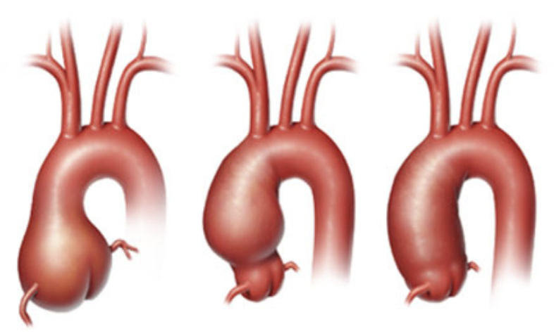
aneurysm type? what parts of the Ao does it involve?
pear-shaped … involves annulus and SOV
dilation at sinotubular junction and beyond
dilation at annulus, SOV, and asc ao
for TTE eval, what view do we look at for aortic aneurysms? which exact parts do we measure? from where to where?
aortic sizes, measured from plax, at end-diastole
measure : annulus, SOV, sinotubular juntion, proximal part of asc ao
can be leading edge to leading OR inner edge to inner
what is marfan’s syndrome?
disorder involving connective tissue, bones, muscles, ligaments, and skeletal structure
what are the echo findings for a pt with marfan’s syndrome? what is the most common?
proximal aorta dilation
multivalvular prolapse
la compression
ai, mr
ao dissection (most common)
lvvo
how common are atherosclerotic aneurysms? how much do they effect thoracic or abdominal ao?
very common, most common
25% are thoracic, other 75% is abdominal
what are the most common sites for aneurysms for thoracic ao?
aortic arch
desc ao
how does the atherosclerotic process work on aortic aneurysms?
weakens ao wall
medial degeneration
localized vessel dilation
how does hypertension effect the aorta?
huge cause for disease of the aorta
weakens the strength of aorta wall
eventual expansion to aneurysm
symptoms of aneurysms (3)
symptoms depend on size and location of the aneurysm
symptoms are a result of compression on other structures
e.g. esophagus, laryngeal nerve, left bronchus
what symptoms arise from aortic aneurysms? (6)
wheezing
coughing
dyspnea
hemoptysis
hoarseness
dysphagia
which imaging modalities can be used for diagnosing anerurysms?
chest xray (good for mediastinal widening)
ct w/ contrast (good)
mri (good, no contrast needed)
angiography (the best! but can miss aneurysm if there is a thrombus)
treatments for aneurysmal aortas?
follow ups
medication
surgery (when over 5-6 cm)
open chest surgery, remove enlarged area of ao and sometimes the valve too, replace with graft and mechanical/biological valve
evar (endovascular aortic aneurysm repair), tube inserted into blood vessel, usually groin, and guides to aorta. catheter places graft in aneurysmic site to reinforce weakened walls
what is a SOV aneurysm? how common is it? is it acquired or congenital? how does it present?
dilation of sov
rare
can be acquired OR congenital
can present ruptured OR unruptured
what are some congenital cause of SOV aneurysms? (4)
bicuspid AoV
connective tissue disease
marfans
ehler danlos
what are some acquired causes of SOV aneurysms? (3)
infective endocarditis (veggies)
atherosclerotic degeneration
iatrogenic post cardiac surgery
if the left SOV had an aneurysm, where would it rupture? complications?
would rupture into the LA or RA
can cause la compression
if the non-coronary SOV had an aneurysm, where would it rupture?
could rupture into LA, RA, LV, or IVS
if the right SOV had an aneurysm, where would it rupture? complications?
could rupture into RA, RV, MPA, or pericardium
can cause:
RVOT obstructions,
RCA dissection or compression and acute MI
compression on conduction system → heart block
tamponade
what do the signs and symptoms (clinical symptoms) of SOV aneurysms depend on? (4)
size
location
compression
rupture
for a pt with a SOV aneurysm, what would you expect from their physical exam?
wide pulse pressure (from AI)
Long diastolic murmur
LVH on EKG
CXR
signs of CHF (fluid in leg, lung, etc)
cardiomegaly (enlarged heart)
unfolding of Ao (not as curvy aortic arch)
what are possible treatments for SOV aneurysms?
surgery (for ruptured and unruptured)
transcatheter repair
what is aortitis?
group of conditions that cause inflammation to walls of aorta
what are the types of aortitis? (4)
infectious
noninfectious
radiation induced
idiotpathic
what are the clinical signs of aortitis?
nonspecific pain
fever
malaise (feeling of bleh)
elevated levels of acute phase reactants
other systemic manifestations
what is acute aortic syndrome? (7)
acute onset of chest pain
disruption of ao wall motion
need for urgent medical attention
Aortic dissection
intramural hematoma penetrating atherosclerotic ulcer (when plaque explodes, clot forms on top causing even more stenosis
traumatic aortic rupture due to blunt deceleration trauma
aortic aneurysm leak and rupture
what is aortic dissection? which gender is more affected?
sudden tear in intima layer of aortic wall, allowing blood to enter the wall and destroy media and further strip intima from adventitia
more common in male
what are the names of the systems used to classify aortic dissections?
deBakey
stanford
how does stanford classify aortic dissections?
by location
type a = ascending ao
type b = any other dissection
how does DeBakey classify aortic dissections?
by location
type 1 = ascending, transverse, and descending ao involved (70%)
type 2 = ascending ao involved, but stops before brachiocephalic (5%)
type 3 = descending ao and may extend into abd ao (25%)
what are the causes of ao dissection? (11)
htn
atherosclerosis
marfan’s
aging
pregnancy
trauma
iatrogenic injury
inflammatory diseases
cocaine
renal disease
strenous physical exercise
what are the symptoms for ao dissection? (6)
excruciating pain
severe at onset
radiate in path of dissection
clammy
shock-like
hypertensive (or hy
what is the echo hallmark for ao dissection?
flap in between the intima and media layer of aortic wall… best seen in short axis
creates false and true lumen
what is the criteria for diagnosing aortic dissection?
oscillation independent of ao
visualized in multiple views
disctintion from other structures (calcification, plaque, wires…)
pericardial effusion
aortic insufficiency
false lumen is larger than true lumen
flap moves towards true lumen in diastole (true lumen expands during systole, vice versa diastole)
spontaneous contrast in false lumen
slow flow in false lumen
more rapid flow in true lumen
contrast agents opacify true lumen first
turbulence at entry site
what complications are associated with false lumens in ao dissections? (3)
compresses true lumen
extends into other branch vessels
thrombosis (esp. aortic wall thickness > 15 mm)
what are the other complications associated with ao dissection? (not dealing w false lumen)
malperfusion (coronary, cerebral, spinal, limb, or abdominal ischemia)
ruptures (pericardial, pleural, peritoneal space, tamponade)
what is the most common cause of aortic isthmus?
trauma from high speed deceleration of mva
what are the views for tricuspid valve? (5)
inflow
psax
apical 4
subcostal 4
non-imaging
what are the causes of tricuspid stenosis? (6)
rheumatic heart disease (most common, 90%)
SLE
carcinoid heat disease
loeffler’s endocarditis
metastatic melanoma
congenital heart disease
t/f : rheumatic TS usually occurs with rheumatic mitral or aortic stenosis
true, isolated rheumatic tv stenosis is VERY rare
what is the NORMAL tv orifice area?
7-9 cm²
what should the severity of tricuspid stenosis be graded on? (3)
valve area
mean PG
ra chamber size
if a pt has tricuspid stenosis, what would you expect from their m-mode? (3)
diminished EF slope (caused RVH and pulm HTN)
anterior displacement of posterior leaflet
thickening
if tricsupid stenosis goes undetected, what could it lead to? (3)
increased chance of mortality when undergoing operations
chronic elevation of RA pressure
low CO (even if left valves are functioning well… bc low venous return)
what are the associated findings of tv stenosis? (6)
RAE
bowing IAS
increased diastolic TV PG
dilated IVC
pulmonary HTN
RVH
what will high RA pressure lead to?
increased venous pressure:
peripheral edema
hepatic congestion
fatigue
chronic RA dilation
decreased SV
a fib, thrombus
what is the 2d criteria for grading TV stenosis?
doming of tv leaflets in diastole
thickening and reduced excursion
smaller orifice
what can mimic TV stenosis?
anything that obstructs RA…
RA tumors
large veggies
large atrial myxomas
what would you expect from a doppler on a pt with TV stenosis? (5)
faster diastolic velocities
decreased ef slope
turbulent flow
prolonged reduction in velocity (normal: fast decline)
increased a wave for hepatic vein
what should TV velocities be?
with tr, tv velocities equal or less than 0.7 m/s
with tv stenosis, velocities are greater than 1.0 m/s
how do we grade tv stenosis?
NOT planimetry
NOT RLLY pht (not proven) … TVA = 190/PHT
quantifiable data (what we mostly grade on):
peak velocities
peak PG
mean PG
if a pt has mild TV stenosis, what would their valve area, mean PG, and pulm artery pressure be?
TVA : >1.5 cm²
mean PG : <5 mmhg
pulm artery pressure : <30 mmhg
if a pt has moderate TV stenosis, what would their valve area, mean PG, and pulm artery pres
TVA : 1-1.5 cm²
mean PG : 5-10 mmhg
pulm artery pressure : 30-50 mmhg
if a pt has severe TV stenosis, what would their valve area, mean PG, and pulm artery pres
TVA : <1 cm²
mean PG : >10 mmhg
pulm artery pressure : >50 mmhg
what is carcinoid heart disease?
result of carcinoid tumors (mostly found in GI tract)
effects right side of heart (TV and PV)
how do carcinoid tumors affect the heart?
tumors produce a vasoactive substance that causes endothelial damage to the right side of heart
primary tumors can be small
at what part in the carcinoid disease does it affect the heart? what is the rate of people with carcinoid disease that get some sort of heart involvement?
heart is effected late into the progression
½
what are the symptoms of carcinoid heart disease? (6)
flushed face
abdominal pain
diarrhea
renal failure
hepatic failure
hepatomegaly (later stages)
what are the cardiac signs of carcinoid heart disease?
elevated venous pressure
diastolic and systolic murmurs (from the regurgitations and stenosis)
what is the 2d appearance of a pt with carcinoid heart disease?
RVE
abnormal septal motion, indicating RVVO
thickened, retracted TV
foreshortened chordae
thickened, retracted PV
TV never fully coapts
what are the doppler signs for carcinoid heart disease?
TR (most prevalent)
increased TV velocities
increased PV velocities
PI
what are the echo findings for carcinoid heart disease?
similar to echo findings for rheumatic TV stenosis
what differentiates the echo findings of carcinoid heart disease from rheumatic disease?
rheumatic has left sided valvular involvement. carcinoid heart disease only effects right side valves.
what mechanisms cause TR? (2)
right heart chamber abnormalities
actual tricuspid valve disease
what causes TR? (4)
pacer wires in right heart
cardiac transplant
TV disease
RV infarction
how is TR caused by RV dilation?
congenital heart disease (e.g. partial anomalous venous return)
RVVO (alters TV apparatus)
how is TR caused by RV infarction?
involved posterior papillary muscle which is attached to posterior and septal leaflet of TV
what tricuspid valve diseases cause TR? (7)
rheumatic TV disease
TV prolapse
endocarditis
ruptured pap/chordae
carcinoid heart disease
trauma
congenital defects (e.g. tv dysplasia and ebstein’s anomaly)