SYS PHYSIO Week 9: Cardiovascular System Continued
 Call Kai
 Learn
 Practice Test
 Spaced Repetition
 Match
 Flashcards
 Knowt Play
1/16

There's no tags or description
Looks like no tags are added yet.
17 Terms
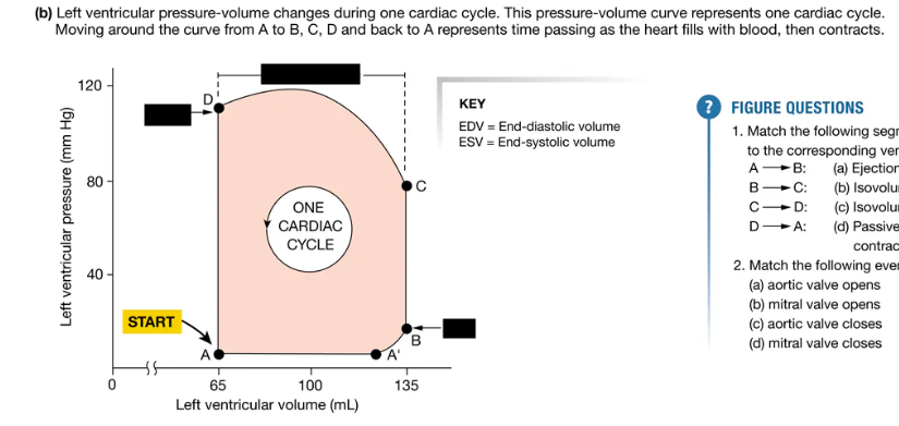
How does the pressure-volume loop relate to the cardiac cycle (i.e., which segment corresponds with which phase)? Label the loop as well
When the heart contracts, the pressure increases and blood flows out of the heart into areas of lower pressure. Figure 14.18b represents pressure and volume changes in the left ventricle, which sends blood into the systemic circulation. The left side of the heart creates higher pressures than the right side, which sends blood through the shorter pulmonary circuit.
The cycle begins at point A. The ventricle has completed a contraction and contains the minimum amount of blood that it will hold during the cycle. It has relaxed, and its pressure is also at its minimum value. Blood is flowing into the atrium from the pulmonary veins.
Once pressure in the atrium exceeds pressure in the ventricle, the mitral valve between the atrium and ventricle opens. Atrial blood now flows into the ventricle, increasing its volume (point A to point B). As blood flows in, the relaxing ventricle expands to accommodate the entering blood. Consequently, the volume of the ventricle increases, but the pressure in the ventricle goes up very little.
The last portion of ventricular filling is completed by atrial contraction (point A’ to B). The ventricle now contains the maximum volume of blood that it will hold during this cardiac cycle, the end-diastolic volume or EDV (point B). In a 70-kg man at rest, end-diastolic volume is about 135 mL. However, EDV varies under different conditions. During periods of very high heart rate, for instance, when the ventricle does not have time to fill completely between beats, the end-diastolic value may be less than 135 mL.
When ventricular contraction begins, the mitral (AV) valve closes. With both the AV valve and the semilunar valve closed, blood in the ventricle has nowhere to go. Nevertheless, the ventricle continues to contract, causing the pressure in this chamber to increase rapidly during isovolumic contraction. Once ventricular pressure exceeds the pressure in the aorta, the aortic valve opens (point C). Pressure continues to increase as the ventricle contracts further, but ventricular volume decreases as blood is pushed out into the aorta (C → D)
The end-systolic volume or ESV (point D) is the minimum volume of blood the ventricle contains during one cycle. An average ESV value in a person at rest is 65 mL, meaning that nearly half of the 135 mL that was in the ventricle at the start of the contraction is still there at the end of the contraction.
At the end of each ventricular contraction, the ventricle begins to relax. As it does so, ventricular pressure decreases. Once pressure in the ventricle falls below aortic pressure, the semilunar valve closes, and the ventricle again becomes a sealed chamber. The remainder of relaxation occurs without a change in blood volume, and so this phase is called isovolumic relaxation (D → A). When ventricular pressure finally falls to the point at which atrial pressure exceeds ventricular pressure, the mitral valve opens and the cycle begins again.


Label the Wiggers Diagram
At what points in the cycle do EDV and ESV occur?
On the Wiggers diagram, match the following events to the lettered boxes:
end-diastolic volume,
aortic valve opens,
mitral valve opens,
aortic valve closes,
mitral valve closes,
end-systolic volume
Why does atrial pressure increase just to the right of point C? Why does it decrease during the initial part of ventricular systole, then increase? Why does it decrease to the right of point D?
Why does ventricular pressure shoot up suddenly at point C?
EDV occurs in step 3, and ESV occurs in step 5.
(a) E, (b) A, (c) D, (d) B, (e) C, (f) F
Atrial pressure increases because pressure on the mitral valve pushes the valve back into the atrium, decreasing atrial volume. Atrial pressure decreases during the initial part of ventricular systole as the atrium relaxes. The pressure then increases as the atrium fills with blood. Atrial pressure begins to decrease at point D, when the mitral valve opens and blood flows down into the ventricles.
Ventricular pressure shoots up when the ventricles contract on a fixed volume of blood.

What are the primary structural and functional differences between arteries, arterioles, capillaries, and veins?
Pressure produced by contraction of the left ventricle is stored in the elastic walls of arteries and slowly released through elastic recoil. This mechanism maintains a continuous driving pressure for blood flow during ventricular relaxation. For this reason, the arteries are known as the pressure reservoir {reservare, to retain} of the circulatory system.
Downstream from the arteries, small vessels called arterioles create a high-resistance outlet for arterial blood flow. Arterioles direct distribution of blood flow to individual tissues by selectively constricting and dilating, so they are known as the site of variable resistance. Arteriolar diameter is regulated both by local factors, such as tissue oxygen concentrations, and by the autonomic nervous system and hormones.
When blood flows into the capillaries, their leaky epithelium allows exchange of materials between the plasma, the interstitial fluid, and the cells of the body. At the distal end of the capillaries, blood flows into the venous side of the circulation. The veins act as a volume reservoir from which blood can be sent to the arterial side of the circulation if blood pressure falls too low. From the veins, blood flows back to the right heart.
Which vessels serve as the pressure reservoir? The primary resistance vessels? The exchange vessels? The volume reservoir (or capacitance) vessels?
Pressure reservoir: the arteries
Primary resistance vessels: arterioles
Exchange vessels: capillaries
Volume reservoir: the veins

How does blood pressure change as we move through the systemic vascular tree from the left ventricle to the right atrium? Label the diagram
It decreases! In the systemic circulation, the highest pressure occurs in the aorta and results from pressure created by the left ventricle. Aortic pressure reaches an average high of 120 mm Hg during ventricular systole (systolic pressure), then falls steadily to a low of 80 mm Hg during ventricular diastole (diastolic pressure). Notice that pressure in the ventricle falls to only a few mm Hg as the ventricle relaxes, but diastolic pressure in the large arteries remains relatively high. The high diastolic pressure in arteries reflects the ability of those vessels to capture and store energy in their elastic walls.

Vasoconstriction and vasodilation occur when the smooth muscle in the walls of blood vessels (which kind of vessels again, primarily?) contracts or relaxes, respectively.
Arteries and arterioles

How does the process of contraction and relaxation occur in smooth muscle? Label the diagram
Contraction:
Contraction begins when cytosolic calcium concentrations increase following calcium entry from the extracellular fluid and calcium release from the sarcoplasmic reticulum 1 → The calcium ions bind to calmodulin (CaM) 2 → The calcium-calmoduiln complex then activates an enzyme called myosin light chain kinase (MLCK) 3 → At the base of the myosin head is a small regulatory protein chain called the myosin light chain. Phosphorylation and dephosphorylation of the myosin light chain control contraction and relaxation in smooth muscle. When calcium-calmodulin activates MLCK, the enzyme phosphorylates the myosin light protein chains 4. → Phosphorylation of myosin enhances myosin ATPase activity. When myosin ATPase activity is high, actin binding and crossbridge cycling increase tension in the muscle 5 →The myosin ATPase isoform in smooth muscle is much slower than that in skeletal muscle, and this decreases the rate of crossbridge cycling. Dephosphorylation of the myosin light chain by the enzyme myosin light chain phosphatase (MLCP) decreases myosin ATPase activity. Interestingly, dephosphorylation of myosin does not automatically result in relaxation. Under conditions that we do not fully understand, dephosphorylated myosin may remain in an isometric contraction called a latch state. This condition maintains tension in the muscle fiber while consuming minimal ATP. It is a significant factor in the ability of smooth muscle to sustain contraction without fatiguing.
Relaxation:
Because dephosphorylation of myosin does not automatically cause relaxation, it is the ratio of MLCK to MLCP activity that determines the contraction state of smooth muscle. MLCP is always active to some degree in smooth muscle, so the activity of MLCK is often the critical factor. As you learned, MLCK activity depends on calcium-calmodulin.
Relaxation in a smooth muscle fiber is a multistep process. As in skeletal muscle, free calcium is removed from the cytosol when calcium-ATPase pumps it back into the sarcoplasmic reticulum. In addition, some calcium is pumped out of the cell with the help of calcium-ATPase and the sodium-calcium exchanger (NCX) 6 → By the law of mass action, a decrease in free calcium cytosolic causes calcium to unbind from calmodulin 7 → In the absence of calcium-calmodulin, myosin light chain kinase becomes inactivate. As MLCK becomes less active, myosin light chain phosphatase dephosphorylates myosin 8 → Myosin ATPase activity decreases 9 → and the muscle relaxes.

There are many ways the body can change vascular smooth muscle contraction/relaxation to create vasoconstriction or vasodilation...
Sympathetic neurohormone epinephrine causes smooth muscle contraction when it binds to alpha-adrenergic receptors but relaxation when it binds to beta-2 adrenergic receptors.
Most smooth muscle neurotransmitters and hormones bind to G protein-linked receptors. The second messenger pathways then determine the muscle response. In general, IP3 triggers contraction and cAMP promotes relaxation.
Pathways that increase IP3 cause contraction several ways:
IP3 opens IP3 channels on the SR to release calcium
Diacylglycerol (DAG), another product of the phospholipase C signal pathway, indirectly inhibits myosin phosphatase activity. Increasing the MLCK/MLCP ratio promotes crossbridge activity and muscle tension.
Signals that cause muscle relaxation work through the following mechanisms:
Free cytosolic calcium concentrations decrease when IP3 channels are inhibited and the SR calcium-ATPase is activated.
Potassium leaking out of the cell hyperpolarizes it and decreases the likelihood of voltage-activated calcium entry.
Myosin phosphatase activity increases, which causes a decrease in muscle tension.
Why does the body care about maintaining MAP at a homeostatic set-point?
What happens if MAP gets too low or too high?
The body cares about maintaining Mean Arterial Pressure (MAP) at a homeostatic set-point because MAP represents the driving force that pushes blood through the circulatory system to perfuse vital organs and tissues.
Think of MAP like water pressure in a municipal plumbing system. If the pressure is too low, water won’t reach the upper floors of a building. If it is too high, the pipes will burst.
Why the Set-Point Matters
The normal homeostatic range for MAP is typically 65 mmHg to 110 mmHg. The body works continuously via feedback loops (like the baroreceptor reflex) to defend this range because it strikes a perfect physiological balance:
It overcomes resistance: It provides just enough pressure to overcome systemic vascular resistance and deliver oxygen and nutrients to delicate cellular networks.
It protects structural integrity: It stays low enough to keep blood vessels from stretching, tearing, or hardening under excessive mechanical stress.
What happens if MAP gets too low?
When MAP drops below 60–65 mmHg, the pressure gradient is no longer strong enough to force blood through organ tissue.
Ischemia and Hypoxia: Tissues are starved of vital oxygen and glucose.
Organ Dysfunction: Brain tissue is highly sensitive; a drop in cerebral perfusion leads to dizziness, loss of consciousness, and rapid neuronal death. The kidneys will also fail to filter waste.
Systemic Shock: Severe, prolonged low MAP plunges the body into cardiovascular shock, resulting in cellular death and irreversible multi-organ failure.
What happens if MAP gets too high?
When MAP chronically climbs above 100–110 mmHg, it exerts a dangerous, constant mechanical strain on the entire cardiovascular tree.
Cardiac Strain: The heart has to pump much harder against the elevated pressure (increased afterload). Over time, this causes the heart muscle to pathologically enlarge (hypertrophy), leading to heart failure.
Vascular Damage: High pressure damages the delicate endothelial lining of the arteries, accelerating plaque buildup (atherosclerosis).
Vascular Ruptures: Weakened, highly pressurized vessels are prone to leaking or bursting, dramatically increasing the risk of blood clots, heart attacks, and hemorrhagic strokes.

Compare and contrast active hyperemia and reactive hyperemia by labeling the diagram


Label the diagrams of arteriolar diameter affecting blood flow

What is the baroreceptor reflex?
The primary reflex pathway for homeostatic control of mean arterial blood pressure is the baroreceptor reflex. Stretch-sensitive mechanoreceptors known as baroreceptors are located in the walls of the carotid arteries and aorta, where they continuously monitor the pressure of blood flowing to the brain (carotid baroreceptors) and to the body (aortic baroreceptors).

Label the CNS control of the heart and blood vessels


Label the Baroreceptor reflex

You should be able to use the baroreceptor reflex to explain the responses to a disturbance in MAP (an unwanted increase or decrease in MAP)...
This difference is important for understanding how the CVCC controls MAP. The sympathetic branch can raise pressure by increasing heart rate, increasing contractility, constricting arterioles, and constricting veins. The parasympathetic branch mainly lowers cardiac output by slowing heart rate. Most vascular smooth muscle receives sympathetic innervation only, so decreases in sympathetic tone are what allow many arterioles and veins to dilate during reflex responses.
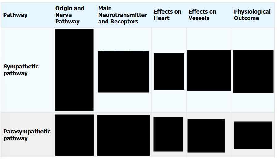
Label the coordinated adjustments of the heart and vessels by the CVCC


Label the coordinated adjustments of the heart and vessels by the CVCC when autonomic output is added
