MAJOR GRAM NEGATIVE BACTERIA
 Call Kai
 Learn
 Practice Test
 Spaced Repetition
 Match
 Flashcards
 Knowt Play
1/49

There's no tags or description
Looks like no tags are added yet.
50 Terms
what is the difference in structure between gram negative and gram positive bacteria

state the gram-negative cell surface antigens
H-antigen
K-antigen
O-antigen
what are the H, K and O antigens
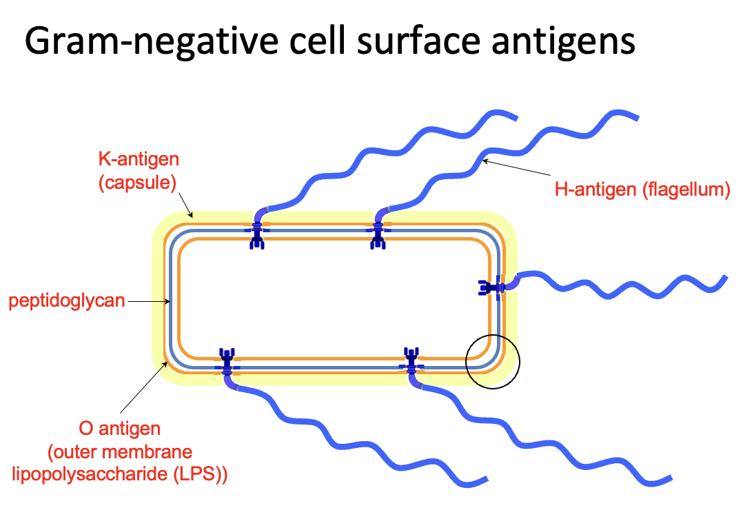
another diagram showing gram negative cell wall structure
capsule extends from surface, loosely attached
LPS attached to outer membrane

do gram negative bacteria retain gram stain
no, they are therefore counterstained to become pink
what do O-antigens do
immune evasion

what does the capsule do
evades antibody binding (immune evasion)

list of medically important gram negative bacteria
Neiserria - meningitis and gonorrhoea
Yersinia pestis
Pseudomonas aeruginosa
—
GI tract - enterobacteria
Salmonella spp. (gastroenteritis)
Shigella spp. (gastroenteritis)
Vibrio cholera
Escherichia coli
Helicobacter pylori
Campylobacter jejuni (food poisoning)
extra gram negative bacteria
Bordetella (whooping cough)
Legionella
Treponema pallidum (syphilis)
Klebsiella (major hospital resistant pathogen)
what shape is Neisseria spp.
gram negative diplococci

where are Neisseria spp. often seen
inside PMNLs of infected individuals
this is an observation diagnostic of infection

which two diseases are Neisseria spp. most associated with
meningitis (N.meningitidis)
gonorrhoea (N.gonorrhoeae)
outline N.meningitidis/ meningococcus
infection of CSF and meninges
commensal carriage in pharynx/ nasopharynx
capsular
serotyping based on polysaccharides
outline N.gonorrhoeae/ gonococcus
STI - genital and oral infection
neonatal transfer manifests as eye infections
lipo-oligosaccharide capsule often sialylated
both N.meningitidis and N.gonorrhoeae are fastidious bacteria, what does this mean?
they need specific nutritional requirements
heated blood (chocolate agar) + CO2

what is meningitis and its symptoms
inflammation of the meninges
intense headache
fever
malaise
photophobia
stiff muscles and neck
what can meningitis lead to
convulsions and death
on darker skin tones, where should a rash be identified as being associated with meningococcal septicaemia
lighter areas: palms, soles of feet, conjunctiva, palate
outline the epidemiology of N.meningitidis
occurs worldwide often in the winter months
2/3 of cases occur in first 4yrs of life
peak prevalence is 1st yr of life
second peak: 16-23yo (onset of large gatherings)
why does meningitis often occur during winter
dryness of air and thus the breakdown in mucosal barriers
this makes it easier for bacteria to access the body
why is the peak prevalence of meningitis in the first year of life
the initial protection from maternal antibodies is lost so the infant becomes susceptible
how is N.meningitidis spread
either directly to subarachnoid space
or
through nasopharyngeal mucosa to enter the bloodstream
points on N.meningitidis pathogenesis
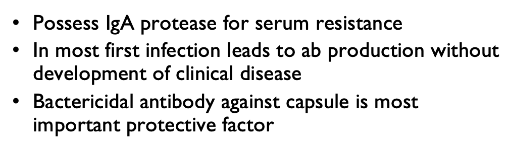

30,000 annual deaths (reduced from 300,000 in 2000)
describe the onset of meningitis
onset of meningitis is very fast
6-24h onset
(therefore speed is the key to successful treatment)
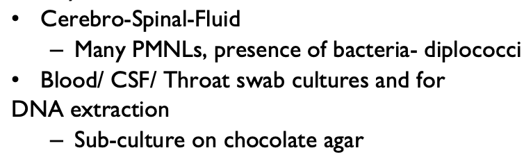
points showing diagnosis of meningitis
what medications are used to treat meningitis
antibiotics
ceftriaxone
cefotaxime + ampicillin
often accompanied by corticosteroids
which antibiotics is prescribed as a prophylaxis for meningitis
ciprofloxacin
what are the chances of passing on meningitis through close contacts
1 in 300
Neisseria summary slide

what other type of bacteria causes meningitis
Haemophilus influenzae (also gram negative)
originally from influenza sufferers
what shape is Haemophilus influenzae
coccobacillus (between a sphere and a rod)


what other conditions can Haemophilus influenzae cause (4)
septicaemia
pneumonia
otitis media (mild infection)
sinusitis (mild infection)
and
meningitis
which strains of H.influenzae are non-capsular
the commensals
how many capsular types of H.influenzae are there
6 capsular types (a-f)
99% of disease is caused by type b capsule type (Hib vaccine combats this)
what is the purpose of the H.influenzae capsule
capsule is a major virulence factor for avoiding C3b (opsonin) binding
incl. fimbriae, IgA proteases
antimicrobial resistance and the WHO list of critical pathogens

outline Pseudomonas aeruginosa
gram negative
common environmental isolate and human commensal
strictly aerobic
rod motile
often multiply to become antibiotic resistant
outline Pseudomonas aeruginosa colonies on agar (appearance and smell)
characteristic green spreading shape
grapey smell
Pseudomonas aeruginosa is a major cause of infection after _____
Pseudomonas aeruginosa is a major cause of infection after burns
why does P.aeruginosa cause infection after burns
well adapted to the warm moist environment in a burn wound
many extracellular proteases breakdown tissues
infection by P.aeruginosa can cause…
septicaemia - ecthyma gangrenosum lesions
how is P.aeruginosa linked to cystic fibrosis patients
P.aeruginosa is the major killer of CF patients - who then develop chronic lung infections and die
name some Klebsiella spp.
Klebsiella pneumoniae
Klebsiella oxytoca
what do Klebsiella spp. cause
hospital sepsis
pneumonia
wound infections (diabetic foot ulcer)
meningitis
antibiotics effective against Klebsiella spp.
colistin
many strains are becoming resistant to last resort antibiotics e.g. carbapenem
where do Klebsiella spp. normally reside
human gut resident
how is Klebsiella spread (infection route)
fecal-oral spread
list of gram negative bacilli (enterobacteria)

summary
