JULIA QUIZ CRAMMMMMMMMM
 Call Kai
 Learn
 Practice Test
 Spaced Repetition
 Match
 Flashcards
 Knowt Play
1/40

There's no tags or description
Looks like no tags are added yet.
41 Terms

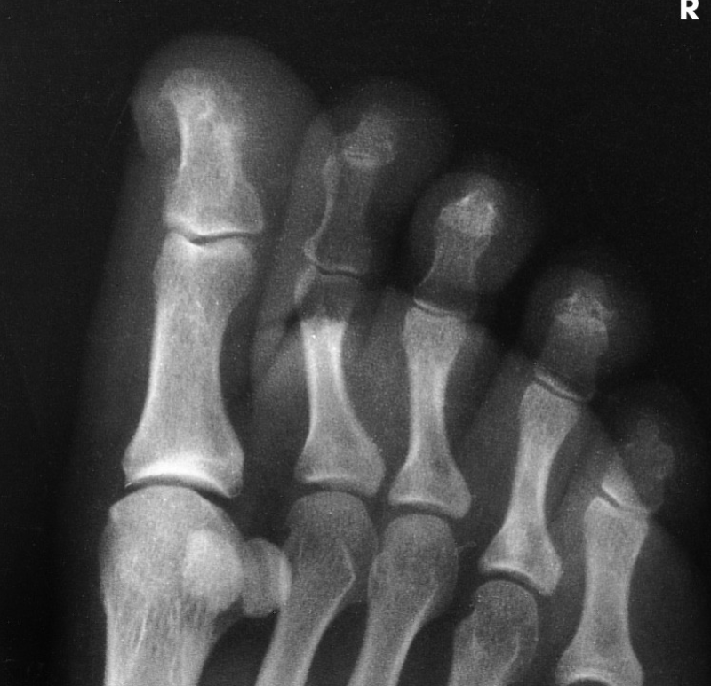
AP/AP AXIAL TOES
Open IP and MTP joint spaces on axial projections
Entire toes, including distal ends of the metatarsals
AP OBLIQUE TOES
Entire toes, including distal ends of the metatarsals
Open IP and 2nd-5th MTP joint spaces

LATERAL TOES
Toe(s) in a true lateral position
Entire toe, without superimposition of adjacent toes
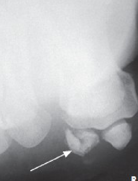
TANGENTIAL PROJECTION SESAMOIDS (Lewis & Holly Method)
Prone: Lewis Method
Seated: Holly method
Sesamoids free of any portion of the first metatarsal
Metatarsal heads

AP/AP AXIAL FOOT
Anatomy from toes to tarsals; may include portions of talus and calcaneus
Overlap of the second through fifth metatarsal bases

AP OBLIQUE FOOT (medial)
Bases of the first and second metatarsals superimposed on medial and intermediate cuneiforms
Tuberosity of the fifth metatarsal

LATERAL FOOT
Entire foot and distal leg
Tibiotalar Joint

AP AXIAL FEET (Weight-bearing Method)
Both feet centered on one image
Anatomy from toes to tarsals; may include portions of talus and calcaneus

LATERAL PROJECTION FEET (Weight-bearing Method)
Entire foot and distal leg
Tibiotalar joint

AXIAL CALCANEUS (Plantodorsal)
Calcaneus & Talocalcaneal (subtalar) Joints
No rotation of the calcaneus demonstrated by Sustentaculum Tali in profile on the medial side

AXIAL CALCANEUS (dorsoplantar)
Calcaneus & Talocalcaneal (subtalar) Joints
No rotation of the calcaneus demonstrated by Sustentaculum Tali in profile on the medial side

LATERAL CALCANEUS
No rotation of calcaneus demonstrated by an open Sinus Tarsi
Tuberosity in profile

AP ANKLE
Medial and lateral malleoli
Tibiotalar Joint space

AP OBLIQUE ANKLE
Distal Tibia, Fibula, and Talus
Proper 45-degree rotation of ankle shown by distal tibiofibular articulation open

AP OBLIQUE ANKLE (MORTISE) 15-20 Degree medial rotation
Distal tibia, fibula, and talus
Proper 15-20 degree rotation of ankle demonstrated by an open Talofibular articulation
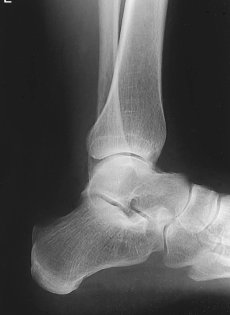
LATERAL ANKLE
5th metatarsal base and tuberosity should be seen to check for Jones fracture
Ankle in true lateral shown by the fibula over the posterior half of the tibia

ANKLE WEIGHT-BEARING METHOD
Medial mortise open
Lateral mortise closed

AP Leg Tib/Fib
Ankle and knee joints on one or more image
Fibular midshaft free of tibial superimposition

Lateral Leg Tib/Fib
Ankle and knee joints on one or more image
Entire leg in true lateral position demonstrated by slight overlap of the tibia on the proximal fibular head

AP Knee
slight superimposition of the fibular head if the tibia is normal
Patella completely superimposed on the femur

AP Oblique Knee (medial)
Tibia and fibula separated at their proximal articulation
Open knee joint

AP Oblique Knee (lateral)
Fibula superimposed over the lateral half of the tibia
Open knee joint

Lateral Projection Knee
Patella in a lateral profile
Open patellofemoral joint space

AP Axial Knee ICF (Beclere)
Open intercondylar fossa
No superimposition of the fossa by the apex of the patella
A
A

Lateral Patella
Open patellofemoral joint space
Patella in lateral profile

Tangential Patella (settegast)
Patella in profile
Open patellofemoral articulation

Tangential Patella Merchant
Patella in profile
Open patellofemoral articulation

Tangential Patella Hughston
Patella in profile
Open patellofemoral articulation
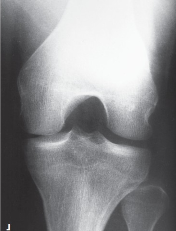
PA axial ICF Holmblad
Open intercondylar fossa
Apex of patella no superimposing the fossa

(more elongated)
PA axial ICF camp-coventry
Open intercondylar fossa
Apex of patella not superimposing the fossa

AP Axial ICF Beclere
Open intercondylar fossa
Apex of patella not superimposing the fossa

What position is this?
Tangential Patella Settegast

What position is this?
Tangential Patella Hughston

What position is this?
AP Axial ICF Beclere

What position is this?
PA Axial ICF camp-coventry
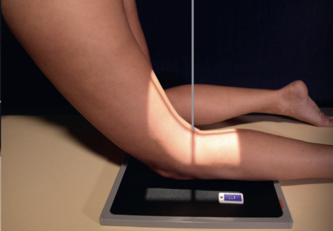
What position is this?
PA Axial ICF Holmblad
What does overrotation on a lateral knee do?
Causes less superimposition
What underrotation on a lateral knee do?
Causes more superimposition

LEWIS or HOLLY?
Lewis

LEWiS OR HOLLY
Holly