Ch. 3 Biologically Important Molecules (done)
 Call Kai
 Learn
 Practice Test
 Spaced Repetition
 Match
 Flashcards
 Knowt Play
1/17

There's no tags or description
Looks like no tags are added yet.
18 Terms
condensation reaction and dehydration reaction
A condensation reaction is a chemical process where two molecules combine to form a single, larger molecule, releasing a small by-product like water, ammonia, or methanol.
dehydration synthesis: the reaction is called a dehydration synthesis if the expelled molecule is water.
Proteins
Proteins are composed of 20 amino acids linked together in polymers
-the composition and sequence of amino acids is what makes each protein unique and fulfill its special role in the cell.
general formula for an amino acid
a-carboxyl group
tetrahedral a-carbon
variable R group
a amino group
the side chain (variable R group) give it the physical and chemical properties that distinguish it from the other amino acids.

Protein structure
There are two common types of covalent bonds between amino acids in proteins:
peptide bonds: covalent bonds that link amino acids together into poly”peptide” chains.
Disulfide bridges: bridges between the cystine thiol R groups.
The peptide bond
peptide bonds: covalent bonds that link amino acids together into poly”peptide” chains.
-peptide bonds are formed between the the carboxyl group of one amino acid and the a-amino group of another amino acid with the loss of water (dehydration synthesis).
The picture on this flashcard shows the formation of a di-peptide.
Dipeptide: two “di” amino acids.
peptide bond formation steps
Nucleophilic amino group (the nitrogen has two electrons) attack electrophilic carbon (the oxygen’s electronegativity makes the carbon electrophilic)
Water leaves “dehydration synthesis” (OH from aa1 leaves and H from NH2 on aa2 leaves)
O=N-H has formed
In a polypeptide chain, the backbone of the polypeptide is the N-C-C-N-C-C pattern.
residue: an individual amino acid that is part of a polypeptide chain
The amino terminus is the first end made during polypeptide synthesis, and the carboxy terminus is the last. Hence, by convention, the amino-terminal residue is always written first.
Question: in the oligopeptide Phe-Glu-Gly-Ser-Ala, which residue has a free alpha-amino group, and which residue has a free alpha carboxyl group?
Answer: the residue that has a free alpha-amino group is Phe, and the residue that has a free carboxyl group is Ala.

Proteolysis or Proteolytic cleavage
Proteolysis: hydrolysis “lysis” of a protein “proteo”.
The protease performs proteolysis on the protein by cleaving the peptide bonds.
Many enzymes only cleave the peptide bond next to specific amino acid
example: Trypsin is a protease that cleaves the carboxyl side of residues arginine and lysine.
Question: Based on the above, if the following peptide is cleaved by Trypsin, what amino acid will be on the new N-terminus and how many fragments will result? Ala-Gly-Glu-Lys-Phe-Phe-Lys?
Answer:
Based on the image, Trypsin cleaves between Lys (Arg) and Gly.
Trypsin will cleave the carboxyl side (last end) of the Lys residue, with Phe on the N-terminus of the new Phe-Phe-Lys fragment. There will be two fragments after trypsin cleavage: Ala-Gly-Glu-Lys and Phe-Phe-Lys


The Disulfide Bond
-the purpose of a disulfide bond is to stablize tertiary protein structure
-the thiols (SH) in the cysteine react to form the disulfide bond
-cysteine becomes cystine (no e in the middle).
Protein structure in three dimensions
-each protein folds into a unique three-dimensional structure that is required for that protein to function properly.
-there are four levels of protein folding that contribute to the final 3d structure.
-denatured proteins are non-functional, improperly folded proteins that still have the peptide bonds.
-proteins are denatured by urea (disrupts H-bonding interactions), extremes of pH, extremes of temperature, and by changes in salt concentration (tonicity).
Primary Structure of a protein: the amino acid sequence
-the simplest level of protein structure is the order of amino acids bonded to each other in the polypeptide chain.
-the linear ordering of amino acid residues is known as primary structure
-Primary structure is the same as sequence.
-the bond which determines primary structure of protein is the peptide bond, simply because this is the bond that links one amino acid to the next in a polypeptide.
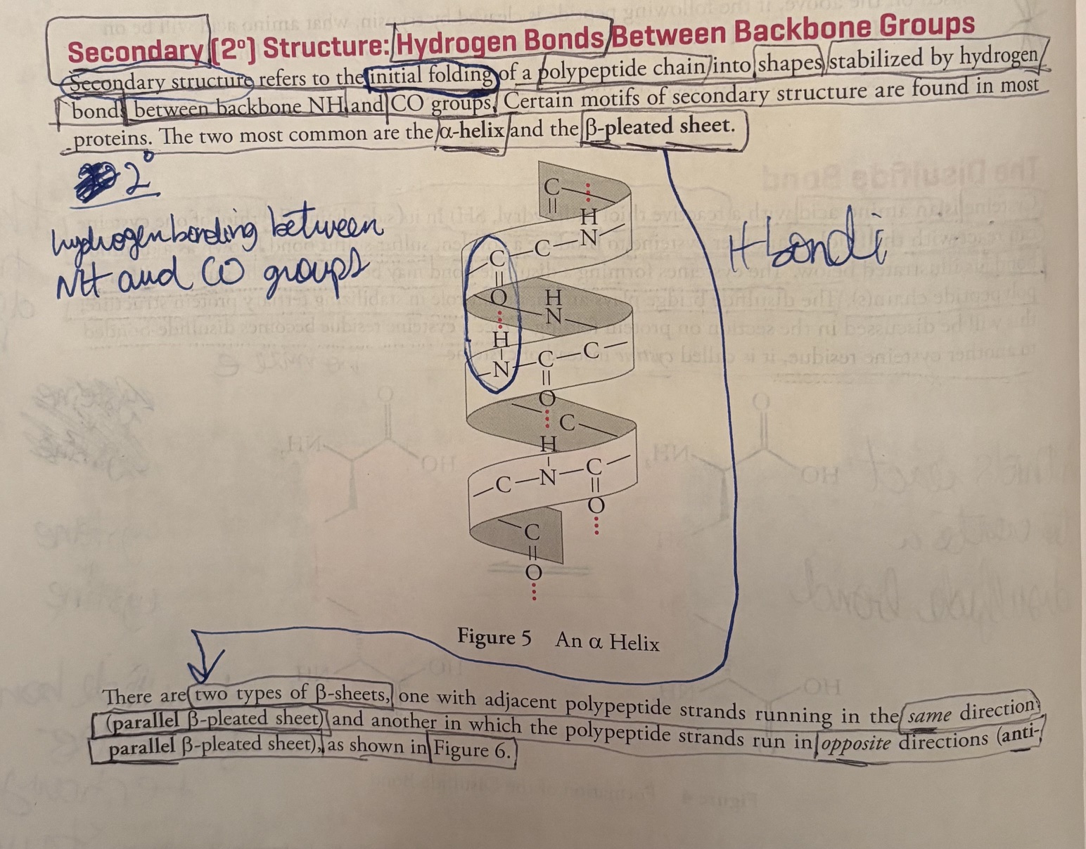
Secondary structure of a protein: hydrogen bonds between backbone groups
-Secondary structure refers to the initial folding of a polypeptide chain into shapes stabilized by hydrogen bonds, between backbone NH and CO groups.
-The two most common secondary structures are the alpha-helix and b-pleated sheet.
-There are two types of B-sheets, one with adjacent polypeptide strands running in the
same direction (parallel B-pleated)
opposite direction (anti-parallel B-pleated), as shown in Figure 6.

opposite direction (anti-parallel B-pleated), as shown in Figure 6.
(this is Figure 6).
anti-parallel: meaning the N-terminus of one strand sits next to the C-terminus of the other.
anti: the N-terminus of one strand sits next to the C-terminus of the other
parallel: the strands and terminus run parallel to each other.
Question: if a single polypeptide folds once and forms a B-pleated sheet with itself, would this be a parallel or antiparallel B-pleated sheet?
It would be an antiparallel beta-pleated sheet. When a single polypeptide chain folds back on itself in a hairpin turn, the two adjacent segments run in opposite directions—meaning the N-terminus of one strand sits next to the C-terminus of the other.

Tertiary (3o) Protein Structure: Hydrophobic and Hydrophilic Interactions
-the level after secondary structure is tertiary structure
-Tertiary structure concerns interactions between amino acid residues located more distantly from each other in the polypeptide chain.
-The folding of secondary structures such as a-helices into higher order tertiary structures is driven by interactions of R groups with each other and the solvent (water).
-Hydrophobic R groups tend to fold into the interior of the protein away from the solvent, hydrophilic R-groups tend to be exposed to water on the surface of the protein.
-Under the right conditions, the forces driving the hydrophobic avoidance of water and hydrogen bonding will fold a polypeptide spontaneously, into the correct conformation, the lowest energy conformation.
Quaternary (4o) Structure: Various Bonds Between Separate Chains
The highest level of protein structure, quaternary structure, describes interactions between polypeptide subunits.
A subunit is a single polypeptide chain that is part of a large complex containing many subunits (a multisubunit complex).
-The arrangement of subunits in a multisubunit complex is what is meant by quaternary structure.
-the interactions between subunits is essential for protein function, as in the cooperative binding of oxygen by each of the four subunits of hemoglobin.
Forces stabilizing quaternary structure
-the forces stabilizing quaternary structure are generally the same as those involved in tertiary structure, non-covalent interactions, van der Waals forces, hydrogen bonds, disulfide bonds, and electrostatic interactions.
-it’s key to understand that the peptide bond is not involved in quaternary structure because the peptide bond defines the primary structure (1o).
Question: what is the difference between a disulfide bridge involved in quaternary structure and one involved in tertiary structure?
Answer: the disulflide bridge involved in quaternary structure are not linked by peptide bonds (because quaternary structure does not involve peptide bonds because peptide bonds define primary structure). Tertiary structure disulfides form between residues in the same polypeptide.
Carbohydrates
-carbohydrates are broken down into CO2 in a process called oxidation.
-oxidation: the process of breaking carbohydrates down into CO2.
-because oxidation releases large amounts of energy, carbohydrates generally serve as the principle energy source for cellular metabolism.
Carbohydrates: Monossacharides and Dissacharides
-Monossacharides: “single sweet unit”, also known as a simple sugar.
-Monossacharides have the general formula: CnH2nOn
-two monossacharides come together form a dissacharide, a few form an oligossacharide, and many form a polypeptide.
-Glycosidic linkage: the bond between two sugar molecules (“glyco”:sugar). Glycosidic linkages are covalent bonds, formed in a dehydration reaction that requires enzyme catalysis.
Some metabotically important Monossacharides
-Fructose
-Glucose
-Ribose
Dissacharides and the alpha or beta-glycosidic bond (Figure 9)
-Sucrose (glucose + fructose)
Sucrose (table sugar) is held together by an α-1,β-2 glycosidic bond
-Lactose (galactose + glucose)
Lactose (milk sugar) is a disaccharide comprised of one galactose molecule and one glucose molecule. These monosaccharides are covalently linked by a β-1,4-glycosidic bond. 4
-Some common dissacharides you might see on the MCAT are sucrose (Glc-a-1,2-Fru), lactose (Gal-B-1,4-Glc), maltose (Glc-a-1,4-Glc) and cellobiose (Glc-B-1,4-Glc).

-Some common dissacharides you might see on the MCAT are sucrose (Glc-a-1,2-Fru), lactose (Gal-B-1,4-Glc), maltose (Glc-a-1,4-Glc) and cellobiose (Glc-B-1,4-Glc).
-Some common dissacharides you might see on the MCAT are sucrose (Glc-a-1,2-Fru), lactose (Gal-B-1,4-Glc), maltose (Glc-a-1,4-Glc) and cellobiose (Glc-B-1,4-Glc).
-Some common dissacharides you might see on the MCAT are sucrose (Glc-a-1,2-Fru), lactose (Gal-B-1,4-Glc), maltose (Glc-a-1,4-Glc) and cellobiose (Glc-B-1,4-Glc).

Polymers made from dissacharides form important biological macromolecules. Glycogen serves as an energy storage carbohydrate in animals and is composed of thousands of glucose units. Starch is the same as glycogen (except that the branches are a little different), and serves the same purpose in plants.
Starch (Amylopectin): Contains α(1→4) linear chains with α(1→6) branch links appearing every 24–30 subunits. Starch also contains amylose, which is entirely unbranched.
Glycogen: Contains the same α(1→4) and α(1→6) bonds, but the α(1→6) branch links happen much more often (every 8–12 units)
Cellulose is a polymer of cellulobiose; but note that cellobiose does not exist freely in nature. It exists only in its polymerized, cellulose form. The glycosidic bonds allow the polymer to assume a long, straight and fibrous shape. Wood and cotton are made of cellulose.
look at the figure.
Lipids
Lipids are oily or fatty substances that play three physiological roles: summarized here and discussed below:
In adipose cells, triglycerides (fats) store energy
In cellular membranes, phosholipids constitute a barrier between intracellular and extracellular environments
Cholesterol is a special lipid that serves as the building block for the hydrophobic steroid.
The cardinal characteristic of the lipid is its hydrophobicity. Hydrophobic means “water-fearing”. It is important to understand the significance of this. Since water is very polar, polar substances dissolve well in water; these are known as “water-loving”, or hydrophilic substances. Carbon-carbon bonds and carbon-hydrogen bonds are nonpolar. Therefore, substances, that contain only carbon and hydrogen will not dissolve well in water. Some examples of this: Table sugar dissolves well in water, but cooking oil floats in a layer above water or forms many tiny oil droplets when mixed with water, but cooking oil floats in a layer exposed to water because they are made of glucose polymerized into cellulose; a nylon jacket does not become wet because it is composed of atoms covalently bound together in a nonpolar fashion. A synonym for hydrophobic is lipophilic (which means “lipid-loving”); a synonym for hydrophilic is lipophilic. We return to these concepts below.
Fatty Acid Structure
Fatty acids are composed of long, unsubstituted alkanes that end in a carboxylic acid. The chain is typically 14 to 18 carbons long, and because fatty acids are synthesized two carbons at a time from acetate, only even-numbered fatty acids are made in human cells. A fatty acid with no carbon-carbon double bonds is saturated with hydrogen because every carbon atom in the chain is covalently bound to the maximum number of hydrogens. Unsaturated fatty acids have one or more double bonds in the tail. These double bonds are almost always (Z) or (or cis).
Why are fatty acid double bonds in cis? In a cis configuration, the hydrogen atoms attached to the double-bonded carbons are on the same side, which forces a rigid bend or "kink" into the hydrocarbon chain.
• Preventing tight packing: Straight chains (like saturated fats or trans fats) can stack tightly together through close dispersion forces. The cis kink prevents fatty acid tails from packing closely.
• Maintaining cell membrane fluidity: Because cis fatty acids cannot pack tightly, they lower the melting point and keep fats liquid at room temperature. In cell membranes, this fluidity is essential for proper cellular function, protein movement, and flexibility across varying temperatures
If fatty acids are mixed into water, how are they likely to associate with each other? The long hydrophobic chains will interact with each other to minimize contact with water, exposing the charged carboxyl group to the aqueous environment.
Figure 12 illustrates how free fatty acids interact with aqueous solution; forming a micelle. The force that drives the tails into the center of the micelle is called the hydrophobic interaction.

Triacylglycerols (TG)
The storage form of the fatty acid is fat. The technical name for fat is triacylglycerol or triglyceride (shown below). The triglyceride is composed of three fatty acids esterified to a glycerol molecule.
Glycerol is a three-carbon triol with the formula HOCH2-CHOH-CH2OH. As you can see, it has three hydroxyl groups that can be esterified to fatty acids. It is necessary to store fatty acids in the relatively inert form of fat because free fatty acids are reactive chemicals.
put figure 13 here.

Lipases hydrolyze fats. Triacylglycerols are stored in fat cells as an energy source. Fats are more efficient energy storage molecules than carbohydrates for two reasons: 1. packing and 2. energy content
Packing: Their hydrophobicity allows fats to pack together much more closely than carbohydrates. Carbohydrates carry a great amount of water-of-solvation (water molecules hydrogen bonded to their hydroxyl groups). Fats pack tighter and store energy more efficiently than carbohydrates because they are hydrophobic and exclude water.
Energy content: fat molecules store much more energy than carbohydrates because fats are more reduced than carbohydrates. C-H bonds in fats store more energy than C-O bonds in carbohydrates. Regardless of what you dissolve it in, fat has more energy carbon-for-carbon than a carbohydrate.
Introduction to lipid bilayer membranes
Membrane lipids are phospholipids derived from diacylglycerol phosphate or DG-P.
Put figure 14 here.

We saw above how fatty acids spontaneously form micelles. Phospholipids also minimize their interactions with water by forming an orderly structure- in this case, a lipid bilayer (below).
Hydrophobic interactions drive the formation of the bilayer,
once formed, it is established by van der waals forces between the tails.
put figure 15 here

Would a saturated or unsaturated fatty acid residue have more van der waals interactions with neighboring alkyl chains in a bilayer membrane? Saturated. (because unsaturated pack less densely, so saturated pack more closely and have more van der waals interactions)
A more precise way to give the answer to the question above is to say that double bonds (unsaturation) in phospholipid fatty acids tend to increase membrane fluidity. Unsaturation prevents the membrane from solidifying by disrupting the orderly packing of the hydrophobic lipid tails. The right amount of fluidity is essential for function. Decreasing the length of fatty acid tails also increases fluidity. At low temperatures, it increases fluidity in the same way as kinks in fatty acid tails; therefore, it is known as membrane antifreeze. At high temperatures, however, cholesterol attenuates (reduces) membrane fluidity. Don’t ponder this paradox too long; remember that cholesterol keeps fluidity at optimum level. Remember, the structural determinants of membrane fluidity are:
degree of unsaturation
tail length
amount of cholesterol
Terpenes
A terpene is a member of a broad class of compounds built from isoprene units (C5H8) with a general formula (C5H8)n
isoprene units (C5H8) are used to build terpenes (C5H8)n
put figure 16 here.

Terpenes may be linear or cyclic, and are classified by the number of isoprene units they contain. For example, monoterpnes consist of two isoprene units, sesquiterpenes consist of three, and diterpenes contain four.
Put Figure 17 here.

Squalene is a triterpene (made of six isoprene units) and a pariticularly important compound, as it is biosynthetically utilized in the manufacture of steroids. Squalene is also a component of earwax
Put Figure 18 here

Whereas a terpene is formally a simply hydrocarbon, there are number of natural and synthetically derived species that are built from an isoprene skeleton and functionalized with other elements (O,N,S,etc.). These functionalized-terpenes are known as terpenoids. Vitamin A (C20H30O) is an example of a terpenoid.
Put figure19 here.

Steroids
Steroids are included here because of their hydrophobicity (making them similar to fats). Their structure is otherwise unique. All steroids have the basic tetracylic ring system (see below), based on the structure of cholesterol.
As discussed eariler, the steroid cholesterol is an important component of the lipid bilayer. It is both 1. obtained from the diet and 2. synthesized in the the liver. Cholesterol is carried in the blood packaged with fats and proteins into lipoproteins. One type of lipoprotein has been implicated as the cause of atherosclerotic vascular disease, which refers to the build-up of cholesterol “plaques” on the inside of blood vessels.
Put figure 20 here
Steroid hormones are made from cholesterol. Two examples are 1. testosterone (an androgen or male sex hormone) and 2. estradiol (an estrogen or female sex hormone).

Phosphorus-containing compounds
Phosphoricc acid is an inorganic acid (it does not contain carbon) with the potential to donate three protons. The pKas for the three acid dissociation equilibria are 2.1, 7.2, and 12.4. Therefore, at physiological pH, phosphoric acid is significantly dissociated, existing largely in anionic form.
Put figure 21 here
Phosphate is also known as orthophosphate. Two orthophoshpate bound together via an anhydride linkage form pyrophosphate. The P-O-P bond in the pyrophosphate is an example of a high-energy phosphate bond. This name is dervied from the fat that hydrolysis store so much energy:
when phosphates are linked together, their negative charges repel
ortophosphates has more reasonance forms and thus a lower free energy than linked phosphates
orthophosphates has a more favorable interaction with the biological solvent (water) than linked phosphate.
The details are not crucial. What is essential is that you fix theimage in your mind of linked phosphates acting like compressed springs, just waiting to lfy open and provide enrgy for an enzyme to catalyze a reaction.

put the picture of figure 22 here.
anhydride linkage (normal)

phosphate anhydride linkage (from the book)

pyrophosphate
Nucleotides
Nucleotides are the building blocks of nucleic acids (RNA and DNA). Each nucleotide contains a ribose (or deoxyribose) sugar group; a purine or pyramidine base joined to a carbon number one of the ribose ring; and one, two or three phosphate units joined to carbon five of the ribose ring. The nucleotide adenosine triphosphate (ATP) plays a central role in cellular metabolism in addition to being an RNA precursor. Significantly more information about the structure of the nucleic acids can be found in MCAT Biochemistry Review. More information on the function of the nucleic acids will be provided in Chapter 4 of this book.
ATP is the universal short-term energy storage molecule. Energy extracted from the oxidation of foodstuffs is immediately stored in the phosphoanhydride bonds of ATP. This energy will later be used to power cellular processes; it may also be used to synthesize glucose or fats, which are longer-term energy storage molecules. This applies to all living organisms, from bacteria to humans. Even some viruses carry ATP with them outside the host cell, though viruses cannot make their own ATP.
put a picture of figure 23 here.
