Biology 162 Practical 2 Flashcards
 Call Kai
 Learn
 Practice Test
 Spaced Repetition
 Match
 Flashcards
 Knowt Play
1/104

There's no tags or description
Looks like no tags are added yet.
105 Terms
skeletal muscle:
makes movement, breathing, speech, and facial expression possible. Almost compleetlu under voluntary control.
Excitability:
Muscle cells produce action potentials
Contractility
Muscle cells can shorten
Extensibility
muscle cells can be stretched or lengthened
muscle fibers
another name for muscle cells
fascicles
bundles of muscle fibers
fascia
an outer membrane that many fascicles wrap around; also holds the fascicles as well as blood and nerves together in one large unit
tendons
anchor muscle tissue to the bones they work to move during contraction
sarcolemma
type of cell membrane that a muscle fiber is surrounded by
sarcoplasm
what the sarcolemma is filled with; type of cytoplasm
myofibrils
are within the sarcoplasm and are bundles of protein
sarcoplasmic reticulum
within the sarcoplasm; a complex network of membrane sacs and tubes
terminal cisternae
the ends of the sarcoplasmic reticulum form flat sacs called:
Cervical Curvature (curve)
curve formed by the C1 through C7 vertebrae
Lumbar Curvature (curve)
curve formed by the T1 through T12 vertebrae
Thoracic Curvature (curve)
curve made by the L1 through the L5 vertebrae
Pelvic Curvature (curve)
curve made up of the sacrum and the coccyx
Atlas
The first of the cervical vertebrae. It articulates the skull superiorly and the axis inferiorly. It also supports your skull.

Axis
The second cervical vertebra. The axis allows you to shake your head back and forth (allows head to move on its axis)

Body
Found on all vertebra except the atlas. The body transits weight down the bodies of the vertebrae.

Superior articular process:
These are the bony projections that point superiorly to articulate with the vertebra above.

Facets
the surfaces where the two bones physically articulate
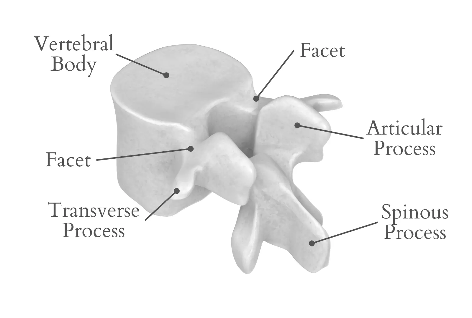
Inferior Articular Process
These are the bony projections that point inferiorly to articulate with the vertebra below.
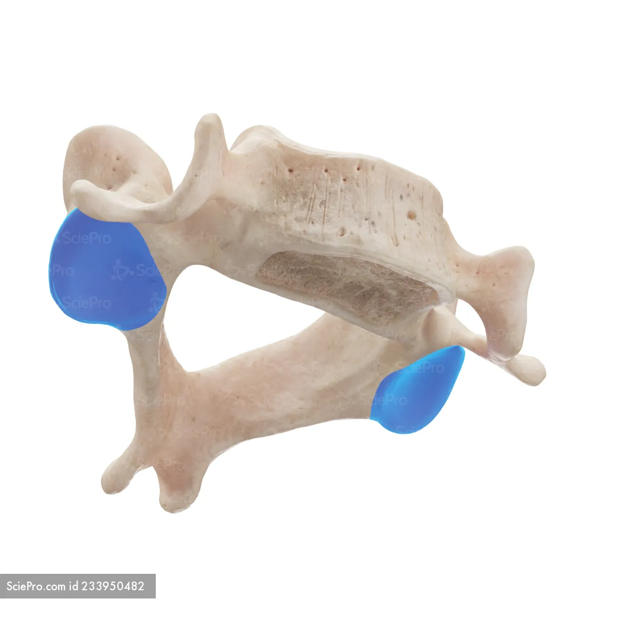
Transverse Process
This is the bony region that extends laterally from the body.

Spinous Process
The somewhat sharp projection that extends posteriorly, opposite from the body of the vertebra.

vertebral arch
the transverse and spinous process arise from this bony arch.

Vertebral Foramen
The opening formed by the vertebral body and arch

Vertebral Canal (spinal canal)
All of the vertebral foramina will line up together and form this long tube. The nerves of the spinal cord will run through here.
Sacrum
Articulates with the fifth lumbar vertebra and wings of the hip bones, or the right and left ilium. The sacrum is actually formed by five vertebrae that have become fused together.

Body (sacrum)
The body of the sacrum is the main thick medial region of the sacrum.
Median Sacral Crest
The median sacral crest is formed from the spinous processes of the fused vertebrae

Sacral Canal
The passageway which corresponds with the vertebral canal

Auricular Surface
On the lateral faces of the sacrum, this forms the joint between the sacrum and ilium.
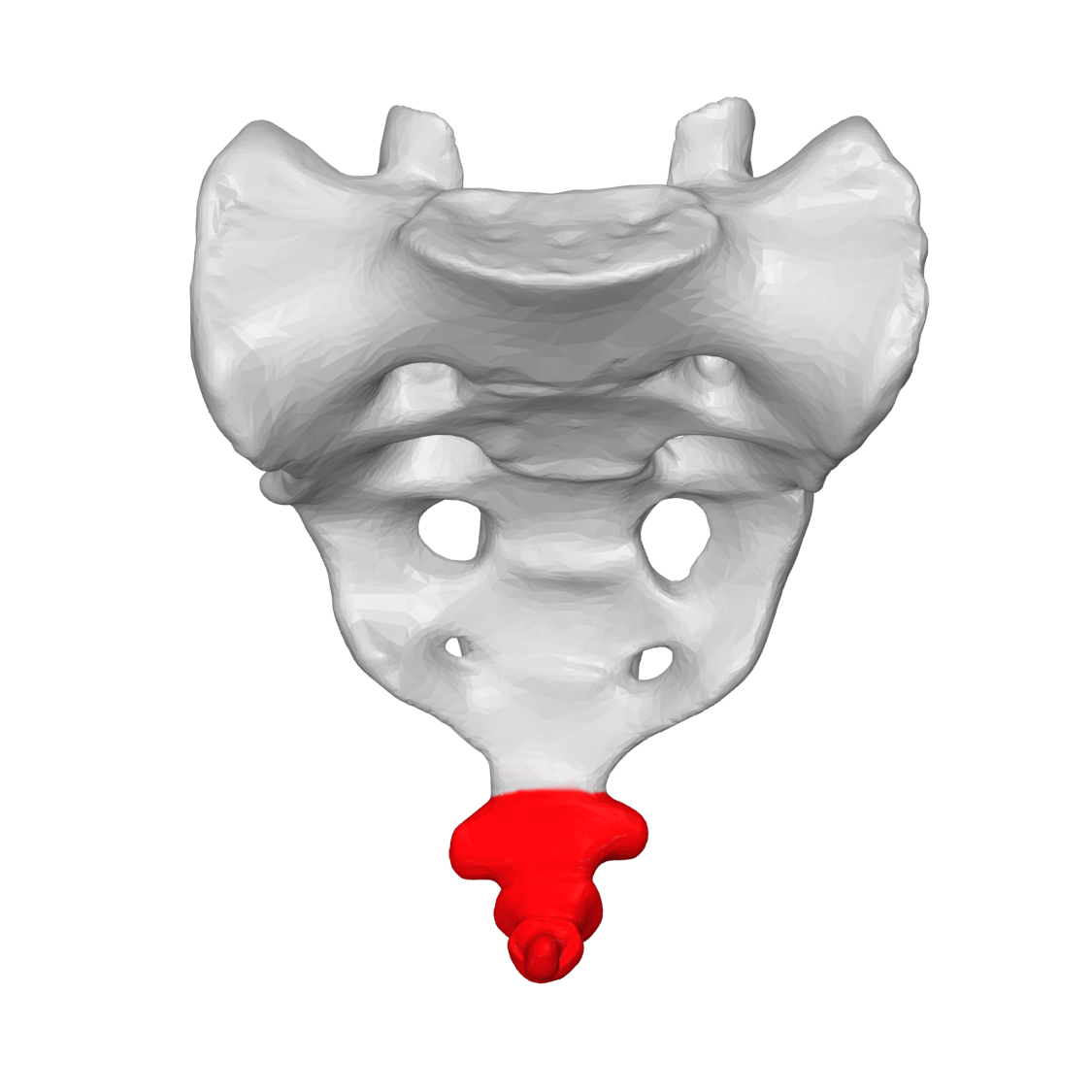
Coccyx
the small, triangular bone at the very bottom of the vertebral column
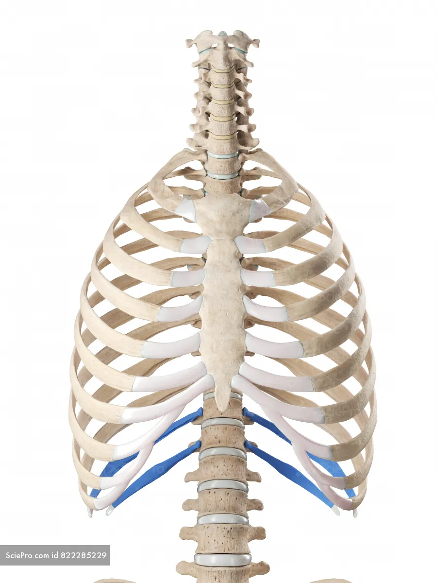
True Ribs
Ribs 1 through 7; articulate directly with the sternum

False Ribs
Ribs 8-12; they either articulate with the sternum indirectly (8-10) or not at all (11-12)
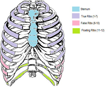
Floating Ribs
another name for ribs 11 and 12 because they do not articulate with the sternum at all.
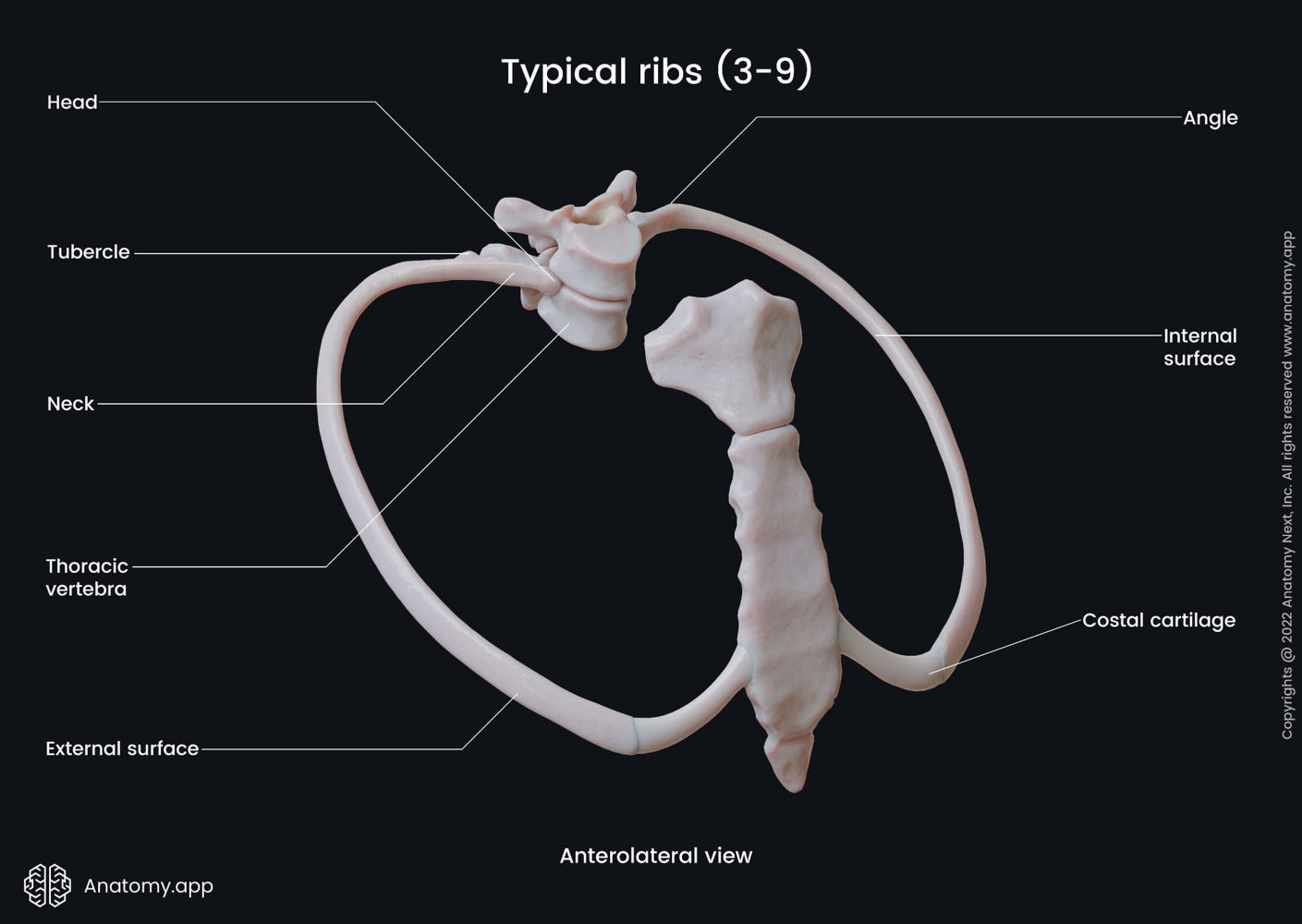
Head (ribs)
The articualr facet on the headof each ribs
Transverse tubules (T-Tubules)
invaginations of the cell membrane and act to connect the terminal cisternae to the cell surface.
Actin
thin filament, is a long strand of globular proteins and looks like a strand of beads.
Myosin
the proteins that make up thick filaments
tropomyosin
a long chain of regulatory proteins that cover the active sites on actin
troponin
a smaller regulator protein that is attached along the lengths of the tropomyosin chains
motor unit
consists of one motor neuron and all of the individual muscle cells it controls
Recruitment
is the activation of additonal motor motor units to increase muscle strength and force of conctraction
Central Fatigue
is cause dby the central nervous system and physiological mechanisms. Manifests as feeling “tired”
Peripheral Fatigue
as problems with the neuromuscular junction between the motor nerve and muscle or problems within the muscle cell at the level of the sarcomere
Isometric contraction
when a muscle may contract but not change length
Concentric Contraction
a muscle will shorten in length when contracting
Eccentric Contraction
a muscle may contract and actually become longer
Agonist muscle
aka the prime mover, and can be thought of as the muscle that is performing the desired action
Antagonist muscle
the musce that is capable of opposing the action
Flexion
describes a decrease in the angle between the body parts
Extension
increases the angle between adjacent body parts
Abduction
describes the movement of a body part away from the midline
Adduction
the movement of of a body part away from the midline of the body
Tubercle (ribs)
this small bumpy ridge is found just distal to the head. Notice: there is a smooth portion, another articular facet, which articulates with the transverse process of the thoracic vertebrae

Body (ribs)
this is the main part of the rib distal to the neck
Angle (ribs)
just distal to the tubercle, the rib makes a sharp bend. This bend is called the angle

Sternum
is sometimes called the “breast bone”. It articulates with the true ribs, and together, they protect the heart and lungs

Body (sternum)
the large middle portion of the sternum

Facets (sternum)
this is where the costal cartilage associated with the ribs articulate

Manubrim
this is the upper part of the sternum
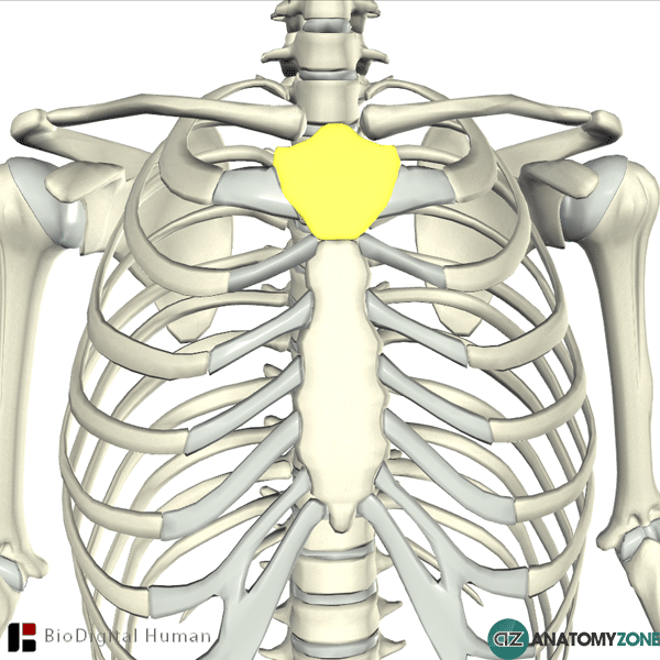
Xiphoid Process
this projection can be found inferior to the body of the sternum
Rectus Abdominis

External Olblique

Internal Oblique

Transverse Abdominis

Linea Alba
connects the xiphoid process to the pubic bone

External Intercostal Muscles
run on either side of your chest form superior-lateral to inferior-medial. This forms a V shape, mirroring the striations of the external oblique muscles in the abdomen. They may assist with inhalation by pulling the ribs upward.

Internal Intercostal Muscles
whose fibers run on either side of your chest like an A shape, from superior-medial to inferior lateral. They may assist with exhalation by pulling ribs downward

Innermost Intercostals Muscles
whose fibers line the inside of your chest and run superior-inferior. This muscle also may assist with exhalation by pulling the ribs downward.

Diaphram
the dome shaped muscle responsible for increasing the volume of your thoracic cavity to help you inhale. It forms the division between the thoracic cavity and the abdominal cavity below.

Sternocleidomastoid
This muscles fibers extend from the mastoid process (the bony lump behind your ears) to the medial clavicle (your collar bone) and manubrium of the sternum

Scapula
is sometimes called the shoulder blade
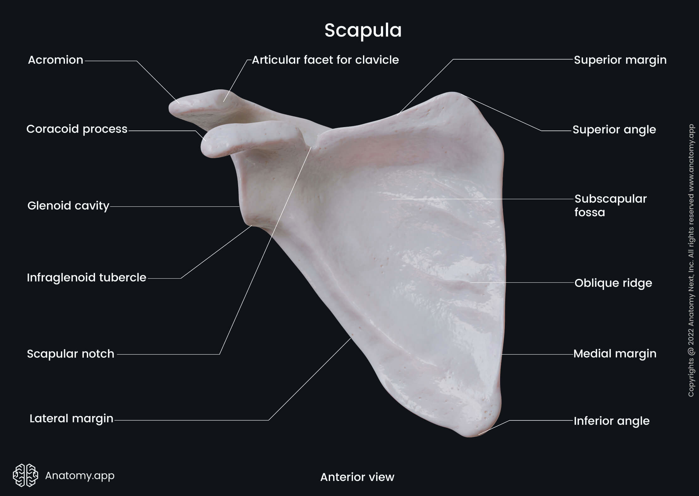
Glenoid Cavity (fossa)
This shallow fossa is where the head of the humerus articulates with the scapula

Coracoid Proces
the projection from the same region of bine supporting the glenoid cavity. This projection looks like a “crow’s beak”, and juts out anteriorly

Acronium Process
A projection of bone that juts anteriorly from the spine of the scapula

Spine of Scapula
The posterior surface of the scapula is divided by a thin “wall” of bone

Subscapular Fossa
The anterior surface of the scapula is a large smooth depression and is not interrupted by any process.
Supraspinous Fossa
Your index finger will fit nicely into the depression fromed by the superior surface of the spinous process and the posterior face of the scapula.

Infrasinous Fossa
The depression formed by the inferior surface of the spinous process and the posterior face of the scapula is the infra-spinous-fossa

Clavicle
aka collar bone

Humerus
bone found in the upper arm

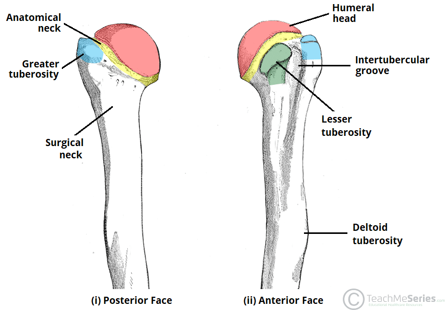
Head (proximal humerus)
This is the smooth round “cap” that articulates with the scapula
Nutrient Foramen
These small holes/tiny openings on the shaft that provide a passageway for blood vessels and small nerves
Greater Tubercle
The lateral and larger bump on the proximal head of the humerus

Lesser Tubercle
The smaller of the bumps mentioned above

Intertubercular Sulcus (bicipital groove)
The small groove between the tubercles allows for the passage of a tendon (from the biceps brachii muscle) and an artery

Deltoid Tuberosity
This is a rough region on the lateral part of the shaft that provides an attachment point for a muscle (the deltoid muscle)

Capitulum
This is the round smooth structure that is part of the distal head of the humerus. The capitulum articulates with the radius bone of the foramen. Some people remember its name because it looks like a little head that you could a '“cap” on.

Trochlea
This is the smooth, spool-shaped structure right next to the capitulum on the distal head of the humerus. The trochlea articulates with the ulna of the lower arm.

Coronoid fossa
The depression on the anterior humerus, just superior to the trochlea, that provides an empty area to accommodate the olecranon process of the ulna when the elbow is extended.

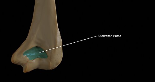
Olecranon Fossa
The depression on the posterior humerus, just superior to the trochlea, that provides an empty area to accommodate the olecranon process of the ulna when the elbow is extended
Medial Epicondyle
This smallish bump can be seen just medially to the trochlea. It provides an attachment point for muscles.

Lateral Epicondyle
This smallish bump can be seen just laterally to the capitulum. It also provides an attachment point for muscles.

Elevation (of the scapula)
when the scapula is raised, or moves vertically.
Depression (of the scapula)
when the scapula is lowered, or moves inferiorly.
Retraction
when the scapula is pulled toward the spine
Protraction
when the scapula is pulled away from the spine