Test 2 Anatomy
 Call Kai
 Learn
Learn Practice Test
 Spaced Repetition
 Match
Match Flashcards
 Knowt Play
1/300

There's no tags or description
Looks like no tags are added yet.
Name | Mastery | Learn | Test | Matching | Spaced | Call with Kai | Chat |
|---|
No analytics yet
Send a link to your students to track their progress
301 Terms

Os coxae
The pelvic girdle is formed by two
formed by fusion of ilium, ischium, the pubis, and the acetabular bone.
pelvic symphysis
cartilaginous joint which either completely or incompletely ossifies in the adult animal
Clinical note: The pelvis is often fractured in car accidents.
Since the pelvis is a box shape, there are usually two fractures. If one site is fixed, the other will usually return to approximate position.

Ilium
most cranial bone of pelvis
coxofemoral or hip joint
acetabulum articulates with femoral head
sacroiliac joint
ilium articulates cranially with sacrum
wing of ilium
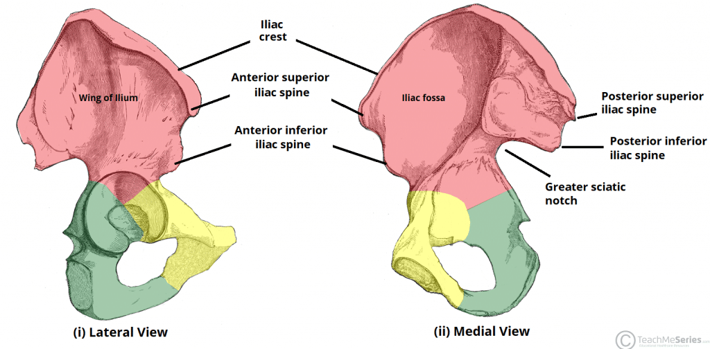
body of ilium

iliac crest

greater ischiatic notch

tuber sacrale

tuber coxae

tuberosity of rectus femoris
??
What bone can be used to obtain a bone marrow sample
iliac crest/dorsal iliac spine
Body of ischium
??
forms lesser ishciatic notch
ischiatic tuberosity

Clinical note: What can be easily palpated in dogs and is a common site for pressure sores
ischiatic tuberosity & tuber coxae
Obturator foramen
occupied by internal and external obturator mm.

ischiatic arch

body of pubis
??
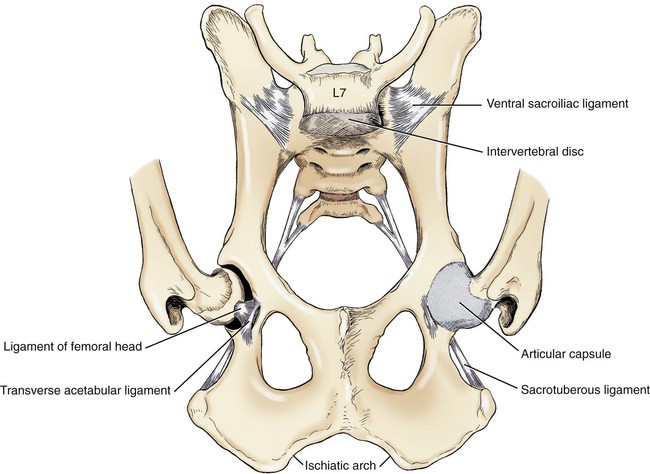
Pubic tubercle
formed by prominences on the cranial border of the right and left bodies on the midline form the pubic tubercle
Pecten
the cranial border of the body lateral to the pubic tubercle is the pecten, which abdominal muscles attach by prepubic tendon.
Iliopubic eminence
pectinius m, attaches here
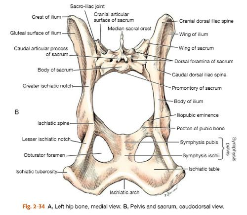
Pelvic symphysis
Clinical note: the pelvic symphysis can often be separated during trauma and is usually accompanied by sacroiliac luxation.
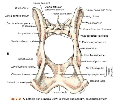
Acetebulum

acetabular fossa
ligament of the head of the femur attaches here

The transverse acetebular ligament join what
two sides of the acetabular notch
What is hip dysplasia
abnormal shallowness of the acetabulum & causes coxofemoral joint to become unstable and arthritic
Cat pelvis’ are what shaped compared to the dog’s being flared
box shaped
femur
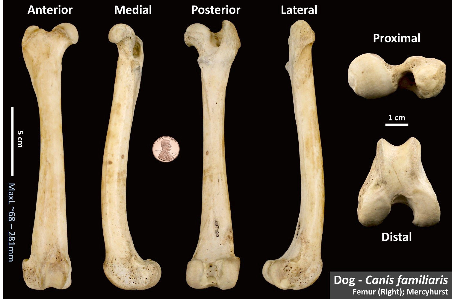
femoral head

fovea of the femoral head

ligament of the femoral head
??
neck of femur
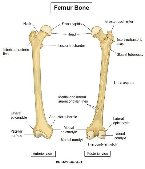
greater trochanter of femur

trochanteric fossa
ridge of bone that forms caudolateral border of the fossa is the intertrochanteric crest
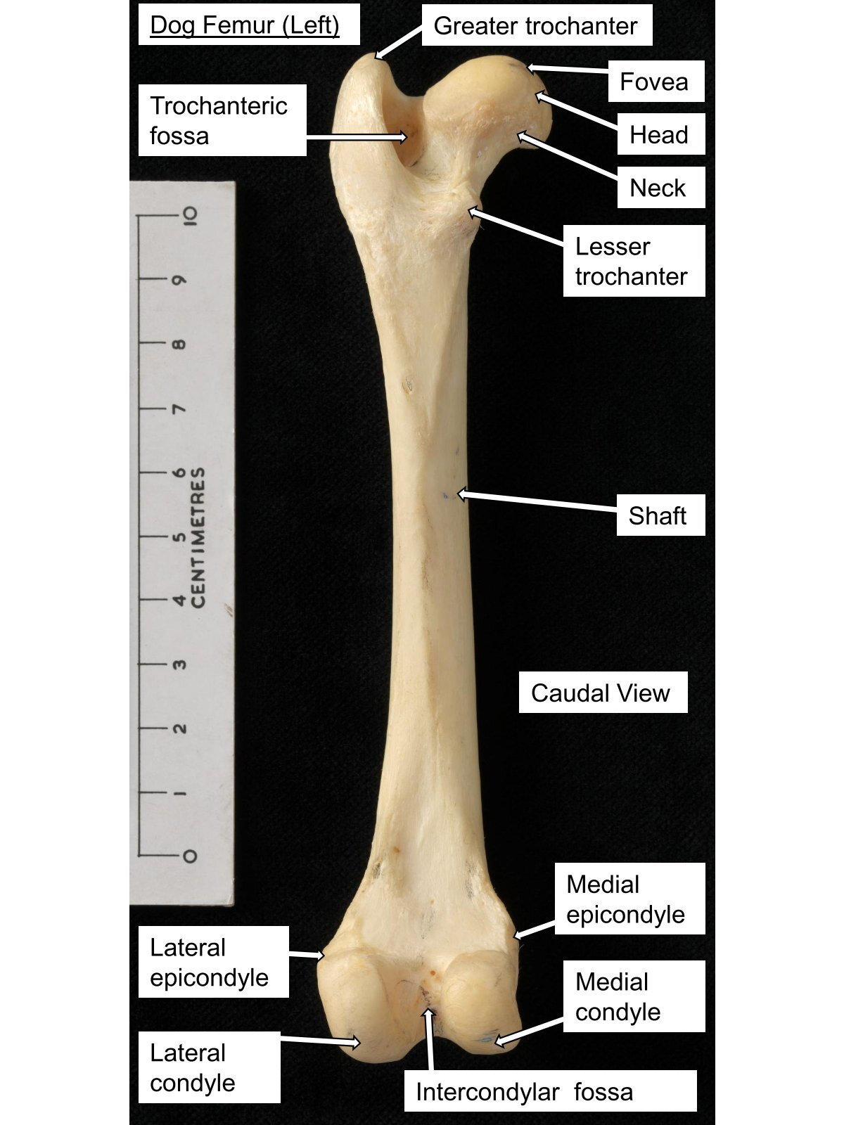
lesser trochanter

third trochanter
attachment site for superficial gluteal m
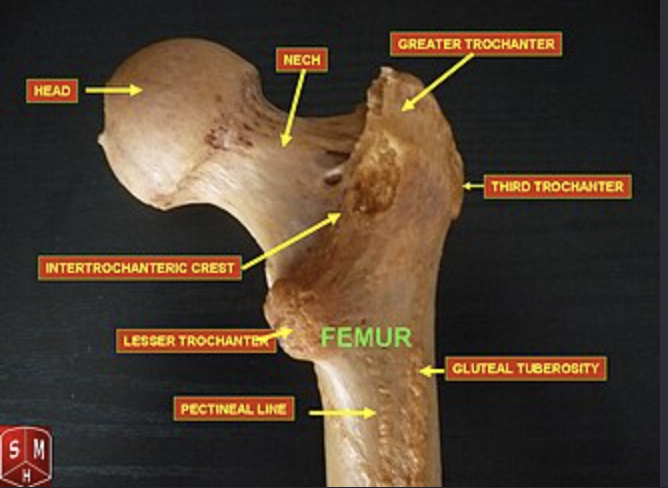
Clinical note: What part of the femur can be used for a bone marrow sample
proximal end of femur or trochanteric fossa
What part of the femur is used as a landmark for a surgical approach to the hip, detecting craniodorsal luxation of the hip and placement of a IM pin
greater trochanter of femur
body of the femur
??
medial & lateral condyles of femur
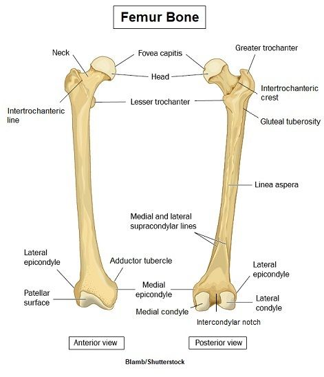
intercondylar fossa of femur
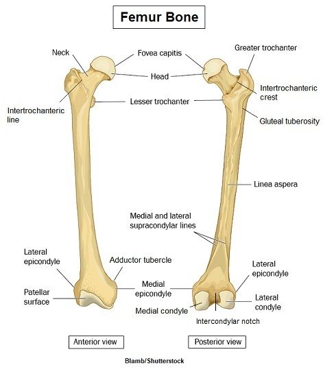
medial and lateral epicondyles of femur
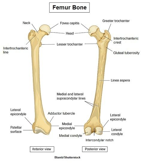
supracondylar tuberoisities of femur
??

medial and lateral trochlear ridges of femur
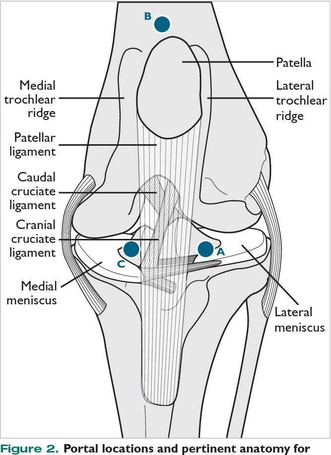
trochlea of femur
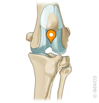
patella
large sesamoid bone in the tendon of the quadriceps femoris muscle

extensor fossa of femur
depression where lateral lip of trochlea and lateral condyle meet

OCD lesions may affect what
lateral condyle of femur
medial and lateral condyles of tibia

extensor groove of tibia

intercondylar eminence of tibia

caudal intercondylar area of tibia

cranial intercondylar area

cranial border of tibia
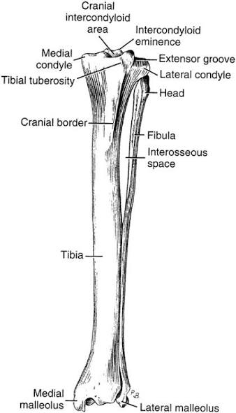
tibial tuberosity
??

patellar ligament
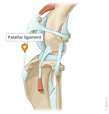
Clinical note: what is easily palpable and a good landmark for surgery
tibial tuberosity
medial malleolus
distal tibia
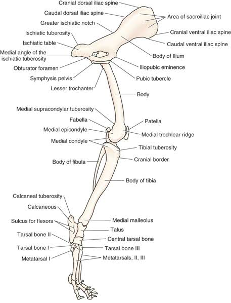
lateral malleolus of fibula

The pes includes
tarsal bones, metatarsal bones, phalanges
talus, calcaneus, central tarsal bone, tarsal bones I-IV

trochlea of talus

tarsocrural joint

tuber calcanei

sustentaculum tali

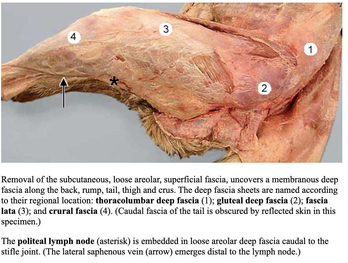
fascia lata
3
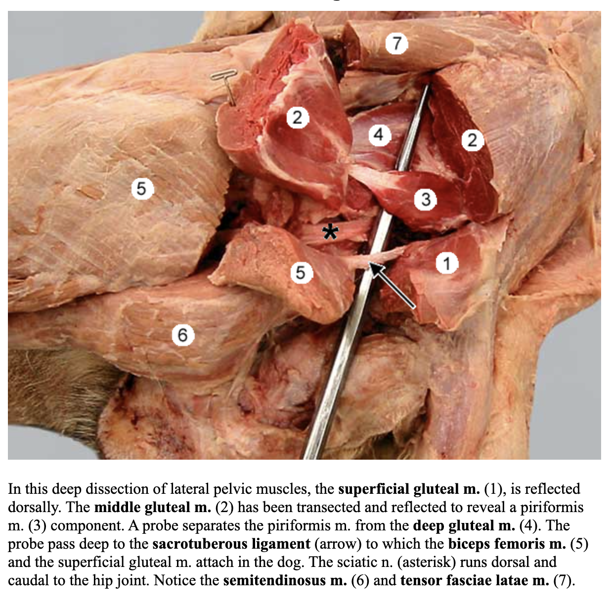
superficial gluteal m.
8
attachments: sacrum, sacrotuberous ligament and third trochanter of the femur
action: extend the hip; minor abduct the limb
blood: Cd. gluteal a.
nerve: Cd. gluteal n.

sacrotuberous ligament

middle gluteal m.
7
attachments: crest, gluteal surface of the ilium and greater trochanter
action: extend the hip and abduction
blood: Cr. gluteal a.
nerve: Cr. gluteal n.

piriformis m.
?
deep gluteal m.
attachments: body of the ilium, ischiatic spine, and greater trochanter
action: extend and abduct the hip
blood: Cr. gluteal a.
nerve: Cr. gluteal n.

gluteofemoralis m. (CAT ONLY)
attachments: caudal vertebrae 2-4 and terminates in fascia lata
action: abduct the hip or move tail laterally
Cats __ have a sacrotuberous ligament
DO NOT
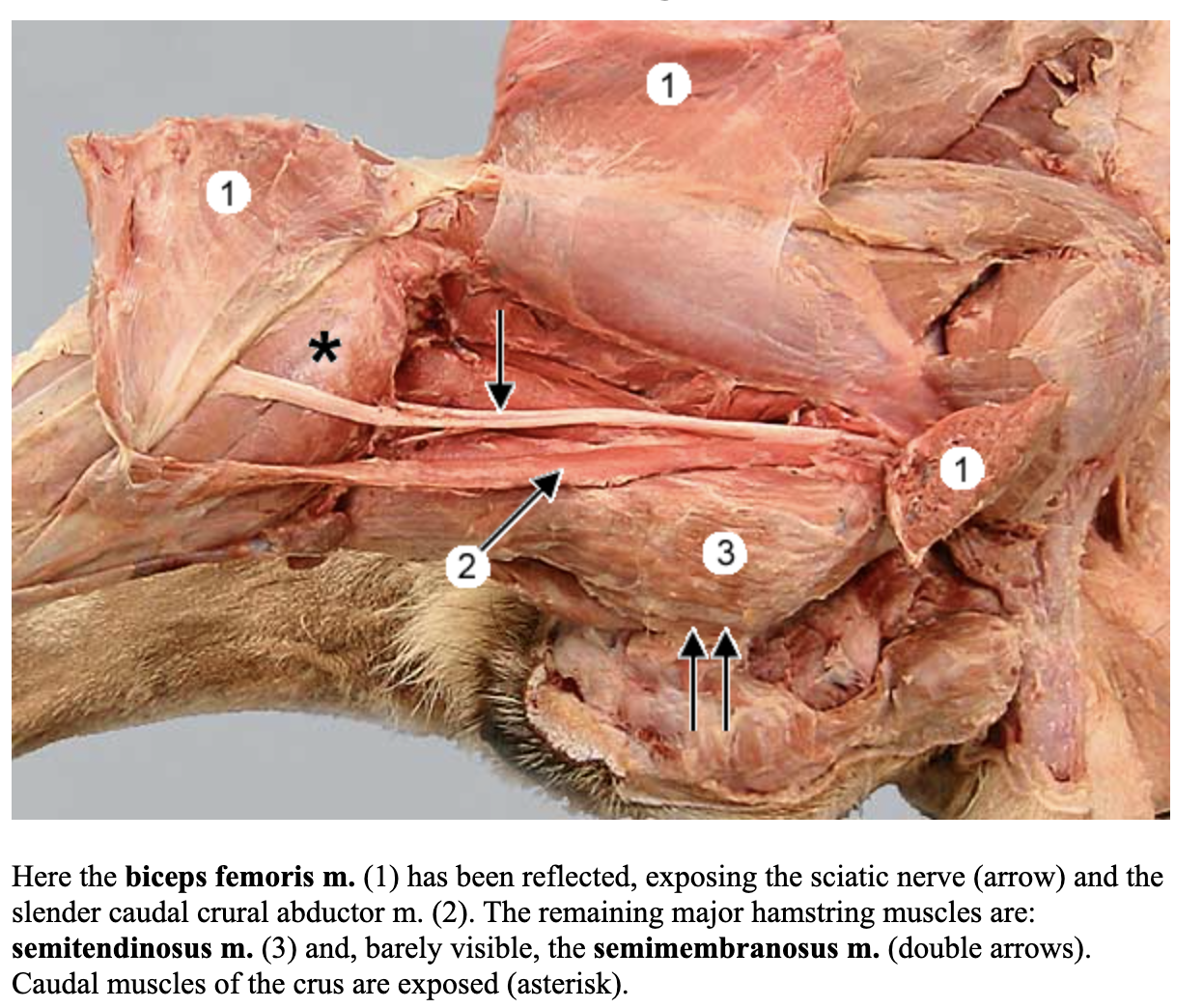
biceps femoris m.
5
attachments: sacrotuberous ligament and ischiatic tuberosity; patellar ligament and tibial tuberosity/body; tuber calcanei
action: extend the hip, stifle, and hock ; flex the stifle- caudal part
blood: deep femoral a.
nerve: sciatic n.
popliteal lymph node
?
sciatic nerve
?
Clinical Note: What is superficially located and should be easily palpated on physical exams
popliteal lymph node
semitendinosus m.
6
attachments: ischiatic tuberosity; medial tibia and tubercalcanei
action: extend the hip and tarsus; flex the stifle
blood: deep femoral a.
nerve: sciatic n.

semimembranosus m.
attachments: ischaitic tuberosity; medial condyle of femur and tibia
action: extend the hip & stifle (can also flex stifle)
blood: deep femoral a.
nerve: sciatic n.
Clinical note: What muscle is very commonly used to give IM injections
caudal muscles of the thigh
sartorius m.
attachments: iliac crest; cranial border of the tibia
actions: flex the hip and stifle; can also extend the stifle
blood: lateral circumflex femoral a.
nerve: femoral n.

pectineus m.
attachments: iliopubic eminence & caudomedial aspect of femur
actions: adduct the limb
blood: deep femoral a.
nerve: obturator n.

femoral triangle
bounded cranially by the sartorius m. and caudally by pectineus & adductor mm.

gracilis m.
1
attachments: pelvic symphysis; cranial tibia and tuber calcanei
action: adduct the limb, extend the hip and the tarsal joints; flex stifle
blood: deep femoral a.
nerve: obturator n.
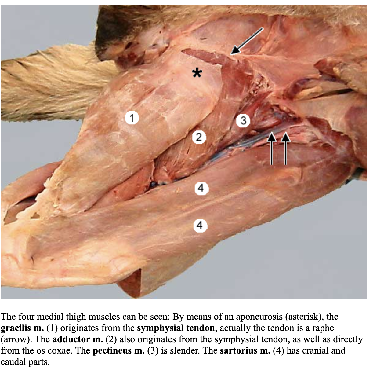
adductor m.
2
attachments: pelvic symphysis & ventral pubis; caudal (medial) aspect of femur
actions: adduct the limb & extend the hip
blood: deep femoral a.
nerve: obturator n.

iliopsoas m.
attachments: lumbar vertebrae, cranioventral ilium, & lesser trochanter of femur
action: flex the hip and lumbar vertebral column
clinical note: what is a common injury in agility dogs causing hind limb lameness but often goes undiagnosed
iliopsoas injury
tensor fasciae latae m.
6
attachments: tuber coxae, patellar ligament/tibial tuberosity by way of fascia
actions: tense the lateral femoral fascia; flex the hip and extend the stifle
blood: lateral circumflex femoral a.
nerve: Cr. gluteal n.

quadriceps femoris m.
4 heads: rectus femoris m, vastus lateralis m, vastus medialis m, vastus intermedius m
attachments:
rectus femoris: tuberosity for the rectus femoris m to tibial tuberosity
vastus lateralis, medialis, and intermedius mm: proximal femur to tibial tuberosity
action: extend the stifle; the rectus femoris m also flexes the hip
blood: lateral circumflex femoral a.
nerve: femoral n.

patella
large sesamoid bone, embedded in the tendon of the quadriceps femoris m. as it crosses the trochlea of femur

patellar ligament
extends from the patella to the tibial tuberosity

Clinical note: What muscle is the primary extensor of the stifle joint & must be functional for the animal to bear weight? What key nerve is it innervated by?
quadricpes femoris m.
femoral n.
muscle also commonly used to give IM injections
lateral saphenous vein

crural extensor retinaculum
3

cranial tibial m.
5
attachments: cranial border of the tibia and proximal MT I & II
action: flex the tarsus
blood: cranial tibial a.
nerve: common fibular n.
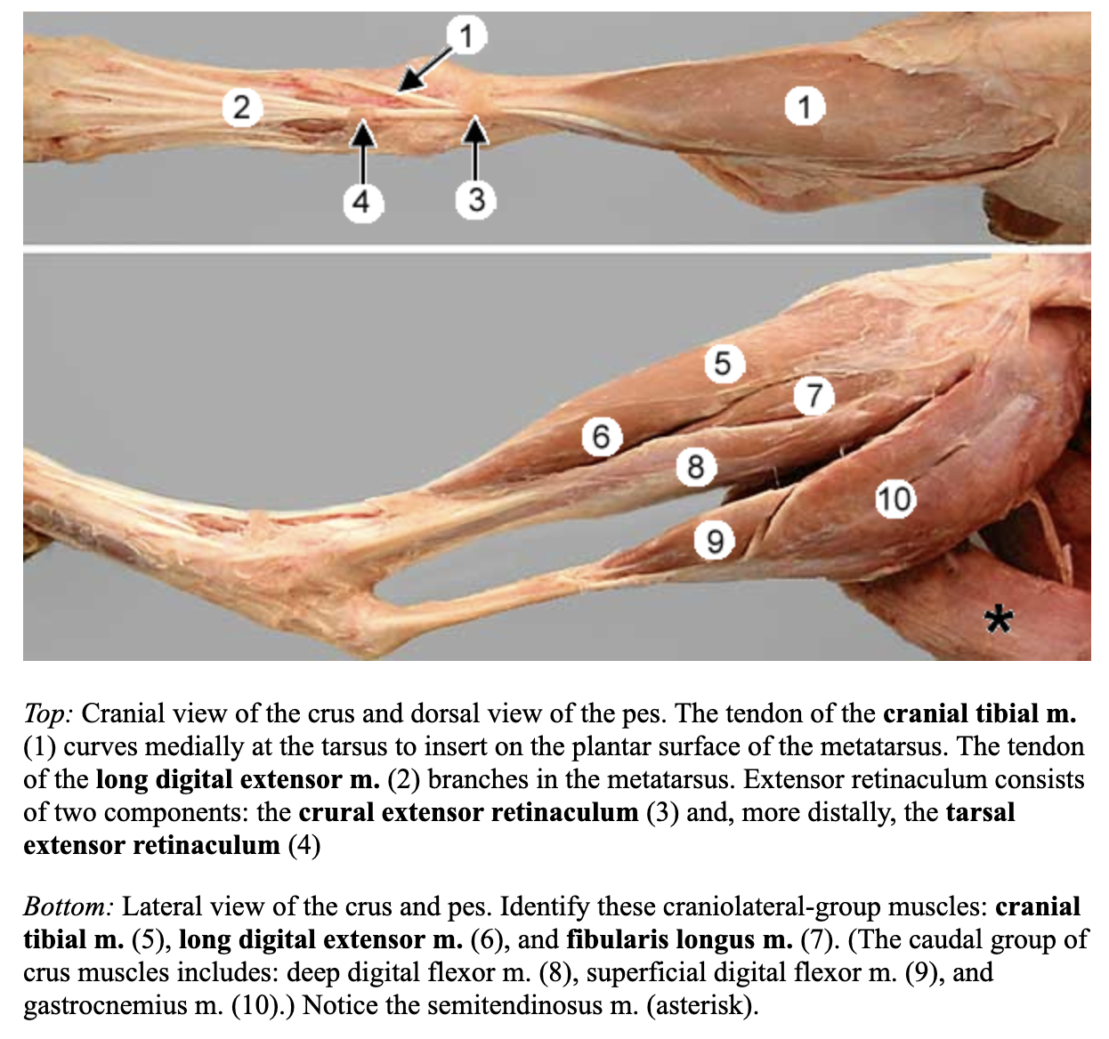
long digital extensor m.
6
attachments: extensor fossa of the femur and distal phalanges of the weight bearing digits (II-V)
actions: flex the tarsus; extend digits II-V
blood: cranial tibial a.
Nerve: common fibular n.

fibularis longus m.
7
attachments: lateral condyle of the tibia, fibular head; plantat aspect of the proximal ends of MT II-V
action: flex tarsus
blood: cranial tibial a.
nerve: common fibular n.

Clinical note: The term fibular/is is also commonly referred to as
peroneal/us clinically
therefore: fibularis longus m is peroneal longus m
fibular n. is the same as peroneal n.