BIOL 2160_CH2_A
 Call Kai
 Learn
Learn Practice Test
 Spaced Repetition
 Match
Match Flashcards
 Knowt Play
1/59

There's no tags or description
Looks like no tags are added yet.
Name | Mastery | Learn | Test | Matching | Spaced | Call with Kai | Chat |
|---|
No analytics yet
Send a link to your students to track their progress
60 Terms
Q1. How do the four primary bacterial shapes differ, and why is morphology useful but insufficient for identification?
Morphology means a bacterium’s visible form, especially its shape and arrangement.
Shape | Description | Picture it as |
|---|---|---|
Cocci (singular: coccus) | Spherical cells | Small balls ⚪ |
Bacilli (singular: bacillus) | Straight rod-shaped cells | Short sticks |
Vibrios (singular: vibrio) | Curved, comma-shaped rods | Commas |
Spirilla (singular: spirillum) | Rigid spiral-shaped cells | Corkscrews |
Why shape helps with identification
The peptidoglycan cell wall helps maintain bacterial shape. A species often has a characteristic form, so shape is a useful first clue when examining an unknown bacterium.
Why shape is not enough
Different species can have the same shape. Seeing a rod does not tell you which rod-shaped species it is.
Appearance can change with growth conditions, cell age, or stress.
Identification rule: Use morphology to narrow the possibilities, then combine it with other evidence, such as staining, metabolism, or DNA sequence data.
Extra distinction: Spirilla are rigid spirals. Spirochetes are also spiral-shaped but flexible
Q2. What is pleomorphism, and why is Mycoplasma pleomorphic and filterable?
Pleomorphism means that cells of the same organism can have different shapes.
Why Mycoplasma is pleomorphic
Most bacteria have a rigid peptidoglycan cell wall that helps hold their shape. Mycoplasma lacks this wall and is enclosed by a flexible cell membrane. As a result, its cells can appear round, elongated, or irregular.
Why it can be filterable
Mycoplasma cells are very small and flexible. Some can squeeze through filters that would retain many typical bacteria. Whether they pass depends on the filter’s pore size and the species.
Cause-and-effect chain:
No rigid cell wall → flexible, variable shape → small cells can pass through some bacteria-retaining filters
Key distinction: Filterable does not mean “a virus.” Mycoplasma is a bacterium; it can grow and reproduce as a cell.
Q3. Compare the arrangements of Streptococcus, Staphylococcus, and Bacillus.
Streptococcus cocci divide in one plane and remain attached as chains. Staphylococcus cocci divide in multiple planes and form irregular grape-like clusters. Some Bacillus rods, including Bacillus anthracis, remain attached end-to-end as chains.
Q4. Distinguish hyphae, mycelia, and trichomes.
The key distinction is a single filament, a network, or a chain of cells.
Term | What it is | Picture it as |
|---|---|---|
Hypha (plural: hyphae) | A thread-like filament that can branch | One branching thread |
Mycelium (plural: mycelia) | A network of hyphae | Many threads woven together |
Trichome | A row of connected cells in a filament, often seen in cyanobacteria | Beads joined in a line |
In a bacterium such as Streptomyces, hyphae grow and branch → the branches form a mycelium. In filamentous cyanobacteria, the connected row of cells is called a trichome.
Q5. How do Thiomargarita namibiensis and Epulopiscium fishelsoni cope with their large size?
Thiomargarita has a huge central vacuole that leaves a thin layer of active cytoplasm near the cell surface, shortening diffusion distances. Epulopiscium has many genome copies distributed through its large cell, supporting local gene expression.
Q6. Why is there a lower limit to bacterial cell size?
A bacterial cell needs a minimum amount of internal space to contain the machinery required to stay alive and reproduce: DNA, ribosomes, enzymes, and other molecules. As the cell gets smaller, its volume shrinks until it becomes difficult to fit and operate that machinery.
One correction to the supplied definition: a bacterial ribosome is generally about 20–30 nm across, not 50 nm. The exact number is less important here than the fact that a ribosome takes up real space—and a cell needs more than one component to function.
Q7. Why might Bismarck brown replace safranin in a Gram stain?
🇬🇧 English Notes1. Role of the counterstain
In a Gram stain, Gram-positive cells retain crystal violet and appear purple. Gram-negative cells lose crystal violet during decolorization, so a counterstain gives them a visible color.
Counterstain | Gram-positive cells | Gram-negative cells |
|---|---|---|
Safranin | Purple | Pink or red |
Bismarck brown | Purple | Yellow to yellow-brown |
2. Why use Bismarck brown?
With safranin, the observer must distinguish pink or red Gram-negative cells from purple Gram-positive cells. This difference may be difficult for someone with color vision deficiency (CVD) to see.
Bismarck brown produces a yellow to yellow-brown counterstain, which can provide clearer contrast with the purple Gram-positive cells.
Q8. How can intercellular channels coordinate cells in bacterial filaments?
A bacterial filament is a chain of connected cells. The partition between neighbouring cells is called a septum. In some filaments, channels through these septa let small molecules move from one cell to the next.
For example, in certain filamentous cyanobacteria, some cells specialize in nitrogen fixation while others perform photosynthesis. Exchange through the channels helps them share needed materials and coordinate their activities.

Q9. How do dead and living magnetotactic bacteria behave in a magnetic field?
Clean Answer
Cell | Aligns with magnetic field? | Swims along it? |
|---|---|---|
Dead | Yes, passively (if magnetosomes remain intact) | No |
Living | Yes, passively | Yes, using flagella |
Exam takeaway: Magnetosomes provide direction; flagella provide propulsion.
Q10. Why is morphology alone insufficient to identify an unknown bacterium?
Many unrelated bacteria are rods or cocci, and conditions such as starvation or antibiotic exposure can alter cell shape. Identification therefore combines morphology with other evidence, such as staining, biochemical tests, and genetic sequences.
Q11. What is the bacterial nucleoid, and how is its DNA compacted?
Q11. Bacterial nucleoid and DNA compaction 🧬
The nucleoid is the region of a bacterial cell that contains its chromosome. Unlike a eukaryotic nucleus, it has no surrounding membrane. The DNA shares the cytoplasm with the rest of the cell.
A bacterial chromosome is much longer than the cell, so it must be folded and organized. Three things help:
Mechanism | How it compacts DNA |
|---|---|
Supercoiling | Twists DNA into tighter coils, reducing the space it occupies. |
DNA-binding proteins | Bend, loop, and hold sections of DNA together. |
Charge-screening ions | Reduce repulsion between the negatively charged parts of DNA, allowing them to sit closer together. |
Picture it: Imagine fitting a very long thread into a small room. You twist it into coils, fold it into loops, and hold the loops in place. The DNA remains accessible, so the bacterium can still read and copy it.
Key distinction: A nucleoid is a DNA-containing region, not a membrane-bound organelle.
Q12. What is transcription–translation coupling in bacteria?
Bacteria lack a nuclear envelope, so ribosomes can begin translating an mRNA while RNA polymerase is still transcribing it from DNA. This places actively translating ribosomes near the nucleoid.
Q13. What are PHB granules, and why are PHAs of commercial interest?
1. What are PHB granules?
Polyhydroxybutyrate (PHB) is a carbon and energy storage polymer produced by some bacteria. When carbon is abundant, cells accumulate PHB as inclusion granules in the cytoplasm.
Location: Cytoplasm
Function: Carbon and energy reserve
Amount: Under some conditions, PHB can exceed 50% of a cell’s dry weight
Structure: The granules are not enclosed by a typical lipid bilayer membrane
2. How are PHB and PHA related?
Polyhydroxyalkanoates (PHAs) are a broad family of microbial storage polymers. PHB is one specific type of PHA.
PHA = polymer family → PHB = one member of that family
3. Why are PHAs commercially interesting?
PHAs can be processed into materials with plastic-like properties. They are produced by microorganisms and can be biodegraded by microbes under suitable conditions, making them candidates for certain bioplastic applications.
Q14. What is the function of sulfur globules in Thiomargarita namibiensis?
Thiomargarita can form elemental sulfur (S⁰) globules while oxidizing sulfide. Stored sulfur can later be oxidized further, providing electrons for energy metabolism when environmental sulfide is scarce.
Q15. What are gas vesicles, and how do they help aquatic bacteria?
Gas vesicles are gas-filled structures surrounded by a protein shell. The shell keeps water out while allowing gases to pass through.
Gas vesicles make the cell less dense, increasing its buoyancy. This helps aquatic bacteria, including some cyanobacteria, position themselves at a depth where conditions such as light and nutrient availability are suitable.
Common confusion: A gas vesicle is not a swimming organelle. It changes buoyancy, allowing the cell to move up or down in the water column.
Q16. How do carboxysomes improve carbon fixation?
Carboxysomes are protein-shell microcompartments containing Rubisco. They concentrate CO₂ around Rubisco, increasing carbon fixation efficiency and reducing its competing reaction with O₂.
Q17. What is an anammoxosome, and what happens inside it?
An anammoxosome is a membrane-bound compartment in anammox bacteria. It houses anaerobic ammonium oxidation, in which ammonium (NH₄⁺) and nitrite (NO₂⁻) are converted to nitrogen gas (N₂). Compartmentalization helps contain reactive intermediates such as hydrazine.
Q18. What are magnetosomes, and how are they organized?
Magnetosomes are magnetic mineral crystals enclosed by membranes inside certain bacteria. The crystals are commonly magnetite (Fe3O4) or greigite (Fe3S4).
In many magnetotactic bacteria, the actin-like protein MamK helps arrange magnetosomes into a chain. The chain acts like a small compass needle: it helps the cell align with Earth’s magnetic field.
Alignment is not movement. The magnetosome chain orients the bacterium; structures such as flagella propel it.

Q19. How does magneto-aerotaxis help magnetotactic bacteria?
Clean Answer
Definition: Magneto-aerotaxis combines magnetic alignment with movement guided by oxygen concentration.
Mechanism:
Magnetosomes align the bacterium with Earth’s magnetic field.
Flagella propel it along that direction.
Oxygen sensing helps it reach a preferred oxygen concentration in aquatic sediment.
Exam takeaway: The magnetic field guides orientation; flagella provide movement; oxygen sensing helps determine whether the bacterium is moving toward a suitable environment.
Q20. How does bacterial compartmentalization compare with eukaryotic compartmentalization?
Bacteria generally lack a membrane-bound nucleus and the extensive endomembrane system of eukaryotes. However, they are not completely unorganized: some have protein-shell microcompartments, membrane-bound compartments, and ordered cytoskeletal structures.
Q21. What is FtsZ, and what does it do during bacterial division?
FtsZ is a bacterial protein related to tubulin, the protein that forms microtubules in eukaryotic cells. FtsZ molecules assemble into a changing structure called the Z-ring at the future division site, usually near the cell’s middle.
What happens during division?
The Z-ring forms: FtsZ assembles on the inner side of the cytoplasmic membrane.
Division machinery gathers: The ring helps recruit and organize other proteins needed for division. Together, these proteins form the divisome.
A septum forms: The divisome directs new cell envelope and peptidoglycan synthesis inward at that site. This inward-growing partition is the septum.
The cell separates: The envelope constricts until two daughter cells form.
Visual sequence: FtsZ assembles at midcell → Z-ring organizes the divisome → septum grows inward → cell divides
Key distinction: FtsZ helps locate and organize division. The Z-ring is dynamic; it is not simply a fixed belt that squeezes the cell in two by itself.
Q22. How does MreB contribute to bacterial cell shape?
MreB helps many rod-shaped bacteria stay rod-shaped. It is an actin-related protein that forms structures just inside the cell membrane and helps organize where peptidoglycan is added to the cell wall.
As the bacterium grows, new wall material must be inserted along its sides in an organized pattern. That lets the cell lengthen while maintaining its rod shape. If MreB is disrupted, wall growth can become disorganized and the cell may lose its normal shape. 🦠
Q23. How does ParM segregate low-copy plasmids?
Low-copy plasmids exist in only a few copies per cell. If they were distributed randomly during division, one daughter cell could receive none.
ParM solves this by forming an actin-like filament between two plasmid-associated complexes. As the filament grows, it pushes the plasmid copies toward opposite sides of the cell. When the cell divides, each daughter cell is more likely to inherit a copy. 🧬
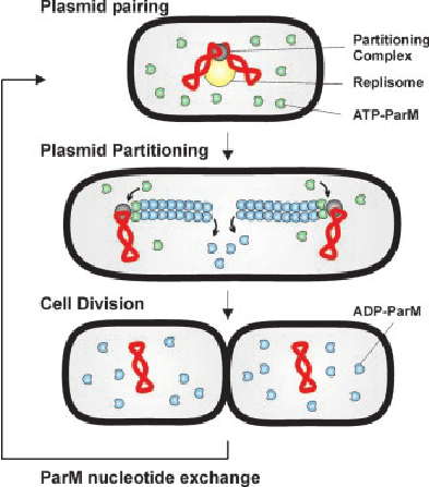
Q24. How can GFP fusions and gene deletion reveal MamK's function?
🔬
The two experiments answer different questions:
Experiment | Question | What researchers observe |
|---|---|---|
MamK–GFP fusion | Where is MamK in the cell? | GFP fluorescence reveals the position of MamK filaments in living cells. |
mamK gene deletion | What happens without MamK? | Researchers check whether the magnetosome chain becomes disorganized. |
GFP is a fluorescent protein. By attaching it to MamK, researchers can track where MamK is located. If deleting mamK disrupts the magnetosome chain, the two results together support the conclusion that MamK helps organize the chain.
Key distinction: Seeing MamK near the chain shows its location; observing a defect after deletion provides evidence for its function.
Q25. What is MamJ's role in magnetosome organization?
Think of MamK as a filament running through the cell and MamJ as a connector that helps hold magnetosomes along it. 🧲
Magnetosomes are membrane-bound structures containing magnetic crystals. In studied magnetotactic bacteria, MamJ links them to the actin-like MamK filament, helping arrange them into a chain. If MamJ is missing, crystals can still form, but the magnetosomes may cluster instead of staying in a regular chain.
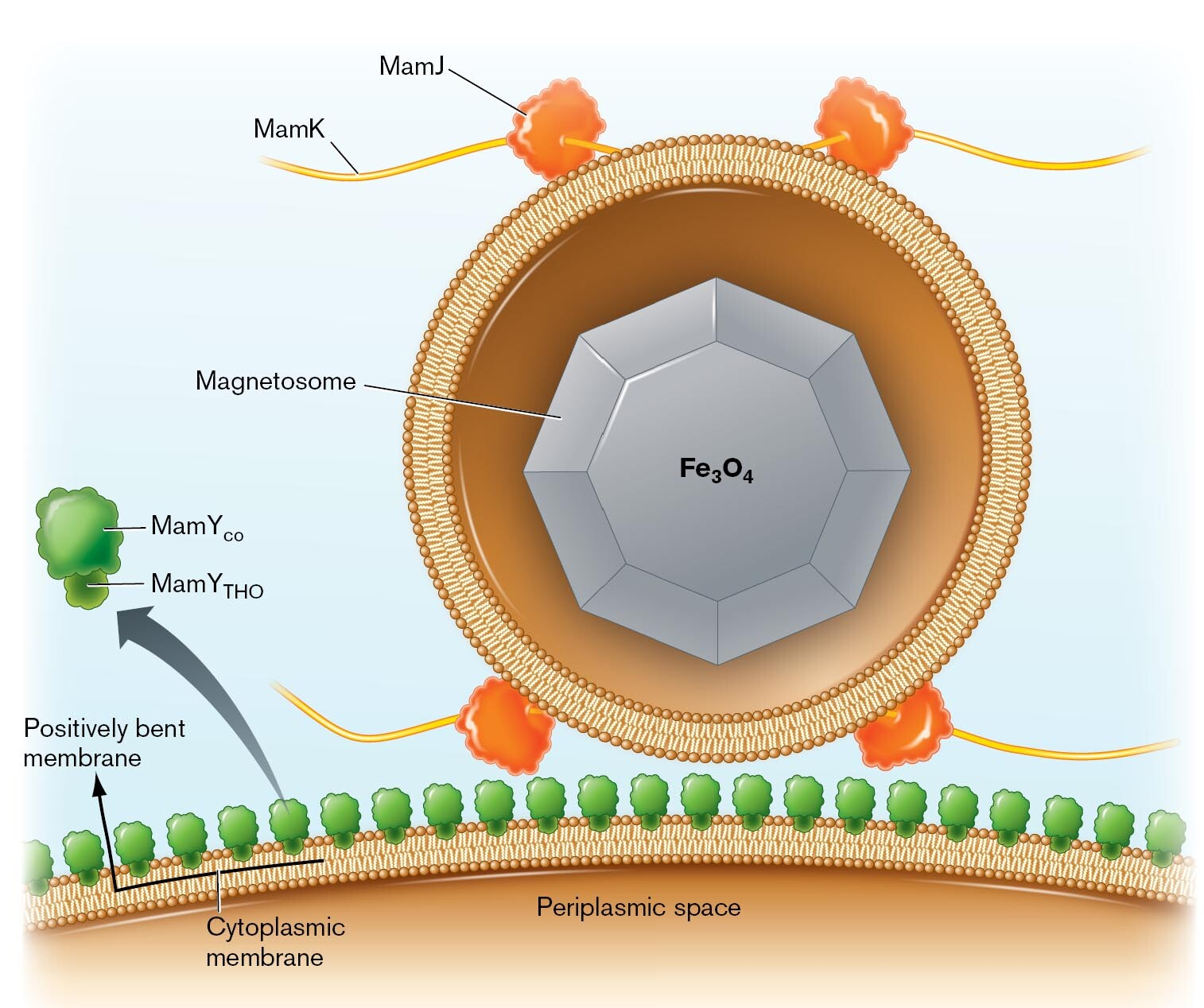
Q26. What happens when temperature-sensitive FtsZ stops working?
At a non-permissive temperature, defective FtsZ cannot properly organize division. Cells may continue to grow and replicate DNA but fail to form division septa, producing elongated filaments.
FtsZ가 작동을 멈추면 세균이 길어지지만 둘로 나뉘지 못해. 🦠
FtsZ는 세균의 가운데에 Z-ring을 만드는 단백질이야. 이 고리가 분열 위치를 정하고, 세포를 둘로 나눌 division septum(분열 격벽)을 만드는 과정을 조직해.
이미지의 temperature-sensitive FtsZ는 허용 온도에서는 작동하지만, non-permissive temperature(작동할 수 없는 온도)에서는 기능을 잃는 변이 단백질이야.
FtsZ 기능 상실 → Z-ring 형성 실패 → septum 형성 실패 → 세포 분열 실패
이때 다른 성장 과정이 계속된다면 세포는 DNA를 복제하고 길어질 수 있어. 그래서 짧은 세균 여러 개 대신 길게 이어진 filament(실 모양 세포)가 관찰돼. “DNA 복제도 반드시 멈춘다”는 뜻은 아니야.
Q27. What shape change may follow loss of mreB in E. coli?
Without functional MreB, organized rod-shaped cell wall growth is disrupted. Cells can lose their elongated form and become rounder or irregular; the exact outcome depends on growth conditions and the mutant's viability.
Q28. How can cytoplasmic cytoskeletal proteins control cell wall synthesis outside the membrane?
The membrane-spanning proteins are the link. FtsZ and MreB sit on the cytoplasmic side of the plasma membrane. They help organize protein complexes that extend through the membrane and connect to peptidoglycan-building enzymes on the other side.
Cytoskeletal protein | Where it helps organize wall synthesis | Result |
|---|---|---|
FtsZ | At the future division site | New wall forms between the two daughter cells |
MreB | Along the sides of many rod-shaped cells | Wall growth helps maintain an elongated shape |
Picture FtsZ or MreB as an inside guide: it does not reach through the membrane to build the wall itself. It helps position the machinery that builds peptidoglycan outside the membrane.
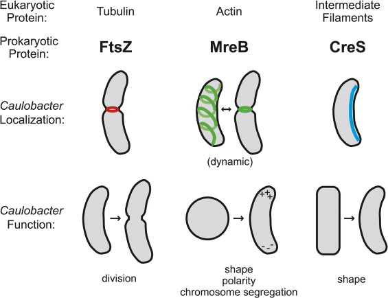
Q29. How are FtsZ, MreB, and ParM related to eukaryotic cytoskeletal proteins?
The key connection is shared protein structure, even though the proteins do different jobs. 🧬
Bacterial protein | Related eukaryotic protein | Main bacterial role |
|---|---|---|
FtsZ | Tubulin | Forms the Z-ring that organizes cell division |
MreB | Actin | Helps maintain cell shape by guiding cell wall construction |
ParM | Actin | Forms filaments that help separate plasmid copies |
A structural homolog has a similar three-dimensional protein structure because of shared evolutionary ancestry. FtsZ is related to tubulin, while MreB and ParM are related to actin. This tells us that dynamic cytoskeletal filaments evolved long before eukaryotic cells appeared.
Q30. Why is active ParM segregation important for low-copy plasmids?
A low-copy plasmid has only a few copies in each bacterial cell. Before the cell divides, those copies must be distributed between the two daughter cells.
The problem with random distribution
If the plasmids simply remain wherever they happen to be, one daughter cell may receive no copy. That cell would lose the plasmid and any genes it carries.
What ParM does
ParM is an actin-like protein that forms a growing filament between plasmid-associated complexes. As the filament grows, it pushes plasmid copies apart toward opposite sides of the cell. When the cell divides, each daughter is more likely to inherit a copy.
Visual sequence: Few plasmid copies → ParM filament grows → copies move apart → cell divides → both daughters inherit the plasmid
Key relationship: The fewer plasmid copies a cell has, the less reliable random distribution becomes, so active segregation matters more.
Q31. How does phospholipid structure create a selective plasma membrane?
1. Amphipathic structure
A phospholipid is amphipathic: it has a hydrophilic, polar head and hydrophobic, nonpolar fatty acid tails.
2. Bilayer formation
In water, phospholipids form a bilayer. Their heads face the watery cytoplasm and the environment outside the cell, while their tails point inward toward each other. This creates a hydrophobic core in the middle of the membrane.
3. Selective permeability
Substance | Can it cross the lipid bilayer easily? | Why? |
|---|---|---|
O₂, CO₂ | Relatively easily | Small and nonpolar |
Ions | No, without help | Their charge is unfavorable in the hydrophobic core |
Glucose and many other large polar molecules | Not readily | Too large and polar to pass through the core efficiently |
Transport proteins provide routes for substances that cannot cross the lipid bilayer readily. This is why the plasma membrane is selectively permeable: different substances cross at different rates, and some require specific proteins.
Q32. What are hopanoids, and what do they do?
1. Definition
Hopanoids are ring-shaped, sterol-like lipids found in the membranes of some bacteria. They play a structural role similar to that of sterols, such as cholesterol, in many eukaryotic membranes.
2. Membrane function
Hopanoids fit among membrane lipids and help stabilize the membrane. They can also influence its fluidity and permeability, helping the membrane maintain suitable properties under changing conditions.
3. Environmental persistence
Hopanoids are chemically stable and can remain in soils and sediments for long periods. This makes them useful as biomarkers in studies of past microbial life.
Q33. Compare facilitated diffusion, symport, antiport, and ABC transport.
These terms describe two different things: where transport energy comes from, and which direction coupled substances move.
Transport type | What moves? | Energy source | Can move a substance against its gradient? |
|---|---|---|---|
Facilitated diffusion | One substance moves down its gradient through a membrane protein | The substance’s own gradient | No |
Symport | Two substances move in the same direction | Often the downhill movement of an ion | Yes, for the coupled substance |
Antiport | Two substances move in opposite directions | Often the downhill movement of an ion | Yes, for the coupled substance |
ABC transport | A transporter moves a substance across the membrane | ATP hydrolysis | Yes |
Imagine an ion moving downhill along its concentration gradient. A symporter can use that movement to carry another substance uphill in the same direction. An antiporter can use it to move another substance uphill in the opposite direction.
The key distinction: symport and antiport name directions, while facilitated diffusion and ABC transport describe how transport is powered. Symport and antiport commonly use an ion gradient, so they are often examples of secondary active transport.
Q34. How does the Sec pathway export a bacterial protein?
Core idea
The Sec pathway moves a protein across the bacterial plasma membrane. The protein generally passes through the membrane channel in an unfolded state.
Step-by-step mechanism
Targeting — signal peptide
An N-terminal signal peptide marks the newly synthesized protein for export.Keeping the protein unfolded — SecB
In the pathway described here, the SecB chaperone helps prevent the protein from folding prematurely in the cytoplasm.Delivery — SecA and SecYEG
SecB delivers the protein to SecA. SecA works with SecYEG, the protein channel in the plasma membrane.Translocation — ATP use
SecA uses energy from ATP hydrolysis to help move the unfolded protein through SecYEG.Processing and folding
A signal peptidase removes the signal peptide. The exported protein can then fold into its functional shape.
Location check: In Gram-negative bacteria, crossing the plasma membrane places the protein in the periplasm. Crossing the outer membrane, if needed, requires an additional step.
Q35. What are the main components of peptidoglycan?
Peptidoglycan contains alternating N-acetylglucosamine (NAG) and N-acetylmuramic acid (NAM) joined by β-1,4 glycosidic bonds. Short peptides attached to NAM crosslink neighboring glycan strands. Crosslink structure varies; E. coli has direct links, while Staphylococcus aureus has pentaglycine bridges.
Q36. How does bactoprenol participate in peptidoglycan synthesis?
Peptidoglycan precursors are assembled inside the cell and attached to bactoprenol, a membrane lipid carrier. Bactoprenol moves the building block across the plasma membrane, where enzymes extend glycan chains and crosslink their peptides.
Q37. Compare lysozyme, lysostaphin, and β-lactam antibiotics.
Peptidoglycan is a mesh around many bacterial cells. It has two main parts: long chains of alternating sugars (NAG and NAM) and short peptide links that connect those chains. These three agents weaken the mesh at different points.
Agent | Target | What it does |
|---|---|---|
Lysozyme | The β-1,4 bond between NAG and NAM | Cuts the sugar backbone of existing peptidoglycan |
Lysostaphin | Pentaglycine bridges found in certain staphylococci | Cuts links between peptidoglycan strands |
β-lactam antibiotics | Penicillin-binding proteins (PBPs), including crosslinking enzymes | Prevent new peptidoglycan from being properly crosslinked |
The key distinction
Lysozyme and lysostaphin cut bonds that are already present. β-lactams instead block enzymes building new crosslinks. This is why β-lactams generally work best when bacteria are actively growing and making cell wall.
To identify the agent in a question, look for its target:
NAG–NAM sugar bond → lysozyme
Pentaglycine bridge → lysostaphin
PBP or crosslinking enzyme → β-lactam
1. What are NAG and NAM?NAG and NAM are two modified sugar molecules:
NAG = N-acetylglucosamine
NAM = N-acetylmuramic acid
They alternate to form a long glycan chain:
NAG — NAM — NAG — NAM — NAG — NAM
The link between neighbouring NAG and NAM sugars is a β-1,4 glycosidic bond. Here, “β-1,4” describes how the two sugar molecules are chemically connected.
Q38. How do β-lactamases cause resistance, and what does clavulanic acid do?
1. How β-lactam antibiotics work
β-lactam antibiotics, such as penicillin and amoxicillin, bind to penicillin-binding proteins (PBPs). These include transpeptidases that cross-link peptidoglycan during bacterial cell wall synthesis.
PBP inhibition → reduced peptidoglycan cross-linking → weakened cell wall
2. How β-lactamases cause resistance
β-lactamases are bacterial enzymes that hydrolyze and open the β-lactam ring of an antibiotic. Once the ring is broken, the drug can no longer effectively inhibit its PBP target.
β-lactamase → broken β-lactam ring → inactive antibiotic
3. What clavulanic acid does
Clavulanic acid inhibits certain β-lactamases. When combined with amoxicillin, it protects the antibiotic from degradation so amoxicillin can inhibit PBPs. The combination is called amoxicillin–clavulanate, or Augmentin.
Limit: Clavulanic acid does not inhibit every type of β-lactamase or reverse every form of β-lactam resistance.
Q39. Compare Gram-positive and Gram-negative cell envelopes.
A cell envelope is the set of layers surrounding a bacterial cell. Picture the layers from the inside outward:
Gram-positive | Gram-negative | |
|---|---|---|
Layer order | Plasma membrane → thick peptidoglycan wall | Inner membrane → periplasm with thin peptidoglycan → outer membrane |
Membranes | One | Two |
Distinctive component | Teichoic acids in the cell wall | Lipopolysaccharide (LPS) in the outer membrane |
Gram stain | Usually purple | Usually pink |
Why the stain differs: The thick Gram-positive wall retains the crystal violet stain during the alcohol wash. The thinner Gram-negative wall does not; the cells then take up the pink counterstain.
Gram-positive: One plasma membrane surrounded by a thick peptidoglycan wall containing teichoic acids.
Gram-negative: An inner membrane, a thin peptidoglycan layer in the periplasm, and an outer membrane containing LPS.
Exam rule: First count the membranes, then locate the peptidoglycan. One membrane + thick wall → Gram-positive. Two membranes + thin wall between them → Gram-negative.
Q40. What are the three regions of LPS, and why do they matter?
LPS has lipid A, a core polysaccharide, and an O antigen. Lipid A anchors LPS in the outer membrane and can trigger strong host inflammatory responses. The core links lipid A to the variable O antigen, which contributes to differences in surface recognition among strains.
Q41. What happens at each step of a Gram stain?
Crystal violet stains cells purple. Iodine helps retain the dye as a crystal violet–iodine complex. Alcohol or acetone decolorizes Gram-negative cells more readily, while Gram-positive cells retain purple dye in their thick peptidoglycan. Safranin then stains decolorized cells pink.
Q42. How do porins and TonB-dependent receptors differ?
Porins allow many small hydrophilic solutes to diffuse through the Gram-negative outer membrane. TonB-dependent receptors bind specific scarce nutrients, such as iron–siderophore complexes, and use energy transmitted from the inner membrane to bring them into the periplasm.
Q43. What is a Type III secretion system (T3SS)?
A Type III secretion system is a needle-like protein apparatus found in some Gram-negative bacteria. It extends from the bacterium toward a host cell and delivers bacterial proteins called effectors into that cell.
Think of the sequence as:
Bacterium contacts host cell → T3SS delivers effectors → effectors change host-cell activity
The effect depends on the effector. For example, an effector may alter the host’s internal structure or interfere with an immune response.
Common confusion: The T3SS shares evolutionary ancestry with parts of the bacterial flagellum’s protein export machinery, but its main job is protein delivery, not swimming.

Q44. What happens when lysozyme removes a Gram-positive wall in isotonic versus hypotonic solution?
Lysozyme breaks down peptidoglycan, the rigid material that supports a Gram-positive cell. Once the wall is removed, only the flexible cell membrane contains the cell. This wall-less cell is called a protoplast.
Surrounding solution | Water movement | Result |
|---|---|---|
Isotonic — similar solute concentration inside and outside | No strong net movement of water | The protoplast may survive temporarily |
Hypotonic — lower solute concentration outside | Water moves into the cell | The unsupported membrane may swell and burst (osmotic lysis) |
The key is water pressure: the cell wall normally resists swelling. Without it, a large influx of water can rupture the membrane.
Q45. Why are Gram-negative bacteria generally resistant to vancomycin?
Vancomycin must reach peptidoglycan precursors to block cell-wall construction. It binds the D-Ala-D-Ala end of these precursors, preventing them from being incorporated properly into the growing wall.
In Gram-negative bacteria, the target lies behind the outer membrane. Vancomycin is usually too large to pass through this barrier, so it cannot reach the target effectively. Gram-positive bacteria have no outer membrane, leaving their wall-building machinery more accessible.
Solving rule: For any antibiotic, ask “Where is its target, and what barrier must the drug cross to reach it?”
Q46. What are the main parts of a bacterial flagellum?
The long helical filament acts as a propeller, the hook connects it to the base, and the basal body anchors it in the cell envelope. A rotary motor turns the structure, usually using an ion gradient for energy.
Q47. Define monotrichous, lophotrichous, and peritrichous.
Monotrichous means one flagellum at a pole. Lophotrichous means a tuft of flagella at a pole. Peritrichous means flagella distributed over the cell surface.
Q48. How do CCW and CW rotation produce runs and tumbles in peritrichous bacteria?
In the classic E. coli example, counterclockwise (CCW) rotation bundles flagella and produces a relatively straight run. Clockwise (CW) rotation disrupts the bundle and causes a tumble that reorients the cell.
Q49. How does positive chemotaxis create a biased random walk?
Bacteria compare chemical conditions over time. When movement leads toward more attractant, signaling prolongs runs and reduces tumbles. When conditions worsen, more frequent tumbles give the cell new directions to try.
Q50. How do spirochetes move through viscous environments?
Spirochetes have periplasmic flagella located between the cell cylinder and outer sheath. Their rotation bends or twists the flexible cell body, producing corkscrew-like movement that works well in viscous material.
Q51. Compare twitching, gliding, and host-actin-based motility.
Motility type | Location | Source of movement | Typical pattern |
|---|---|---|---|
Twitching | Solid surface | Extension and retraction of Type IV pili | Short, jerky movements |
Gliding | Solid surface | Species-dependent bacterial surface motility mechanisms | Smooth movement |
Host-actin-based | Inside a host cell | Polymerization of host actin behind the bacterium | Intracellular movement and cell-to-cell spread |
1. Twitching motility
A Type IV pilus extends, attaches to a surface, and retracts. Retraction pulls the bacterial cell forward. Repeated cycles produce a jerky movement.
Examples: Pseudomonas aeruginosa and Neisseria spp.
2. Gliding motility
Bacteria move smoothly across a solid surface without rotating flagella. Gliding does not have one universal mechanism; the machinery differs among bacterial groups.
Examples: Myxobacteria and some filamentous cyanobacteria.
3. Host-actin-based motility
Intracellular bacteria induce the host cell to polymerize actin behind them. The growing actin tail pushes the bacterium through the host cytoplasm and can help it spread into neighboring cells.
Examples: Listeria monocytogenes and Shigella spp.
Q52. Compare adhesive fimbriae and conjugative sex pili.
Both are protein structures that extend from a bacterial surface, but they help the cell connect to different things.
Feature | Adhesive fimbriae | Conjugative sex pili |
|---|---|---|
Main function | Attach the bacterium to a surface or host cell | Establish contact with another bacterium for DNA transfer |
Usual appearance | Numerous, short fibres | Usually fewer and longer |
What they connect | Bacterium → surface | Donor bacterium → recipient bacterium |
Outcome | Helps the cell remain attached, including during biofilm formation | Helps initiate conjugation, the transfer of DNA between cells |
How a sex pilus helps conjugation
A donor cell extends a sex pilus and contacts a recipient cell.
The cells are brought into close contact.
Conjugation machinery transfers DNA from the donor to the recipient.
Common confusion: The sex pilus helps establish contact. The DNA is transferred by the conjugation machinery; the pilus itself should not be pictured simply as a hollow straw carrying DNA.
Q53. How can a Caulobacter stalk help in a nutrient-poor environment?
A Caulobacter stalk is a thin extension of the cell envelope. Its tip has a sticky holdfast, which anchors the bacterium to a surface. 🦠
In nutrient-poor water, the stalk gives the cell more surface exposed to its surroundings. That may help nutrient uptake, while attachment keeps the cell in a useful location. The word may matters: extra surface area alone does not prove that nutrients enter through the stalk.

Q54. What is a capsule, and how can it protect a pathogen?
1. Definition
A capsule is a well-organized layer attached to the outside of some bacterial cells. It is usually made of polysaccharides. Capsules and loosely attached slime layers are both forms of the glycocalyx.
2. Protective functions
Avoiding phagocytosis: The capsule covers the bacterial surface, making it harder for phagocytic immune cells to attach to and engulf the bacterium.
Preventing desiccation: Its water-attracting material helps the cell retain moisture.
Supporting colonization: In some species, it helps the cell adhere to surfaces and contributes to biofilm formation.
Example: The capsule of Streptococcus pneumoniae helps the bacterium evade host defenses.
가장 중요한 병원성 기능은 phagocytosis(식세포작용) 회피야. 예를 들어 Streptococcus pneumoniae의 협막은 숙주가 세균을 제거하기 어렵게 만든다.
Q55. What are the main stages of biofilm formation?
Q55. Main stages of biofilm formation 🦠
A biofilm is a community of microorganisms attached to a surface and surrounded by a self-produced extracellular matrix.
Stage | What happens |
|---|---|
1. Initial attachment | Free-living (planktonic) cells contact a surface. Early attachment may be reversible. |
2. Stable attachment and microcolonies | Cells attach more firmly, divide, and form small groups called microcolonies. |
3. Matrix production and maturation | Cells produce an extracellular matrix containing polysaccharides, proteins, and extracellular DNA. The biofilm grows into a structured community. |
4. Dispersal | Some cells leave the biofilm and may attach to a new surface. |
Visual sequence: Free-living cells → surface attachment → microcolonies → mature biofilm → dispersal
Why a mature biofilm behaves differently
Cells in different parts of the biofilm experience different amounts of oxygen, nutrients, and waste products. These local chemical gradients can make some cells grow more slowly. Together with the matrix and other protective responses, this can make a biofilm harder to remove or treat than free-living cells.
Q56. What is an S-layer, and what can it do?
An S-layer (surface layer) is a regularly repeating sheet of protein or glycoprotein subunits on the outside of a microbial cell. Picture many identical tiles assembled into a thin outer coat. 🧩
Its role depends on the species:
Protection: Acts as a barrier against some external threats.
Shape and support: Adds structural stability; in some archaea, it is a major part of the cell wall.
Attachment: Helps the cell interact with surfaces or other organisms.

Q57. What are bacterial species, strains, and type strains?
These terms describe different levels of identification:
Term | Meaning | Think of it as |
|---|---|---|
Species | A named group of closely related bacteria | The broad group |
Strain | A particular genetic variant or isolate within a species | One version of that species |
Type strain | The designated reference strain to which the species name is permanently linked | The name’s reference point |
How they fit together
Two bacteria can belong to the same species but be different strains. Their genomes and traits may differ; for example, one strain may carry a gene that the other lacks.
A type strain is one specific strain chosen when a species is formally named. Researchers use it as a taxonomic reference when deciding what that species name refers to. It does not have to be the most common or most typical strain.
Relationship: Species → contains multiple strains → one designated strain serves as the type strain.
Q58. Why was Woese's comparison of 16S rRNA important?
Q58. Why Woese’s 16S rRNA comparison mattered 🧬
16S rRNA is an RNA component of the small ribosomal subunit in bacteria and archaea. Woese and colleagues compared its sequence across microorganisms to infer evolutionary relationships: organisms with more similar sequences generally share a more recent common ancestor.
Why use 16S rRNA?
It is widespread: Bacteria and archaea need ribosomes, so they have small-subunit rRNA.
Some regions are conserved: They change slowly, allowing comparisons across distant groups.
Other regions vary: Their differences help distinguish more closely related groups.
What did the comparisons reveal?
Microbes that looked similar or had similar metabolisms were not necessarily close relatives. The sequence evidence showed that Archaea are a major lineage distinct from Bacteria. This helped establish the three-domain framework: Bacteria, Archaea, and Eukarya.
16S rRNA sequencing also let researchers identify many microbes directly from environmental samples, even when they could not grow them in the laboratory.
Key idea: Shape and metabolism describe what an organism looks like or does; rRNA sequences provide evidence about its evolutionary history. For eukaryotes, the corresponding small-subunit rRNA is 18S rRNA.
Q59. Compare Proteobacteria and Firmicutes (Bacillota).
Proteobacteria (Pseudomonadota) | Firmicutes (Bacillota) | |
|---|---|---|
Typical structure | Gram-negative: thin peptidoglycan layer and an outer membrane | Gram-positive: thick peptidoglycan layer and no outer membrane |
Metabolism | Highly diverse | Also diverse; some members form endospores |
Examples | Escherichia, Vibrio, Rhizobium | Bacillus, Clostridium, Streptococcus |
Exception to remember | Members do not all share one metabolic strategy | Mycoplasma has no cell wall, so it does not stain Gram-positive |
Why is Mycoplasma confusing?
Bacillota is an evolutionary group, whereas Gram-positive describes a cell-wall structure and staining result. Most familiar Bacillota are Gram-positive, but Mycoplasma lacks a cell wall. Its membership in Bacillota therefore does not mean it stains Gram-positive.
Proteobacteria (Pseudomonadota): Generally Gram-negative bacteria with diverse metabolic strategies. Examples include Escherichia, Vibrio, and Rhizobium.
Firmicutes (Bacillota): A group containing many low-G+C, Gram-positive bacteria, including Bacillus, Clostridium, and Streptococcus. It also includes wall-less Mycoplasma.
Exam takeaway: Compare their typical cell envelopes, then mention Mycoplasma as an exception to the usual Bacillota pattern.
Q60. What distinguishes Cyanobacteria, Actinobacteria, Spirochaetes, and Planctomycetes?
Cyanobacteria perform oxygenic photosynthesis and are related to the ancestors of chloroplasts. Actinobacteria include high-G+C, often branching bacteria such as Streptomyces. Spirochaetes are flexible spiral bacteria with periplasmic flagella. Planctomycetes include bacteria with unusual internal structures, including anammoxosomes in anammox species.