Anatomy Lab Quiz #3
 Call Kai
 Learn
 Practice Test
 Spaced Repetition
 Match
 Flashcards
 Knowt Play
1/117

Earn XP
Description and Tags
Bones/Muscles of Upper Extremity, CNS
118 Terms
what bones make up the pectoral girdle?
clavicles & scapula
what is the purpose of the pectoral skeleton?
attaches upper limbs to axial skeleton; attachment site for muscles that move upper limbs
what do the clavicles articulate with?
sternum (medially) & scapula/acromion (laterally)

function of the clavicles
anchors muscles, act as brace to hold scapula & arms out laterally

scapula
between ribs 2 & 7, flat & triangular, 3 borders & 3 angles
how do you tell the anterior from the posterior on the scapula?
scapular spine is posterior, anterior surface is always smooth
how do you tell the medial from the lateral on the scapula?
medial is long, smooth border; acromion is lateral

acromion
continues w/ scapular spine posteriorly

glenoid cavity/fossa
“socket” part of the ball and socket joint, articulates with the head of the humerus
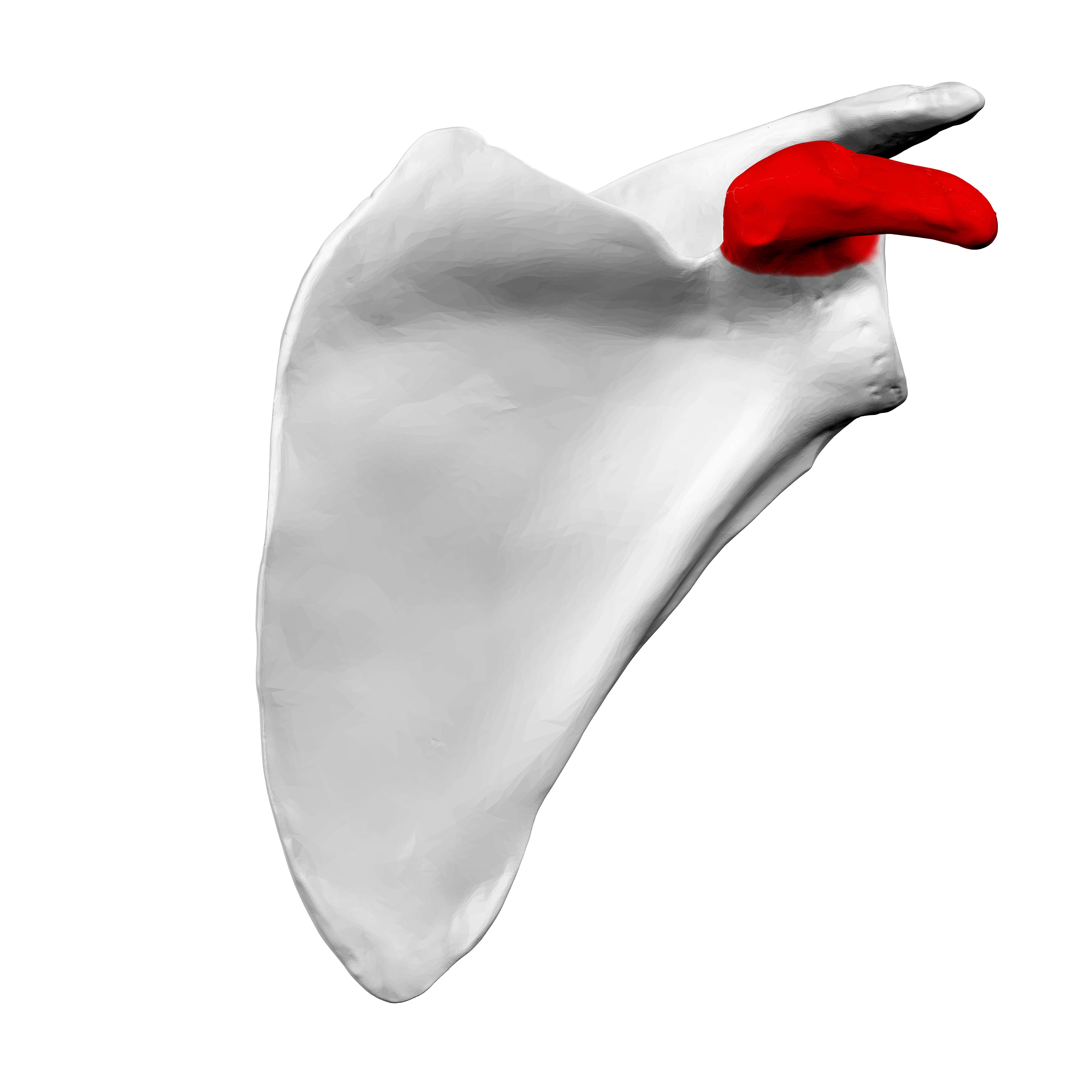
coracoid process
hooked, superior lateral side of scapula

suprascapular notch
indentation at top of scapula

supraspinous fossa
scooped out area above scapular spine
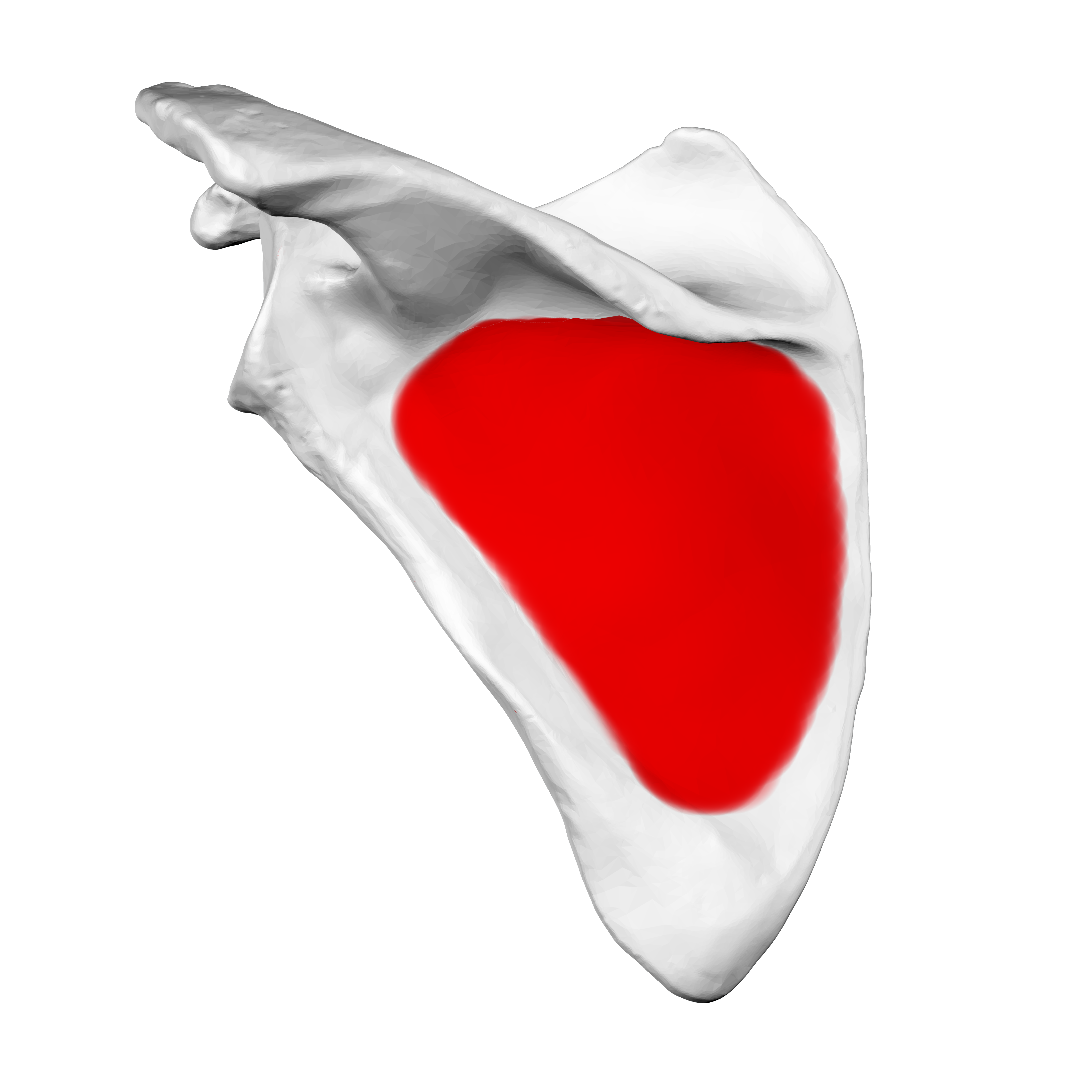
infraspinous fossa
scooped out area below scapular spine
how do you tell anterior from posterior on the humerus?
bowtie at distal end is anterior
how do you tell medial from lateral on the humerus?
the head of the humerus always points medially

greater tubercle of humerus
bigger bump

intertubercular sulcus
groove between the bumps

head of humerus
articular surface, “ball” of ball and socket joint
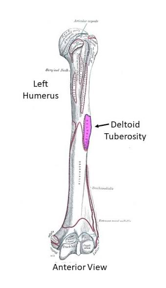
deltoid tuberosity
bump made by deltoid muscle, midway down bone

coronoid fossa
scooped out area that bone fits into

capitulum
articulates w/ radial head, for rotation
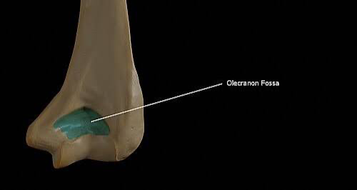
olecranon fossa
scooped out area the olecranon process (ulna) fits into

medial epicondyle
bone that’s on top of the condyle

trochlea
“pulley” articulation w/ the ulna (flexion & extension)
how do you tell medial from lateral at the elbow joint?
the ulna is always medial (has the styloid process if anterior & projects up if posterior)

coronoid process
beak-like projection of ulna
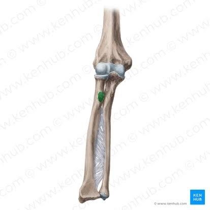
radial tuberosity
bump on radius, on medial side of radius

interosseous membrane
membrane between bones

styloid process
pen-like protrusion
ball-and-socket joint of the shoulder
made up of glenoid fossa of scapula & head of humerus
multiaxial
flexion/extension, abduction/adduction, rotation
elbow joint
made of the trochlea of humerus & trochlear notch of the ulna
uniaxial
flexion/extension

proximal interphalangeal joint (PIP)
hinge joint, uniaxial (flexion/extension)

metacarpophalangeal joint (MCP)
condyloid joint, biaxial (flexion/extension & abduction/adduction)

distal interphalangeal joint (DIP)
hinge joint, uniaxial (flexion/extension)

interphalangeal joint (IP)
hinge joint, uniaxial (flexion/extension)

carpometacarpal joint
saddle joint, specific to thumb
what muscles produce flexion?
ones that cross the anterior surface of a joint
what muscles produce flexion?
ones that cross the posterior surface of a joint
what muscles produce abduction?
ones that cross the lateral surface of a joint
what muscles produce adduction?
ones that cross the medial surface of a joint

subclavius
“under the clavicle”, stabilizes scapula, attach on scapula

pectoralis minor
pulls scapula forward (anterior) & downward (inferior), attach on scapula
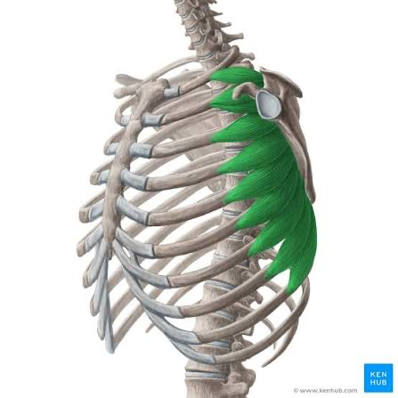
serratus anterior
used in pushing/punching & horizontal scapular movements, attaches to scapula

trapezius
trapezoid-shaped, elevates & rotates scapula, attach on scapula

levator scapula
elevates & adducts scapula, attach on scapula

rhomboid major/minor
rotate scapula, attach on scapula

deltoid
prime mover of shoulder for abduction, attach on humerus

pectoralis major
prime mover for shoulder flexion, adduction, attach on humerus

latissimus dorsi
puller upper extremity into extension, attaches on humerus

supraspinatus (green)
above scapular spine, rotator cuff muscle, attaches on humerus

infraspinatus
below scapular spine, rotator cuff muscle, attaches on humerus

teres minor
rotator cuff muscle, attaches on humerus

subscapularis
under scapula, rotator cuff muscle, attaches on humerus

biceps brachii
prime mover in elbow flexion, attach on the forearm
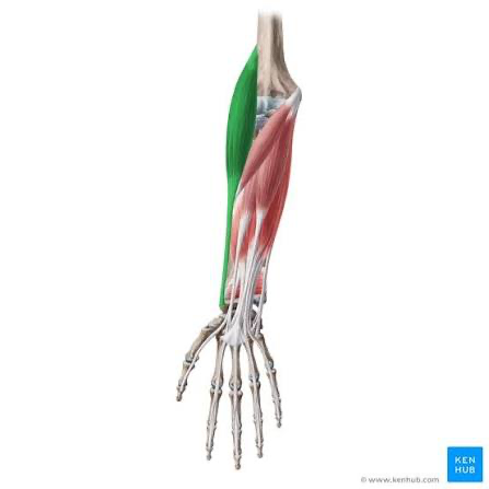
brachioradialis
helps w/ forearm/elbow flexion, attach on the forearm

brachialis
deep to biceps brachii, forearm/elbow flexion, attach on the forearm
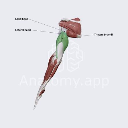
triceps brachii
prime mover for elbow extension, attach on the forearm

anconeus
tiny, helps stabilize elbow during extension, attach on the forearm

pronator quadratus
prime mover in pronation, attach on the forearm
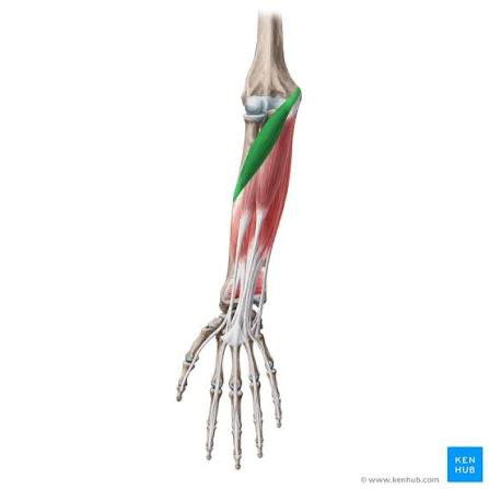
pronator teres
adds to pronation, attach on the forearm

supinator
supination/turns palm up, attach on the forearm

flexor carpi radialis
causes wrist flexion, lateral deviation, on thumb-side
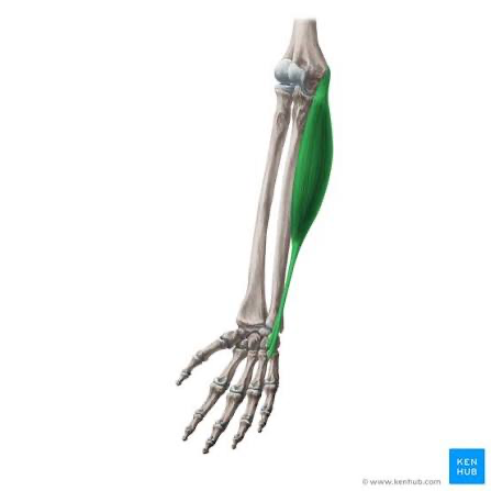
flexor carpi ulnaris
wrist flexion, medial deviation, on pinky-side

flexor digitorum superficialis
flexion of wrist and digits 2-5

palmaris longus
weak wrist flexion

flexor pollicis longus
causes thumb flexion

flexor digitorum profundus
causes distal finger flexion
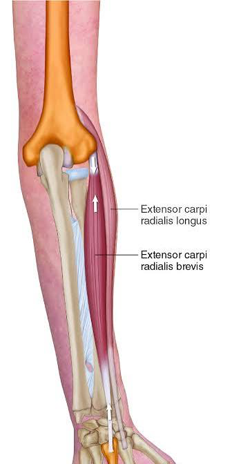
extensor carpi radialis longus/brevis
causes wrist extension, thumb-side

extensor digitorum
prime mover of finger extension

extensor indices
extension of index finger
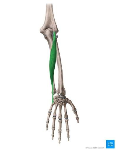
extensor carpi ulnaris
causes wrist extension

extensor pollicis longus/brevis
causes thumb extension

abductor pollicis longus
causes thumb abduction

hypothenar eminence
includes muscles that flex, abduct, and oppose the 5th digit (flexor digit minimi brevis, abductor digiti minimi, opponens digiti minima)
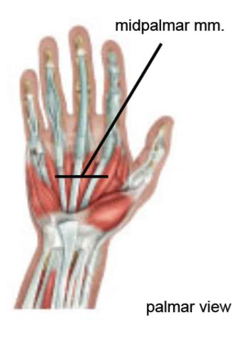
midpalmar muscles
includes deep muscles that flex, abduct, adduct the fingers (lumbricals, interossei)

thenar eminence
includes muscles that flex, abduct, adduct, and oppose the thumb (adductor pollicis, flexor pollicis brevis, abductor pollicis brevis, opponens pollicis)

central sulcus
separates the frontal lobe anteriorly from the parietal lobe posteriorly

precentral gyrus
houses the primary motor cortex and controls voluntary movement throughout the body

postcentral gyrus
houses the primary somatosensory cortex

corpus callosum
thick, C-shaped bundle of nerve fibers, acts as a neural bridge that connects the left and right cerebral hemispheres
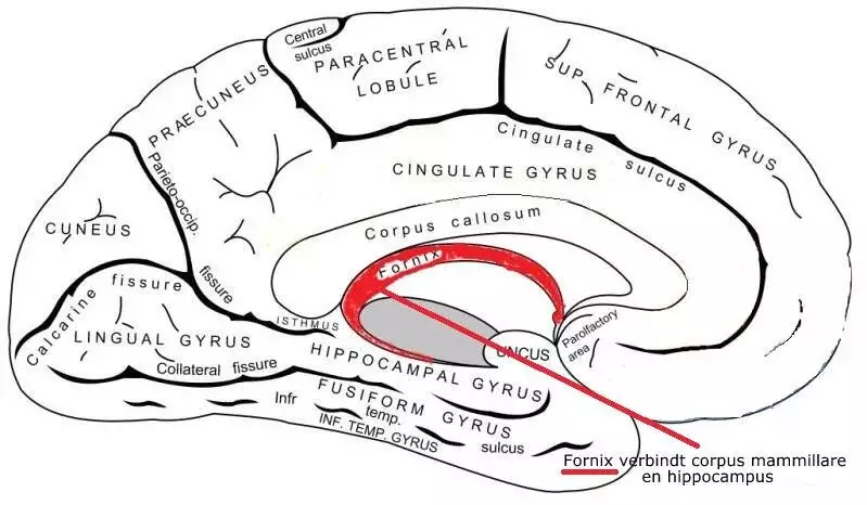
fornix
C-shaped, arching bundle of nerve fibers (white matter) in the brain that acts as the primary output tract of the hippocampus
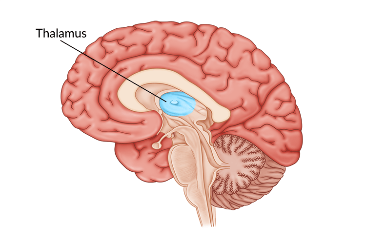
thalamus
mass of gray matter located deep in the center of the brain above the brainstem, acting as the primary information relay station and sensory gateway to the cerebral cortex

hypothalamus
just below the thalamus and above the brainstem. It acts as the body's main control center, linking the nervous system to the endocrine system to keep internal bodily functions balanced
optic chiasm
at the base of the brain where the optic nerves from each eye meet and partially cross over

pituitary gland
located at the base of the brain, just beneath the hypothalamus; controls other hormone-producing glands in the body and regulates essential functions like growth, metabolism, reproduction, and water balance

pons
acts as a major neural bridge connecting the higher brain and cerebellum to the spinal cord and medulla oblongata

medulla oblongata
lowest part of the brainstem, connecting the brain to the spinal cord; regulates vital involuntary processes, including heart rate, blood pressure, and breathing; manages protective

cerebellum
motor control & coordination, balance & posture, motor learning, cognitive & sensory roles

arbor vitae
branched, tree-like network of white matter located inside the cerebellum

mammillary body
located at the posteroinferior aspect of the hypothalamus within the diencephalon of the brain; essential for the processing, consolidation, and recall of episodic and spatial information
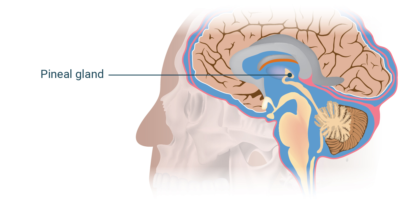
pineal gland
in the epithalamus region of the brain, main job is to synthesize and secrete melatonin
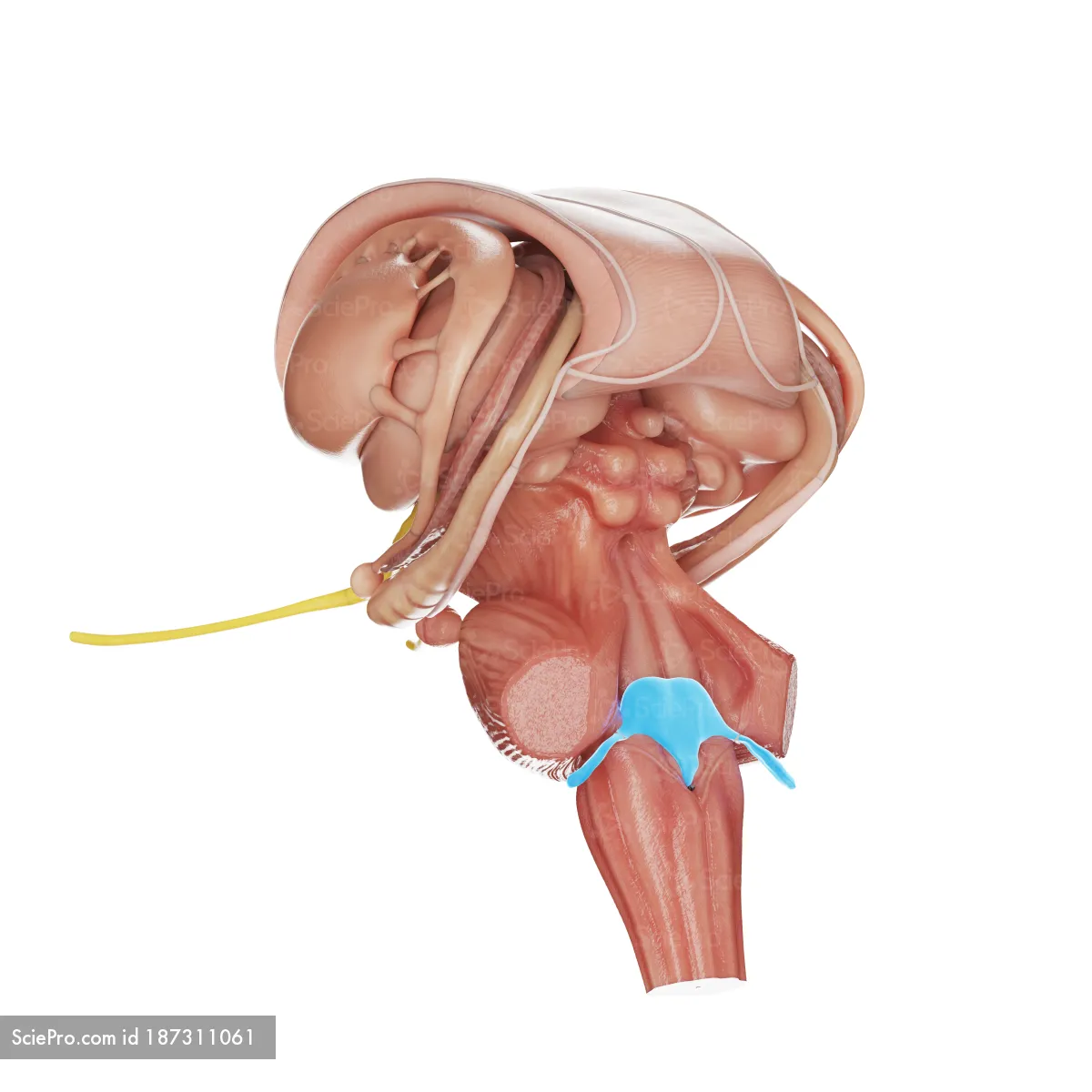
choroid plexus of the 4th ventricle
produces cerebrospinal fluid
what type of material composes the gray matter of the CNS?
cell bodies & unmyelinated axons
what type of material composes the white matter of the CNS?
myelinated axons
dorsal root ganglion
located on the dorsal (back) root of each spinal nerve just outside the spinal cord; collects & transmits sensory information (fine touch, pressure, vibration, temperature, & pain) from the body's periphery to the central nervous system

3 meningial layers that cover the brain & spinal cord
dura mater (outermost, thickest)
arachnoid mater (web-like)
pia mater (innermost, thin & clear)
pyramids of medulla
ridges on the ventral surface of the medulla, composed of neurons leaving the CNS

lateral ventricles (in green)
pair of large, C-shaped fluid-filled cavities located within each cerebral hemisphere of the brain

3rd ventricle (in red)
narrow, slit-like cavity located along the midline of the brain's diencephalon, positioned between the left and right thalami

4th ventricle (in green)
located within the brainstem, dorsal to the pons and upper medulla oblongata and ventral to the cerebellum