H6 Mosher- Chromatin structure and gene expression, including epigenetics

0.0(0)
Studied by 0 people
call kaiCall Kai
learnLearn
examPractice Test
spaced repetitionSpaced Repetition
heart puzzleMatch
flashcardsFlashcards
GameKnowt Play
Card Sorting

1/5

encourage image

There's no tags or description

Looks like no tags are added yet.

6 Terms

1
New cards

how do the different histone proteins assemble? how does this associate with DNA?

  • conserved ‘histone fold’ shape- small helix-turn-large helix-small helix

  • H2A and 2B form a dimer x 2

  • H3 and 4 form a tetramer

  • the histone octamer is one H3-H4 tetramer (binds to ~1 loop of DNA first) and two H2A-H2B dimers

  • ~147 nucleotide DNA wraps around this core particle almost twice

  • N-terminal histone tails extrude out from the DNA- can be modified for DNA marking

  • H1 is the linker histone protein- this can bind the linker DNA (between two nucleosomes) and clamp the entry and exit DNA together for higher compaction eg. more abundant in heterochromatin


<ul><li><p>conserved ‘<strong>histone fold</strong>’ shape- small helix-turn-large helix-small helix</p></li><li><p>H2A and 2B form a dimer x 2</p></li><li><p>H3 and 4 form a tetramer</p></li><li><p><strong>the histone octamer is one H3-H4 tetramer</strong> (binds to ~1 loop of DNA first) <strong>and two H2A-H2B dimers</strong></p></li><li><p><strong>~147 nucleotide DNA </strong>wraps around this core particle <strong>almost twice</strong></p></li><li><p>N-terminal histone <strong>tails </strong>extrude out from the DNA- can be modified for DNA marking</p></li><li><p><strong>H1 is the linker histone protein</strong>- this can bind the linker DNA (between two nucleosomes) and clamp the entry and exit DNA together for higher compaction eg. more abundant in heterochromatin</p></li></ul><p></p><p></p><p></p>
2
New cards

how do nucleosomes assemble?

  • nucleosomes complexes on DNA take up a ‘beads on a string’ or 10nm fibre structure (when actively transcribed)

  • this is assembled into a 30nm fibre (hypothesised by solenoid or zigzag structure)

  • chromatin is bound to the nuclear matrix by proteins, to make chromatin loops (unknown how)

  • within the interphase nucleus, the chromosomes have distinct territories because they stay bound to the matrix; they aren’t all intermixed

  • this is compressed down further to produce meiotic/mitotic chromosomes
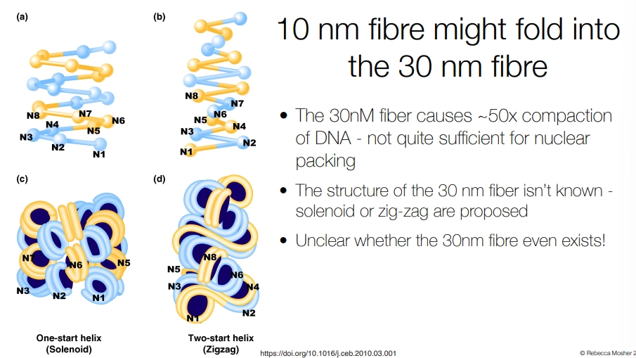

<ul><li><p><strong>nucleosomes </strong>complexes on DNA take up a ‘beads on a string’ or <strong>10nm fibre structure</strong> (when actively transcribed)</p></li><li><p>this is assembled into a <strong>30nm fibre</strong> (hypothesised by <strong>solenoid </strong>or <strong>zigzag </strong>structure)</p></li><li><p>chromatin is bound to the <strong>nuclear matrix</strong> by proteins, to make <strong>chromatin loops </strong>(unknown how)</p></li><li><p>within the interphase nucleus, the chromosomes have distinct <strong>territories </strong>because they stay bound to the matrix; they aren’t all intermixed</p></li><li><p>this is compressed down further to produce meiotic/mitotic chromosomes</p></li></ul><p></p>
3
New cards

what is heterochromatin?

  • some sections of chromatin, called heterochromatin, stain more densely/darkly than others, called euchromatin

  • these are mostly distributed at the pericentromere, but can also have occasional knobs or blobs

  • most genes are in the euchromatin, while the heterochromatin contains more ‘junk’ DNA eg. transposons, that the host doesn’t want to transcribe

  • heterochromatin is resistant to gene expression

  • we observe position effect variegation (PEV)- the position of a gene in a chromosome determines whether it is constantly or partially expressed

    • eg. the white gene in drosophila eyes causes red pigment to be produced, but when exposed to X-rays, inversions bring it into the pericentromeric heterochromatin

    • this suppresses white gene transcription, causing less red pigment to be produced = variegation

    • Su(var) genes encourage heterochromatin, decreasing transcription of the white gene so that variegation is increased (su(var)- mutation suppresses variegation)

    • E(var) genes suppress heterochromatin


<ul><li><p>some sections of chromatin, called <strong>heterochromatin</strong>, <strong>stain more densely/darkly </strong>than others, called <strong>euchromatin</strong></p></li><li><p>these are mostly distributed at the <strong>pericentromere</strong>, but can also have occasional knobs or blobs</p></li><li><p>most <strong>genes are in the euchromatin</strong>, while the heterochromatin contains more <strong>‘junk’ DNA</strong> eg. <strong>transposons</strong>, that the host doesn’t want to transcribe</p></li><li><p><strong>heterochromatin is resistant to gene expression</strong></p></li><li><p>we observe <strong>position effect variegation</strong> (PEV)- the position of a gene in a chromosome determines whether it is constantly or partially expressed</p><ul><li><p>eg. <strong>the white gene in drosophila eyes</strong> causes <strong>red </strong>pigment to be produced, but when exposed to<strong> X-rays</strong>, inversions bring it into the <strong>pericentromeric heterochromatin</strong></p></li><li><p>this <strong>suppresses </strong>white gene transcription, causing less red pigment to be produced = <strong>variegation</strong></p></li><li><p><strong>Su(var)</strong> genes <strong>encourage </strong>heterochromatin, decreasing transcription of the white gene so that<strong> variegation is increased (su(var)<sup>-</sup> mutation suppresses variegation)</strong></p></li><li><p><strong>E(var) </strong>genes <strong>suppress </strong>heterochromatin</p></li></ul></li></ul><p></p>
4
New cards

how is heterochromatin maintained, using the example of the drosophila white eye gene?

  • the H3K4 demethylase Su(var) demethylates the lysine at position 4 in histone H3, which is otherwise a chemical marker for being euchromatin, as it recruits chromatin remodelers etc. (H3K4me and H3K9me are mutually exclusive)

  • the H3K9 methyltransferase Su(var) methylates the lysine at position 9 in histone H3, which is a chemical marker for being heterochromatin

  • two heterochromatin protein 1 Su(var)s recognise and bind two adjacent nucleosomes with H3K9me modifications, dimerise, and form a binding platform for effector proteins eg. HDAC:

  • the histone deacetylase Su(var), HDAC, removes acetylation on lysine residues in histones (acetylation of positively charged lysine makes them neutral = weakened DNA ionic interactions)

  • this increased positive charge tightens bonding with histones, maintaining heterochromatin

  • maintaining heterochromatin = white+ gene transcribed less = less red pigment = more variegation

  • Su(var)- variants = decreased heterochromatin = white+ gene transcribed more = more red pigment = suppressed variegation
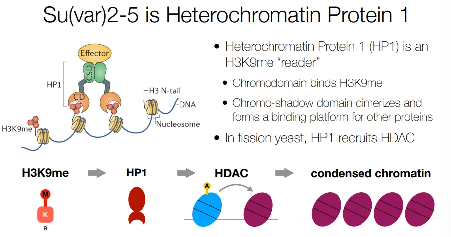

<ul><li><p><strong>the H3K4 demethylase Su(var) demethylates the lysine at position 4 in histone H3</strong>, which is <strong>otherwise </strong>a chemical marker for being euchromatin, as it recruits chromatin remodelers etc. (H3K4me and H3K9me are mutually exclusive)</p></li><li><p><strong>the H3K9 methyltransferase Su(var) methylates the lysine at position 9 in histone H3</strong>, which is a <strong>chemical marker </strong>for being <strong>heterochromatin</strong></p></li><li><p>two <strong>heterochromatin protein 1</strong> Su(var)s recognise and bind two adjacent nucleosomes with <strong>H3K9me</strong> modifications, <strong>dimerise</strong>, and form a <strong>binding platform </strong>for effector proteins eg. <strong>HDAC</strong>:</p></li><li><p>the <strong>histone deacetylase </strong>Su(var), HDAC, <strong>removes acetylation </strong>on lysine residues in histones (acetylation of positively charged lysine makes them neutral = weakened DNA ionic interactions)</p></li></ul><ul><li><p>this <strong>increased positive charge tightens bonding </strong>with histones, <strong>maintaining heterochromatin</strong></p></li><li><p>maintaining heterochromatin = white<sup>+</sup> gene transcribed less = less red pigment = more variegation</p></li><li><p>Su(var)<sup>- </sup>variants = decreased heterochromatin = white<sup>+</sup> gene transcribed more = more red pigment = suppressed variegation</p></li></ul><p></p>
5
New cards

what are the structural and maintenance differences between heterochromatin and euchromatin?

heterochromatin:

  • demethylation at H3K4

  • methylation at H3K9 is bound by heterochromatin protein 1

  • this is a binding site for histone deacetylase = condensed chromatin

  • also has abundant H1 linker protein

  • this leads to trasncription repression


euchromatin:

  • methylation at H3K4

  • demethylation at H3K9 = no HP1 binding = uncondensed chromatin

  • also has low H1 linker protein

  • this permits transcription


<p><strong>heterochromatin:</strong></p><ul><li><p>demethylation at H3K4</p></li><li><p><strong>methylation at H3K9</strong> is bound by <strong>heterochromatin protein 1</strong></p></li><li><p>this is a binding site for <strong>histone deacetylase</strong> = condensed chromatin</p></li><li><p>also has<strong> abundant H1 </strong>linker protein</p></li><li><p>this leads to trasncription repression</p></li></ul><p></p><p><strong>euchromatin:</strong></p><ul><li><p>methylation at H3K4 </p></li><li><p><strong>demethylation at H3K9</strong> = no HP1 binding = uncondensed chromatin</p></li><li><p>also has <strong>low H1 </strong>linker protein</p></li><li><p>this permits transcription</p></li></ul><p></p>
6
New cards

how is heterochromatin maintained in DNA replication?

  • when DNA is unwound to create a replication bubble, the histones are evicted

  • the H3-H4 tetramers are randomly transferred to either daughter strand (red)

  • this dilutes the heterochromatin markers on H3 by 50%

  • the newly-produced nucleosomes (grey) that are added are reinforced with markers by reader and writer domains


<ul><li><p>when DNA is <strong>unwound </strong>to create a replication bubble, the histones are <strong>evicted</strong></p></li><li><p><strong>the H3-H4 tetramers are randomly transferred to either daughter strand</strong> <em>(red)</em></p></li><li><p>this <strong>dilutes </strong>the heterochromatin markers on H3 by 50%</p></li><li><p>the <strong>newly-produced nucleosomes</strong><em> (grey)</em> that are added are <strong>reinforced </strong>with markers by <strong>reader and writer</strong> domains</p></li></ul><p></p>



