Anatomy and Physiology - Immune System
 Call Kai
 Learn
 Practice Test
 Spaced Repetition
 Match
 Flashcards
 Knowt Play
1/44

There's no tags or description
Looks like no tags are added yet.
45 Terms
Immune system
Not composed of organs, but cellular and molecular structures located throughout the body
Function: provide immunity, or protection from harm
Infectious agents (pathogens) can damage or kill a host: bacteria, viruses, fungi, protozoans, and multicellular parasites
Prokaryotic cells
Simple, small cells that lack a nuclear envelope (and so do not have a distinct nucleus); free-floating circular DNA
No membrane-bound organelles
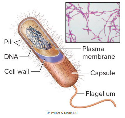
Typically have a cell wall
Prokaryotes are always unicellular
Eukaryotic cells
Complex, large cells with linear DNA stored in the nucleus
Have membrane-bound organelles
May or may not have a cell wall
Eukaryotes can be multicellular or unicellular

Bacteria (pathogen)
Prokaryotes, unicellular microbes—some have a sticky polysaccharide capsule and/or hairlike pili; some produce enzymes and/or toxins
Enclosed by a cell wall

Viruses (pathogen)
Pieces of DNA or RNA in a protein shell (capsid); some are membrane-enclosed
Viruses are not cells—they cannot reproduce by themselves and are much smaller; only about one-hundredth of a micrometer
Are obligate intracellular parasites: must enter a cell to reproduce → direct infected cell to make copies of nucleic acid and capsid → the virus or immune response may kill the host cell


Fungi (pathogen)
Eukaryotic cells with membrane and cell wall
Include molds, yeasts, multicellular fungi that produce spores
Release proteolytic enzymes, inducing inflammation


Protozoans (pathogen)
Eukaryotic cells without a cell wall that interfere with normal cellular functions
Those that cause disease are called unicellular parasites
Disease examples: malaria and trichomoniasis


Parasitic worms (pathogen)
Nonmicroscopic multicellular parasites (eukaryotes)
Take nourishment from host they live in to grow in size
Ex: tapeworm


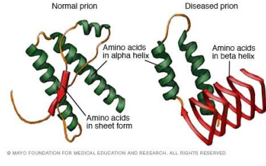
Prion
Fragments of infectious proteins
Neither cells nor viruses
Cause disease in nervous tissue
For example, Variant Creutzfeldt-Jakob disease (“mad cow disease”) can be spread from cows to humans by consuming infected meat
Immune response
Collective term for all physiologic processes of the immune system components
Can be innate or adaptive, depending on the type of immunity
Primary location of immune cells
Blood: all 5 leukocytes circulate here
Secondary lymphoid structures (ex: lymph nodes, spleen, tonsils): T-cells, B-cells, macrophages, dendritic cells, NK cells
Select organs (ex: lungs): macrophages
Skin and mucosal membranes (ex: respiratory tract): dendritic cells
Connective tissue throughout body: mast cells
Cytokines
Small proteins that regulate immune activity, produced by cells of innate and adaptive immune system
Chemical messengers released from one cell that bind to receptors of target cells: can act on cell that released it (autocrine); on local cells (paracrine); or on distant cells after circulating through blood (endocrine)
Have short biological half-life
The means of communication and weapons available to fight infection
Interleukin (cytokine)
IL-#
Regulates immune cells
Source:
T-lymphocytes
Macrophages
Endothelial cells
Other various cells
Tumor necrosis factor (cytokine)
TNF-(Greek letter)
Destroys tumor cells; may have other functions as well
Source:
T-lymphocytes
Macrophages
Mast cells
Dendritic cells
Colony-stimulating factor (cytokine)
(First letter of cell(s) it regulates)-CSF
Stimulates leukopoiesis in bone marrow to increase synthesis of a specific type (colony) of leukocytes
Source
T-lymphocytes
Monocytes
Interferon (cytokine)
IFN-(Greek letter)
“Interferes with” replication of pathogens that enter cells (intracellular pathogens)
Source:
Infected cells
NK cells
T-lymphocytes
Innate immunity
Born with this, no previous exposure, immediate response
Provided by multiple components that protect against a wide array of substances
Recognize microbes as foreign because of receptors; pattern recognition receptors (TLRs, or toll-like receptors) are proteins on immune cell surface that bind to common microbe surface patterns (PAMPS, or pathogen-associated molecular patterns)
Innate immunity components
Skin and mucosal membranes: prevent entry
Nonspecific internal defenses
Most immune cells (ex: macrophages, NK cells)
Antimicrobial proteins (ex: interferon, complement)
Physiologic responses (ex: inflammation, fever)
Adaptive immunity
Provided by lymphocytes that are activated to replicate and respond when stimulated by a specific antigen; response takes days
T-cells: cell-mediated immunity
B-cells: antibody-mediated immunity → become plasma cells: synthesize and release antibodies

Antigen
Unique molecule (such as a protein or polysaccharide) able to bind to a component of the adaptive immune system
Examples: virus capsids, cell walls of bacteria or fungi, bacterial toxins, tumor antigens of cancer cells
Lymphocytes able to distinguish foreign vs. self-antigens (those of a person’s own body)
T-lymphocytes or antibodies contact a region of the antigen called the antigenic determinant (epitope)

Immunogens
Antigens that induce an immune response (i.e., is immunogenic)
Immunogenicity affected by how foreign it is, its size, complexity, and quantity
Hapten
Small substance that attaches to another molecule to initiate an immune reaction
Too small to function as an antigen alone
Can become immunogenic when attached to a molecule in the host
May trigger an immune response, such as a hypersensitivity reaction to certain drugs or chemicals
Skin barriers (1st line of defense - preventing entry)
Few microbes can penetrate intact skin
Physical barriers of epidermis and dermis
Epidermis: keratinized stratified squamous epithelium; exfoliates to remove potential pathogens
Dermis: connective tissue contains hyaluronic acid in dermis, which limits microbe spread
Skin secretions (1st line of defense - preventing entry)
Skin releases antimicrobial substances from sweat and sebaceous glands
Sweat helps wash away microbes; contains dermcidin, lysozyme, defensins
Sebum is low in pH (acidic) and interferes with microbial growth; contains lactate and fatty acids
Mucous membranes (1st line of defense - preventing entry)
Line body openings
Produce mucus, containg antimicrobial substances (defensins, lysozyme, IgA)
Cilia in respiratory tract sweep/remove trapped microbes
GI tract utilizes saliva and stomach acid to kill ingested microbes
Urinary tract flushes microbes away
Reproductive tract contains acidic secretions
Microbiome (1st line of defense - preventing entry)
Commensal microbiota; are microorganisms that reside on body surfaces (ex: skin, mucous membranes like on GI tract)
Nonpathogenic
Interfere with attachment of potentially pathogenic organisms

Phagocytic cells (2nd line of defense - nonspecific internal defenses)
Engulf unwanted substances by phagocytosis
TLRs on cell surface bind to PAMPS
Macrophages
Neutrophils
Dendritic cells


Neutrophils and macrophages (2nd line of defense - nonspecific internal defenses)
Phagocytic; destroy engulfed particles
Intake vesicle (phagosome) fuses with lysosome = phagolysosome
Digestive enzymes break down the unwanted substances
Respiratory burst produces reactive oxygen-containing molecules that help destroy the microbes
Degraded residue is released by exocytosis
Macrophages can also perform antigen presentation

Dendritic cells (2nd line of defense - nonspecific internal defenses)
Phagocytic; destroy engulfed particles and then present fragments
Antigens are presented on dendritic cell surface to T-lymphocytes
Necessary for initiating adaptive immunity

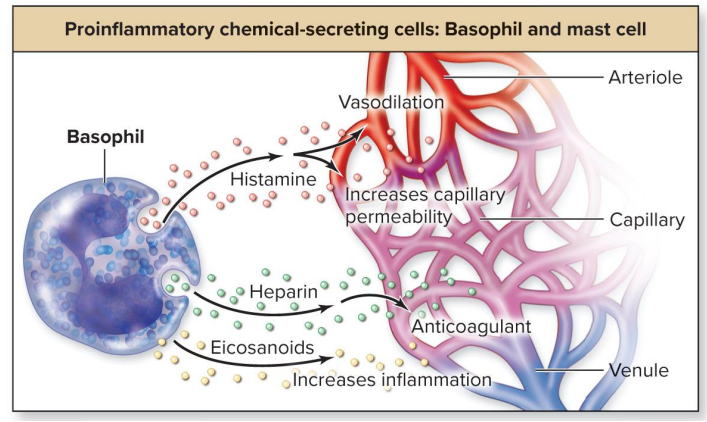
Proinflammatory chemical-secreting cells (2nd line of defense - nonspecific internal defenses)
Basophils and mast cells (resemble basophils; found in connective tissue, close to small blood vessels)
Release histamine → arteriole vasodilation and ↑ capillary permeability (classic allergy symptoms)
Release heparin → anticoagulant
Release eicosanoids (ex: leukotrienes) → ↑ venule inflammation

Apoptosis-initiating cells (2nd line of defense - nonspecific internal defenses)
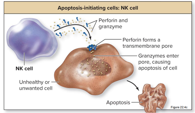
NK (natural killer) cells destroy unhealthy/unwanted cells by initiating apoptosis (programmed cell death, not pathologic death)
Form in bone marrow, circulate in blood, and accumulate in secondary lymphoid structures
Perform immune surveillance—patrol body, detect unhealthy cells


How NK cells initiate apoptosis
Release cytotoxic chemicals (perforin and granzymes)
Perforin forms a transmembrane pore (perforating the cell)
Granzymes enter cell through pores → apoptosis (shriveling)

Parasite-destroying cells (2nd line of defense - nonspecific internal defenses)
Eosinophils degranulate, releasing enzymes and other cytotoxic substances, including proteins that form transmembrane pores in parasite
Participate in immune responses of allergy and asthma
Engage in phagocytosis of antigen-antibody complexes

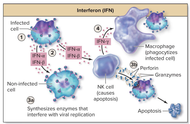
Interferons (2nd line of defense - nonspecific internal defenses)
Antimicrobial proteins
A class of cytokines that nonspecifically interferes with spread of intracellular pathogens (for example, viruses, intracellular bacteria)
IFN-α and IFN-β produced by leukocytes and virus-infected cells
Bind to neighboring cells and prevent their infection by triggering synthesis of enzymes that destroy viral nucleic acids → synthesis of viral proteins inhibited
Stimulate NK cells to destroy virus-infected cells (killing itself)
IFN-g produced by T-lymphocytes and NK cells
Stimulates macrophages to destroy virus-infected cells

Interferon pathway of a virus-infected cell
Body cell is infected with a virus
Virus-infected cell releases IFN-α and IFN-β
a. IFNs alert neighboring body cells to synthesize enzymes that interfere with viral replication, protecting them from infection
b. IFNs also stimulate NK cells to destroy virus-infected cells by apoptosis, killing the original infected cell that released them
NKs release IFN-g that stimulates macrophages to eliminate the infected cell
Complement system (2nd line of defense - nonspecific internal defenses)
Antimicrobial proteins
Group of over 30 plasma proteins identified as “C#”
Work along with (“complement”) antibodies
Synthesized by liver, continuously released in inactive form
Activation occurs by enzyme cascade
Complement activation follows pathogen entry; 3 pathways
Classical pathway (complement activation)
Antibody attaches to foreign substance, then complement binds to antibody
Alternative pathway (complement activation)
Complement binds to polysaccharides of bacterial or fungal cell wall
Mannose-binding lectin pathway (complement activation)
Lectin protein binds to carbohydrate on some microbes, and complement binds to the lectin

Complement system functions
Increases inflammation by activating various cells (mast cells, basophils, neutrophils, macrophages)
Opsonization: complement protein (opsonin, or “red flag”) binds to pathogen to enhance likelihood of phagocytosis
Eliminates immune complexes: complement links immune (antigen-antibody) complexes with erythrocytes, which are later stripped of these by macrophages within the liver and spleen
Induces cytolysis: collectively, complement proteins form a protein pore in the microbe’s plasma membrane (called membrane attack channels/MAC) → fluid enters → cell lyses/bursts


Inflammation (2nd line of defense - nonspecific internal defenses)
An immediate, local, nonspecific response of vascularized tissue to injury and infection
Release of inflammatory and chemotactic factors
Vascular changes result, including arteriole vasodilation, ↑ in capillary permeability, and stimulation of capillary endothelium of injured tissue to display cell-adhesion molecules (CAMs)/selectins
Recruitment of leukocytes through margination, diapedesis, and chemotaxis
Delivery of plasma proteins to site, including antibodies, complements, clotting proteins, kinins
Exudate is formed when increased fluid, immune cells, and proteins leave blood capillaries and enter the interstitial space, helping to “wash” the area of injurious or infectious agents

Chemotaxis (recruitment of leukocytes)
Migration of immune cells along a chemical gradient/in response to a chemical
Diapedesis (recruitment of leukocytes)
Leukocytes (amassed by margination) squeeze out thru endothelial cells of the vessel to site of infection/injury
Aided by vasodilation
Margination (recruitment of leukocytes)
CAMs on leukocytes adhere to CAMs on capillary endothelium
First neutrophils, then macrophages
Exudate (product of inflammation)
Mix of fluid, immune cells, and proteins; ex: pus
Exudate leaves the blood
Circulates through the infected/injured interstitial tissue, picking up infectious agents, dead cells, and cellular debris
Increased interstitial HP pushes it into lymphatic capillaries, becoming new lymph
Filters through lymph nodes