Treatment Planning
 Call Kai
 Learn
Learn Practice Test
 Spaced Repetition
 Match
Match Flashcards
 Knowt Play
1/23

Earn XP
Description and Tags
Name | Mastery | Learn | Test | Matching | Spaced | Call with Kai | Chat |
|---|
No analytics yet
Send a link to your students to track their progress
24 Terms
Identify the main components of a linear accelerator.
Gantry, Gantry/treatment head, Gantry stand, treatment couch/bed, Imaging devices
Describe how photons are produced within the linear accelerator.
Electrons are produced.
They are accelerated to very high speed through the wave guide.
Magnet forces electrons to change directions towards the patient.
Electrons strike a tungsten metal target where ionizing radiation (photons) are produced.
The positive nuclei in tungsten attract negatively charged electrons.
As the electrons are pulled towards the nuclei, they are forced to change direction and slow down.
The lost kinetic energy is converted into an x-ray photon.
The x-ray photons continue through the treatment head, while the electrons are absorbed within the target.
Identify the primary collimator, X and Y jaws, and multi-leaf collimators (MLCs) within the treatment head.

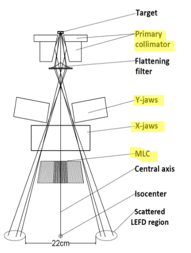
Describe the basic role of the primary collimator, jaws, and MLCs in shaping the photon beam.
Primary collimator → 1st component in the gantry head that influence the shape of the photon beam; thick piece of metal with a cone-shaped opening that allows some photons to pass though while absorbing photons travelling outside the opening (fixed for everyone)
X and Y jaws → 2nd component in the gantry head that influence the shape of the photon beam; pairs of thick rectangular metal blocks organized into 2 upper and 2 lower blocks that can move in and out to change the size of the treatment field (bigger or smaller); they trim the cone treatment field into a rectangle/square shape.
Multi-Leaf Collimator (MLCs) → final component at the bottom of the gantry head that shapes the photon beam; consists of many thin metal leaves which can move independently to shape the beam to closely match the tumour.
Define a treatment field.
The shaped area of radiation (photon beam) that is delivered to the patient.
Which feature on the gantry head allows for greater flexibility when shaping and positioning treatment fields?
The collimator can rotate independently of the gantry.
What is a field light and how does it work?
Light source located above the jaws in the gantry head that projects an outline of the treatment field onto the patient’s skin. Because it is located above the jaws, the jaws and MLCs shape the light in the shape of the treatment field.
What is an ODI and what is it for?
Optical Distance Indicator - a ruler that is projected onto the patient’s skin that allows therapists to measure the distance b/w the treatment machine and the patient’s surface.
Why is the ODI important?
Radiation therapy treatments are planned using specific geometric distances. The ODI helps verify that the patient is positioned correctly relative to the treatment machine each day.
What is SSD?
Source to Surface Distance technique - the distance from the source to the patient’s surface measured 100cm for each treatment field.
What are the limitations of the SSD technique?
Most modern rad-t treatments involve using multiple treatment fields to treat a patient —> the human body is irregularly shaped so the distance from the machine to the patient’s skin changes with every single gantry angle —> so RTs would need to reposition the patient b/w each treatment field to ensure the correct SSD is maintained.
limitations due to this:
increased treatment time
increased patient discomfort
increased risk of error
reduces clinical efficiency
What is an isocenter and why is it useful?
The point in space where the rotation axis of the gantry, collimator, and treatment couch intersect. This is designed to be 100cm. It is useful because of the Source to Axis (SAD) technique: the isocenter is placed w/in or close to the tumour so that the distance from the gantry source to the axis is 100cm no matter what gantry angle is used.
Is SSD still relevant?
Yes, the SAD (source to axis distance) technique is used for treatment planning but the SSD (source to surface distance) technique is used as a part of it to read the distance on the patient’s skin. The SSD will be smaller than 100cm and change depending on the angle.
What are lasers used for in the treatment room?
They project onto the patient’s skin which help RTs position patients accurately. 3 lasers converge at the isocenter - used as a reference.
Where are lasers placed?
Most treatment rooms contain:
ceiling laser
wall mounted lasers (one on each side of the room)
They are named after the anatomical planes: sagittal, front (coronal), and transverse (axial)
What is a control panel?
It is located on both sides of the treatment couch; it is used to position and reposition patients throughout their treatment appointment. It contains buttons that allow therapists to move the treatment couch in different directions:
vertically (up and down)
longitudinally (towards and away from the gantry)
laterally (left and right)
rationally (turning the couch)
What is a hand pendant?
The hand pendant is a detachable control device used to move the treatment couch, the gantry, and internal parts of the gantry head (same controls as treatment table) but the hand pendant is detachable, therapists can move around the room while controlling the treatment couch and gantry, while maintaining a clear view of the patient.
What do the buttons on the hand pendant represent?

Can you take images with the gantry? How? Why is it useful?
Yes, images are taken everyday on most patients, before treatment. Image panels are attached to the gantry. They can be extended into position when image is required. These images help verify the patient positioning and treatment accuracy.
What are the different types of images the panels can take?
KV Image - uses kilo voltage (kV) x-rays (lower energy x-rays) (e.g. CT); produce images with better contrast
MV Image - uses mega voltage (MV) x-rays (higher energy x-rays) (lower image quality)
How does the gantry take images for each type of imaging?
The KV uses in the imaging panels (on the left is the source and right is the detector panel - x-ray comes from source and detector gives us the image).
The MV has its source in the gantry head and the detector panel is under the treatment table - same x-ray process but way smaller amount for imaging)
Which is more advantageous and why?
MV because the image is taken from the gantry head - as this is also where the treatment comes from, you can use it to take a direct picture of the treatment field (can just use an image instead of a field light to check treatment field)
Where are the emergency stop buttons and what do they do?
There are several including one on each side of the treatment couch on the panels, on room walls, on each side of the gantry body. They immediately cut power to the machine which halts machine movement and treatment delivery. These features help protect patients and staff if an unexpected situation occurs.