BMS 300- Unit 1
 Call Kai
 Learn
Learn Practice Test
 Spaced Repetition
 Match
Match Flashcards
 Knowt Play
1/152

There's no tags or description
Looks like no tags are added yet.
Name | Mastery | Learn | Test | Matching | Spaced | Call with Kai | Chat |
|---|
No analytics yet
Send a link to your students to track their progress
153 Terms
Cellular functions take place in an
Aqueous environment
Dipole Moment
A measure of the separation of partial positive and partial negative charges within a molecule, determined by the magnitude of the charges and the distance between them.
Hydrogen Bonding in Water
The electrostatic attraction between a hydrogen atom covalently bonded to an electronegative atom (such as oxygen) and a small electronegative atom on a neighboring polar molecule.
Hydrophilic Interactions
Interactions involving polar or charged molecules that dissolve readily in water by forming ionic or hydrogen bonds with water molecules.
Hydrophobic Interactions
Interactions involving nonpolar, uncharged molecules (such as fats or oils) that do not dissolve in water due to an inability to form favorable bonds with water molecules.
Amphipathic Molecules
Molecules containing both hydrophilic (polar/charged) and hydrophobic (nonpolar) regions, such as phospholipids.
Phospholipid structure
•Modified triglyceride
•R group
•Has amphipathic properties
Micelle
Single layer lipid sphere
Liposome
Two layer liped sphere (more stable)
Integral membrane proteins can serve
–In cellular communication
–as ionic pathways
–To maintain ion concentrations
Peripheral proteins
Sit loosely on the inner or outer surfaces
How does the body extract energy from food?
–Anaerobic and aerobic pathways
–Beta-oxidation
–Deamination
Metabolism
Sum of cellular reactions required to sustain life
Our bodies extract energy (contained in the bonds between atoms of a molecule) and convert it to a form that our cells can use.
Cells extract usable energy from carbohydrate via four main pathways
•Glycolysis
•pyruvate to acetyl CoA
•the citric acid cycle
•the electron transport chain
In glycolysis how does the body decide to convert pyruvate to lactate or acetyl CoA?
When limited oxygen is avalible pyruvate is shuanted to form lactate.
When oxygen is readily avalible and energy is needed, pyruvate is converted to acetyl CoA.
Citric Acid Cycle
• Acetyl-CoA combines with oxaloacetate to form citric acid
•Oxaloacetate is regenerated
• One glucose molecule produces two acetyl-CoA molecules; the citric acid cycle will occur twice
• NADH and FADH2 deliver their cargo of high-energy electrons to the electron transport chain (ETC)
Electron transport chain
• As electrons are passed down the chain, they move from a higher to a lower energy level, releasing energy.
• Some of the energy is used to pump H+ ions out of the matrix and into the intermembrane space, establishing an electrochemical gradient.
• H+ ions flow down the gradient, powering ATP synthase
The electron transport chain (ETC) consists of a series of protein complexes & sits in the
Inner mitochondrial membrane
A drug selectively permeabilizes the inner mitochondrial membrane, allowing protons (H+) to leak freely down their electrochemical gradient into the mitochondrial matrix bypassing ATP synthase. Assuming glucose is readily available, what will be the physiological status of Glycolysis versus the Electron Transport Chain (ETC) in these cells?
Glycolysis will accelerate, and the ETC will continue to pump protons.
Carbohydrates Metabolism - Steps

Extracting Energy from Fat
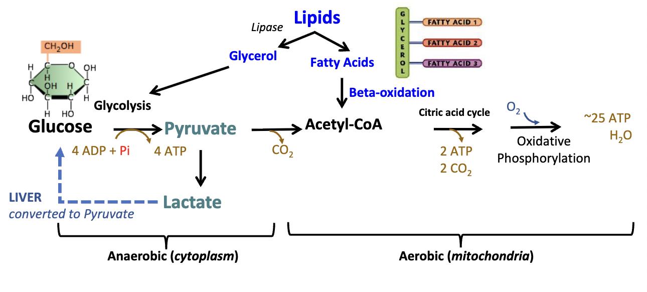
Starts with lipid digestion into glycerol and free fatty acids (FFA)
Lipids break down into glycerol (to glycolysis anaerobic in the cytoplasm) and fatty acids (beta-oxidation aerobic in the mitochondria)
Beta-oxidation of fatty acids
• Breakdown (beta-oxidation) of fatty acids takes place inside the mitochondria
• Acetyl-CoA, which enters the Krebs (TCA) cycle to generate NADH and FADH2 for transferring pairs of high-energy electrons to ETC to fuel ATP synthesis
Extracting Energy from Proteins
• During starvation, the body breaks down protein and extracts energy from the amino acids
• Deamination strips down the amino acid to a "carbon skeleton“ while producing a nitrogen byproduct that becomes urea
• The carbon skeleton structure determines where it enters the catabolic pathways
Nucleus
Contains DNA
Nuclear membrane is continuous with Endoplasmic Reticulum
Nuclear pores allow selective transport
Complex of proteins that span nuclear membrane
Orientation & Base Pairing of DNA
Strands of a DNA double helix always run in opposite directions
Antiparallel orientation
Bases-pairs interact via hydrogen bonds
A – T: two H-bonds
C – G: three H-bonds
Transcription
The process by which information in a DNA strand is copied into an mRNA
Initiated by transcription factors binding to a promoter region
What does RNA polymerase do in transcription?
Unwinds DNA
Reads template strand 3’ to 5’
Synthesizes RNA strand 5’ to 3’
Catalyzes phosphodiester bonds
mRNA is complementary to DNA sequence (U replacing T)
Post-transcriptional Modifications
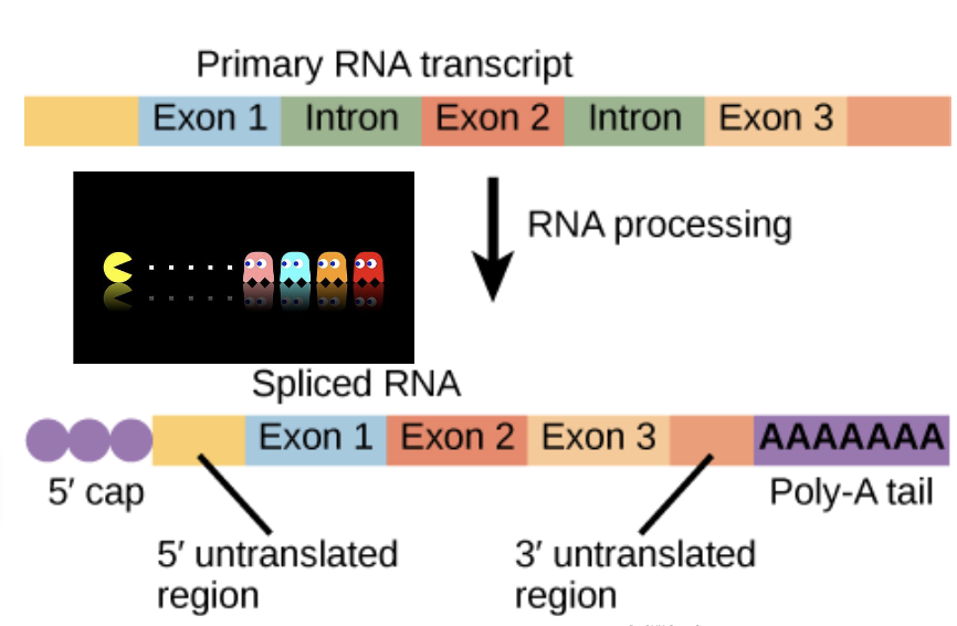
5’ Capping
Protects from 5’-3’ exonuclease degradation
Ribosome binding site
3’ poly adenylation
Protects from 3’ to 5’ exonuclease degradation
Splicing
Removes non-coding regions
Small nuclear RNAs (snRNAs) are components of the spliceosome that aid in splicing
Alternative splicing: multiple proteins are possible from one gene
RNA
RNA is single-stranded, but it can fold into secondary structures
Hydrogen bonds between bases
Complex structures that function as ribozymes – catalytic activity
Found in spliceosomes, which edit mRNA
Found in ribosomes, which aid protein synthesis
Messenger RNA (mRNA)
Forms a template for protein synthesis
Transfer RNA (tRNA)
Carries amino acids to ribosomes for protein synthesis
Brings amino acids to the ribosome for the growing polypeptide chain
Ribosomal RNA (rRNA)
Structural core of ribosomes
Small nuclear RNAs (snRNA)
Involved in processing of DNA and RNA in nuclei
Where are mRNA, tRNA, and rRNA made?
Nucleus
Where do mRNA, tRNA, and rRNA function?
Cytosol
Translation
The process by which a protein is synthesized from the information contained in a molecule of messenger RNA (mRNA)
Small Ribosomal subunit
Binds and reads mRNA
Large subunit
Docking site for tRNAs & Catalyzes peptide bonds between amino acids released from tRNAs
Aminoacyl-tRNA
Synthetase facilitates linking tRNA to new amino acid
Charging tRNA
Aminoacyl-tRNA synthetase facilitates linking a tRNA to a new amino acid
ATP cleavage provides energy to link the amino acid to tRNA via an ester bond
tRNA can drop off an amino acid at a ribosome
Post-translational Modifications (PTM)
Some are reversible and some are irreversible
Mostly addition of a group or molecule
Can also remove a group or cleave a protein
PTMs are important for proper protein functions in cells
Failure to properly modify proteins causes problems in multiple organ systems
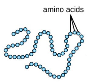
Primary Protein Structue
Sequence of a chain of amino acids
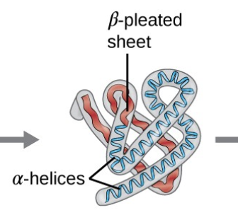
Secondary Protein Structure
Local folding of the polypeptide chain into helices or sheets

Tertiary Protein Stucture
3D folding pattern of a protein due to side chain interactions
are formed mainly by hydrophobic interactions between amino acid side chains
Quaternary Protein Structure
Protein consisting of more than one amino acid chain
Defined by the interactions between different polypeptides (subunits)

Amino Acids
Proteins are constructed of long chains of amino acids.
All amino acids contain a carboxyl group and an amino group linked by a carbon
an R group is attached to this carbon.
The R group is characteristic of each of the 20 amino acids
Some R groups are hydrophobic while others are hydrophillic

Peptide bonds
Amino acids are linked one to another by the formation of a peptide bond
Peptide bonds form when the carbon on the carboxyl group forms a covalent bond with the nitrogen in the amine group in the adjacent amino acid

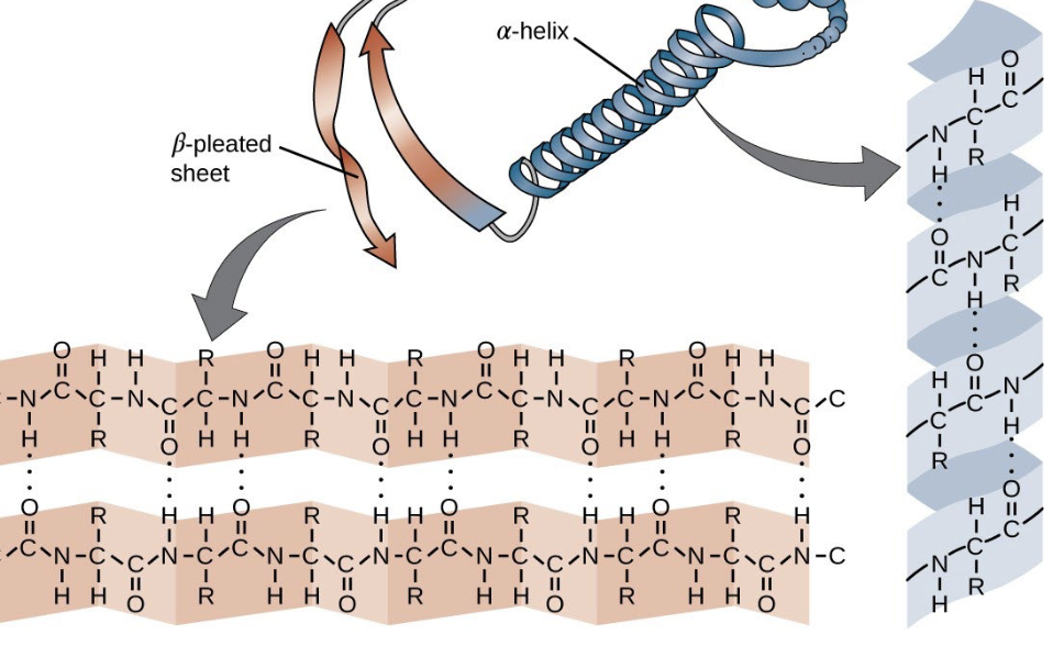
α-helices
polypeptide backbone forms a very compact corkscrew
i + 4 H bonding
Hydrogen bonding between the carbonyl oxygen (C= O) of one amino acid and the hydrogen on the amide (N—H ) of a nearby amino acid
Side chains face outward from the helix axis

β-sheets
More extended than an alpha helix
Hydrogen bonds are formed between the peptide bond C=O and N—H groups of polypeptides that lie side by side
The charge and orientation of R groups on amino acids are important in determining all of the following protein characteristics except
The secondary structure within the protein
Polypeptide backbone Determinants of Shape
• Type of Amino Acids
• Order of Amino Acids
• interactions/bonds between Amino Acids
• Environmental factors – temperature, pH, etc.
Prions
Protein Infectious Agents (PRP)
In the cell membrane, ____________________ normally form the ion
channels
Alpha Helices
Smooth Endoplasmic Reticulum
Lacks ribosomes
Location of functions including cellular detoxification (abundant in liver cells) storage of calcium ions (sarcoplasmic reticulum in muscle cells).
Rough Endoplasmic Reticulum
ER is responsible for proteostasis (protein homeostasis)
Biosynthesis, folding, maturation, stabilization, and trafficking of transmembrane and secretory proteins
The rough ER contains ribosomes on its surface
With the assistance of chaperones, nascent proteins fold and undergo other functional modifications, including glycosylation, disulfide bond formation, and oligomerization.
Properly folded and modified proteins are then packaged into vesicles to be shipped to the Golgi apparatus and other locations in the cell.
Chaperones identify improperly folded proteins and facilitate degradation in the cytosol by proteasomes
SR-SRP complex process
1. SR-SRP complex brings a hydrophobic polypeptide to the ER
SR-SRP complex leaves. The hydrophobic polypeptide is looped into the translocon, and it binds a recognition site.
The polypeptide loop pushes open the plug
The signal peptide leaves the translocon by a lateral gate, where the signal peptidase degrades it
The polypeptide is released into the ER lumen at the end of translation for PTM
Insertion of multi-pass trans- membrane proteins in RER
The signal recognition particle (SRP) and its receptor (SR) are required to initiate the translocation of the first transmembrane domain
The threading of subsequent transmembrane domains is managed by
the ribosome- translocon assembly
the hydrophobicity of the translated domain.
Golgi apparatus
• The Golgi apparatus is a major sorting and dispatch station for the products of the ER
• Vesicles enter via the cis face and are transported through membrane- enclosed cisternae.
Protein modifications include phosphorylation, glycosylation, and tags that define their cellular destinations
1. Lysosomes –contain proteolytic and degradative enzymes
2. Plasma membrane -receptors, channels, Single or multi-pass proteins
3. Extracellular fluid (Secreted proteins)- hormones, antibodies
Recap of organelles involved in the central dogma
Nucleus: DNA replication, RNA transcription, and RNA editing
Rough ER: Following cotranslational translocation, proteins are further processed
Golgi apparatus: Proteins are further processed and sent to various destinations (cell membranes, secreted out of the cell or degraded)
The presence of a signal peptide in a protein sequence indicates that it will LEAST likely be processed in the
Cytoplasm
Hydrogen bonding is NOT required for?
The primary structure of proteins
What type of bond is responsible for the dipole moment of a water molecule?
Polar bonds
Which of the following are responsible for hydrophobicity?
Nonpolar particles
What is the result of translation?
Protein
Consider the primary structure of proteins. What type of bonds connects two amino acids together?
Covalent bond
In eukaryotic cells, enzymes that form phosphodiester bonds between nucleotides function in the:
nucleus
What is the name of the motor protein associated with actin filaments?
Myosin
What is the function of glycolysis?
Breakdown of a 6-carbon glucose to two 3-carbon pyruvate molecules
Botulinum toxin is a protease that cleaves vesicular associated membrane protein (VAMP) into smaller pieces, rendering it nonfunctional. What will occur due to exposure to botulinum toxin?
Disruption of the formation of the SNARE complex
ER-associated degradation (ERAD) pathway
Degrades troubled proteins by ubiquitin- proteasome system (UPS)
Cell recognizes protein as misfolded
Protein is ubiquinated
Protein is retrotranslocated from ER cytosol to cytoplasm
Protein is degraded by proteosome
The Unfolded Protein Response(UPR)
if the ERAD cannot handle the misfolded proteins, the cell activates the unfolded protein response or UPR to adjust the protein folding pathways in the cell.
inhibit protein translation
Increase folding capacity of the ER by causing more chaperones to enter the ER
What is the last resort for ER stress due to unfolded proteins?
Autophagy
What is a Vesicle?
Enclosed lipid bilayer
Contains cytoplasm
Carries materials
Formed by budding off an existing membrane
plasma membrane
organelle membrane
How to vesicles get transported?
Through coated vesicles
COPII
From ER to Golgi
COPI
From Golgi to ER membrane
Clathrin
From the plasma membrane
• an example of receptor- mediated endocytosis
Exocytosis –SNARE Complex Steps
v-SNAREs in the vesicle bind to
t-SNARES in the target membrane
Water is squeezed from between the two membranes
Stalk formation
Hemi-fusion
Fusion

Trafficking of Vesicles and Cytoplasmic Proteins
Requires roads and vehicles
Roads are the cytoskeleton
Vehicles are the motor proteins
Cytoskeleton Intermediate filaments
Structureal only
Cytoskeleton microtubules
Structural and transport
Long-distance transport
Cytoskeleton F - actin
Structural and transport
Short-distance transport
Actin filaments
Double stranded; made of globular (G) actin
Polar
• plus end – toward membrane
• minus end – toward nucleus

Microtubules
Tube-like, made from dimers
Polar
• plus end – toward the membrane
• minus end – toward nucleus

Motor Proteins/Molecular Motors
ATPases
Bind and cleave ATP → ADP
Energy released powers movement along cytoskeleton
Motor protein types:
Kinesin
Dynein
Myosin
Kinesin moves towards the
Plus (+) end on microtubules
Dyenin moves towards the
Minus (-) end on microtubules
Myosin V moves towards the
Plus (+) end on F-actin
Myosin VI moves towards the
Minus (-) end on F-actin
How are cells connected to each other and the extra-cellular matrix?
• Gap junctions
• Tight junctions
• Anchoring junctions
How do we classify epithelial tissue?
• Cell shape
• Number of layers
Gap junction
Mediates cell-to- cell communication
They are aggregates of intercellular channels that permit direct cell–cell transfer of ions and small molecules
Connect the cytoplasm in adjacent cells
Couple cells both electrically and metabolically
Found in most cells (e.g., bone, nerves, muscles, etc.
Gap-junctional channels are composed of hexamers of integral proteins
Connexins
Isoforms
Same type of protien seen in multiple tissues
Different combinations of connexins create channels that differ in
permeability and regulation
• Regulation can be mediated by changes (pH, [Ca 2+ ] ) or signals (neurotransmitters)
• Turnover of connexons is rapid
New connexons are inserted into the plasma membrane by
Exocytosis
