Red Meat Pathology 2: Pigs
 Call Kai
 Learn
 Practice Test
 Spaced Repetition
 Match
 Flashcards
 Knowt Play
1/32

There's no tags or description
Looks like no tags are added yet.
33 Terms
Describe the appearance and features of enzootic pneumonia.

Describe the appearance of mild to very severe enzootic pneumonia in pigs.

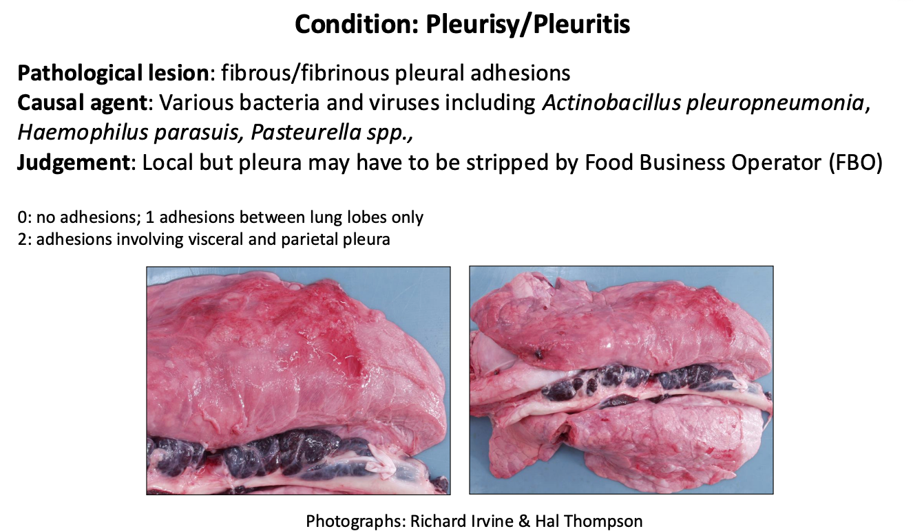
Describe the appearance of pleurisy/pleuritis.
Inflammation on surface of the pleura, can affect the thoracic wall
Describe the appearance of contagious pleuropneumonia.
Necrotic tissue inside the lobes

Describe the appearance of acute, resolving, and evolution of pleuropneumonia into an abscess.

Describe the appearance of viral pneumonia.

Describe the appearance of pyaemia.
Embolic etiology, check other tissues as likely more than one are affected
Root cause is generally from other infection throughout the body
Pyaemia in pigs (often associated with generalized abscessation) is a severe form of blood poisoning where pus-forming bacteria enter the bloodstream and travel to various internal organs, tissues, or the spine, causing multiple abscesses

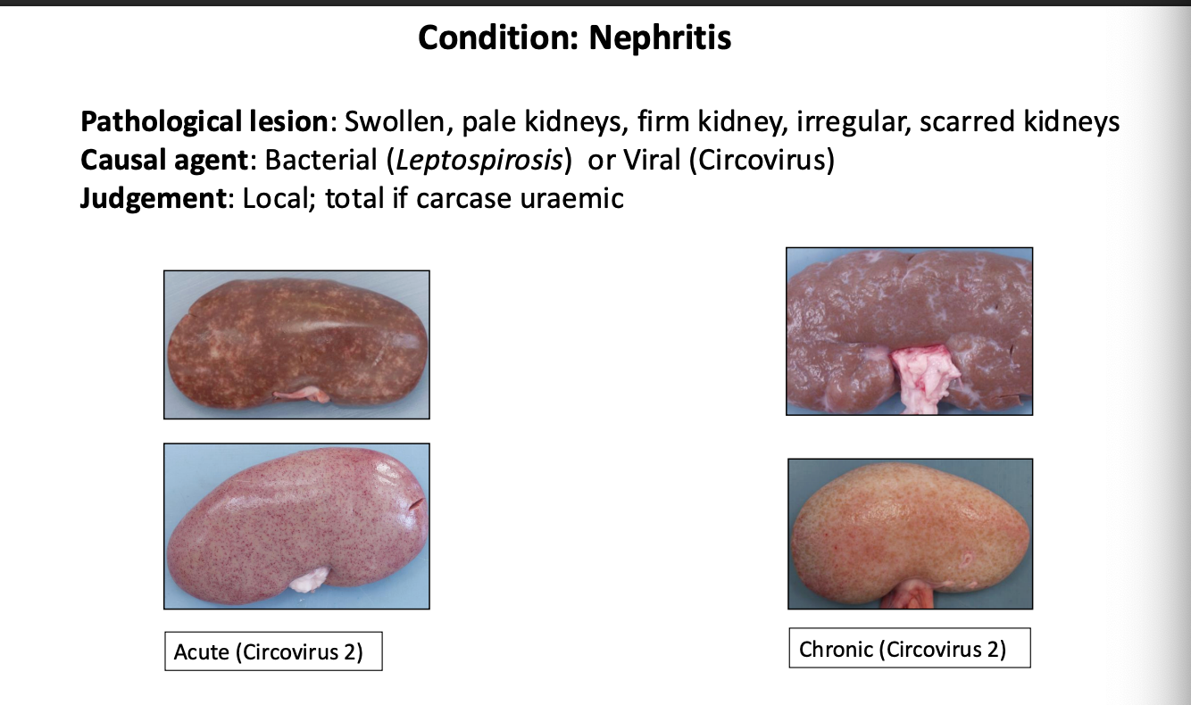
Describe the appearance of nephritis.
Describe the appearance of renal cysts.

Describe the appearance of milk spots (On liver).
• Pathological lesion: diffuse white foci invading the liver parenchyma.
• Causal agent: fibrotic tracts left by migrating Ascaris suum larvae en route to the lungs
• Judgement: local rejection

Describe the appearance of pericarditis.
Inspect other tissues as well

Describe the appearance of peritonitis.

What is Glasser’s Disease?
Glässer's disease is an infectious bacterial disease in pigs caused by Glaesserella parasuis (formerly known as Haemophilus parasuis)
Cause & Transmission: The bacterium normally lives harmlessly in the upper respiratory tract of healthy pigs. Stressful events—such as weaning, mixing groups, transport, or coinfections with viral diseases (like PRRS or swine flu)—trigger the bacteria to spread systemically
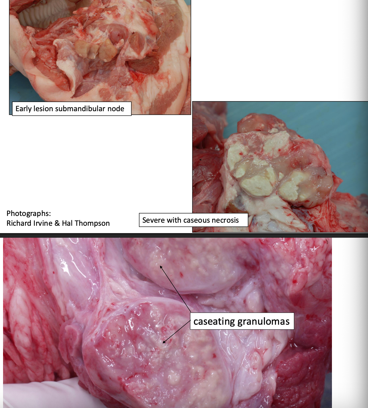
Describe the features of Granulomatour lymphadenitis.
Condition: Granulomatous lymphadenitis
Pathological lesion: Local: Single enlargement of submandibular lymph node with white fleshy granulomas visible on cut surface
Generalised: Lesions in bronchial and mesenteric lymph nodes, lung, liver, spleen
Causal agent: Mycobacterium avium and intracellulare BUT infection with Mycobacterium bovis group should be considered if outside pigs exposed to cattle or badgers
Judgement: Local; total if generalised and full inspection by DVO
Describe the TB judgement for bovine and suidae.
Article 33, MOC
Practical arrangements for official controls for tuberculosis during post-mortem inspection from ore mas take precautions 9o are the is of on tentiation or ther carcases, the slaughter line and staff present in the slaughterhouse.
All meat from animals in which post-mortem inspection has revealed localised lesions similar to tuberculoid lesions in a number of organs or a number of areas of the carcase shall be declared unfit for human consumption. However, where a tuberculoid lesion has been found in the lymph nodes of only one organ or part of the carcase, only the affected organ or part of the carcase and the associated lymph nodes shall be declared unfit for human consumption.
Describe the classic appearance of organs affected by lymphoma.
Pathological lesion: Massive enlargement of carcase lymph nodes. Splenomegaly and hepatomegaly
Causal agent: Sporadic. Lymphoma will account for 95% of tumours in slaughter pigs
Judgement: Total

Describe the pathology of Trichinella spiralis.
• Nematode Trichinella spiralis
• Zoonotic parasitic disease
• humans eating undercooked meat - especially pork, charcuterie, very important source of infection to humans
• most mammals are susceptible
• decreased incidence in last 50 years by limiting access of farmed pigs to wildlife and rodents

Describe the control measures used to prevent infection with trichinella.
• prevent ingestion by any animal of viable trichinella cysts in muscle (trichinae)
• Good management
Controlling rodents
preventing cannibalism
ie tail biting
preventing access to wildlife carcasses
• inspection of meat at slaughter
trichinoscope / digestion method
**Cannot see lesions grossly - need to use trichinoscope to digest the muscle and look at it microscopically
What is a “nurse cell” in regards to trichinella?
A nurse cell in Trichinella infection is a modified, living host skeletal muscle cell that the parasitic nematode (Trichinella spiralis and related species) transforms into a permanent "home" and life support system
Keeps the nematode alive, encased in a capsule until it is eaten to continue its lifecycle
Describe how testing for trichinella is performed?
Artificial Digestion: The muscle sample is chopped or blended and placed in a warm solution of pepsin and hydrochloric acid, which mimics mammalian gastric juices. Acid digests the muscle fibers and connective tissue, freeing the worm. Sediment is examined under a microscope to identify the worm if present.
• Groups to test
all breeding pigs (boars and sows)
pigs from any system other than controlled housing
• slaughterhouses can set up their own testing
• carcases tested day of slaughter
if trichinella not found carcases can be released
Describe the appearance of arthritis.
• 1 or 2 swollen joints = local trimming
• 3 swollen joints = Total condemnation

What are the main portals of entry for arthritis into the joint?

Describe the appearance of acute traumas in porcine carcasses.


What is the underlying pathology of tail damage and causes in pigs?

Describe the main appearance of aural haematomas.

Describe the appearance of papular dermatitis (Mange).
Degree of severity is highly variable

Describe the appearance of septicaemia.
Areas of necrosis due to underlying vascular compromise - appearing black to green

Describe the appearance of African Swine Fever.
Is there any treatment available?
The causative agent of African swine fever is the African swine fever virus (ASFV).
• Asfavirus
• No treatment available, no vaccines
• Risks related to attenuated vaccines
• are Keetection and rapid control of spread
• disease of domestic and wild pigs, whose mortality rate can reach 100%
• NOT zoonotic

Describe the appearance of aujesky’s disease - psuedorabies.
Young pigs are most susceptible - usually die after neurological symptoms
What is the national serum survey?
A national serum survey for Aujeszky’s disease (also known as pseudorabies) is a systematic blood-testing program used by countries or regions to check their pig populations for antibodies against the Aujeszky's disease virus (ADV).

Describe the appearance of emaciation.

Describe the appearance of anaemia.
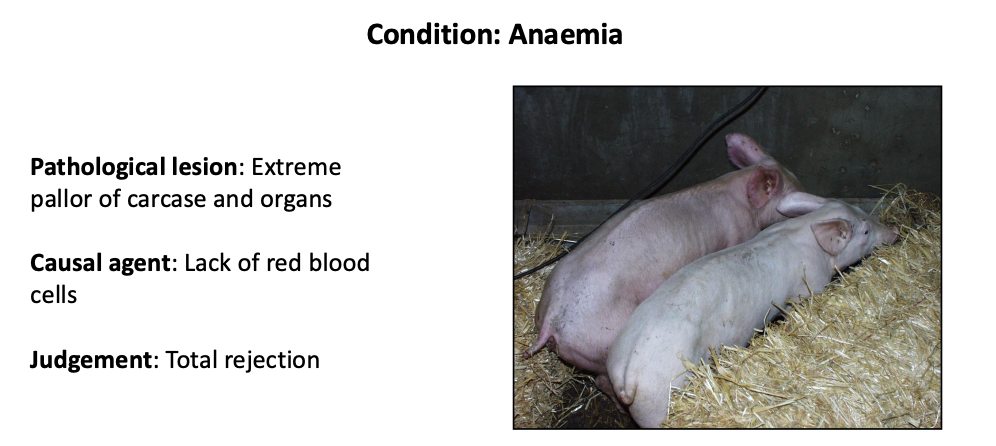
Describe the common causes of anaemia.
Pigs (Especially neonatal) are very sensitive to iron deficiency
Striking color changes with changes in iron levels
