Micro Lab Midterm Exam
 Call Kai
 Learn
 Practice Test
 Spaced Repetition
 Match
 Flashcards
 Knowt Play
1/129

There's no tags or description
Looks like no tags are added yet.
130 Terms
Aseptic technique
Preventing contamination
Culture
Medium that contains living microbes
Neutrophiles
Organisms that prefer pH levels between 5.5 and 8.5
Dichotomous
Divided into two parts
What are the 3 basic stains?
Methylene blue
crystal violet
safranin
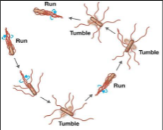
Active Movement
Independent movement over greater distances
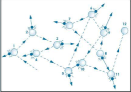
Brownian Movement
Random collisions between cells and water
Nutritional components
Types of carbon source (citrate) or sugar (glucose, Lactose, maltose, etc)
Christmas Tree Streaking
IT IS NOT AN ACTUAL CHRISTMAS TREE!!!

Inhibitors
High NaCl concentration, bile, salts, selective dyes
Differential Media
Uses biochemical and physiological properties of bacteria to identify them
Key
Consist of a series of choices that lead the user to the correct name of a given item
Flagella
- Structures used by cells to move in aqueous environments
- Act like propellers in bacteria
- Basal body “run” — Hook “tumble” — Filament “run”

Endospores
- Lack ATP
- Environmentally stable, dormant
- Found intracellularly
- Spore coat is resistant to staining
- Malachite Green

Inoculation
Transferring microorganisms into or onto a medium
Aerotolerance
The ability or inability to live in the presence of oxygen
Aerobic
Living in the presence of oxygen
Anaerobic
Living without air
What are the 2 types of media to test Aerotolerance?
- Fluid thioglycolate broth
- Trypticase soy agar deep stain
Fluid Thioglycolate Broth
Used red dye to show O2 presence
Trypticase Soy Agar Deep Stab
Uses depth of agar to provide O2 - free environment
Pure culture
One type of microorganism
Mixed culture
More than one type of microorganism
Magnification
Making an image appear bigger
Resolution
Ability to distinguish two close objects clearly

Contrast
Difference between an object and its background

Immersion oil
Because the glass microscope slide and immersion oil have the same refractive index, the oil keeps the light rays from refracting
USED ONLY IN 100X!!!
Inoculating loop
Transfer microorganisms, especially when working with liquid cultures or streaking plates
Inoculating needle
Transfers microorganisms and it is useful for inoculating agar deeps
Compound light microscope
Uses lenses and visible light to magnify specimens
Total magnification
eyepiece magnification × objective magnification
4x (red) = 40x
10x (yellow) = 100x
40x (blue) = 400x
100x (black) = 1000x
What are the 5 culture methods?
Streak plate
Spread plate
Broth
Agar slant
Agar deep
Streak plate
Separates bacteria to obtain isolated colonies

Spread plate
Spreads a sample across the agar surface

Broth
Liquid growth medium
Parts of the Microscope

Microorganisms
Are free living, they do not reside on or in a specific plant or animal host and are not known to cause diseases.
- They are nonpathogenic
- They are saprophytes and perform the important ecosystem role of decomposing organic matter
Ubiquity of Microorganisms

Liquid media
A liquid nutrient solution used to grow microorganisms. It is also called broth and does not contain solidifying agar.

Agar slant
Solid medium in a tube, with a slanted surface
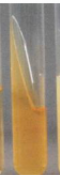
Agar deep tube
Solid medium in a tube, useful for growth patterns below the surface

Agar Plate (solid)

Semi-solid
can be either solid or liquid

What are the 8 Colony Morphology Characteristics?
1. Pigmentation
2. Size
3. Surface
4. Appearance
5. Opacity
6. Shape/Form
7. Elevation
8. Margin
Colony morphology
The visible appearance of bacterial colonies growing on solid agar
Form
Overall shape
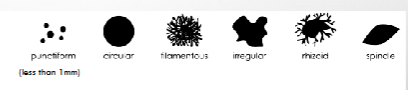
Margin
Edge of the colony
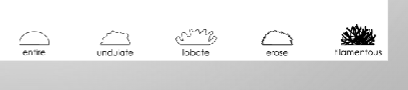
Appearance

Elevation
Height or profile

Size
How large it is

Pigmentation
Color

Surface/texture
Smooth, rough, wrinkled, etc
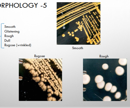
Opacity
Transparent, translucent, or opaque

Obligate aerobe
Requires oxygen, growth occurs at the top

Obligate anaerobe
Oxygen is harmful; growth occurs at the bottom

Facultative anaerobe
Can grow with or without oxygen; growth occurs throughout, usually heavier at the top

Aerotolerant anaerobe
Does not use oxygen for respiration but tolerates it'; growth is fairly uniform
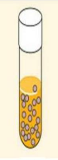
Microaerophile
Requires oxygen at a lower concentration than atmospheric oxygen; growth forms a band below the surface
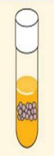
Why do we stain bacteria?
Most bacteria are nearly colorless, so they are difficult to see clearly under a microscope. Staining increases contrast and helps you observe their shape and arrangement.
Simple stain
- Uses a positively charged basic dye.
- Colors the bacterial cells.
- Examples: methylene blue, crystal violet, safranin.
- Commonly uses heat fixation.
Hans Christian Gram (1884)

Negative stain
- Colors the background rather than the cells.
- Cells appear clear or lightly colored against a dark background.
- Usually air-dried, not heat-fixed.
- Helps preserve the natural appearance of cells.

Cocci
Round cells

Bacilli
Rod-shaped cells

Spirilla/spirochetes
Spiral-shaped cells

Diplo-
Pairs

Strepto-
Chains

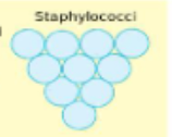
Staphylo-
Clusters
Tetrads
Groups of four

Streptobacilli
Chains of rods

Monotrichous

Lophotrichous

Amphitrichious

Peritrichous

What are the 6 steps to Heat Fixation?
1. Prepare the smear.
2. Allow it to air-dry.
3. Heat-fix the smear.
4. Apply the stain.
5. Rinse and dry as directed.
6. Observe under the microscope.
Crystal violet
Stains all peptidoglycan cell walls
Gram’s iodine
- Modorant
- Fixes crystal violet to cell walls
Ethanol (decolorizer)
Removes the purple stain from Gram-negative cells when used correctly.
Safranin
- Counterstain
- Stains unstained cell walls
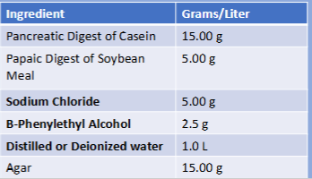
Phenylethyl Alcohol Agar (gram positive)
inhibits growth of gram negative and some gram positive
Gram-positive
- PURPLE
- Thick peptidoglycan layer retains the crystal violet–iodine complex.

Gram-negative
- PINK
- Loses the purple complex during decolorization and takes up safranin.

Mobility
An organism's ability to move independently.
True motility
Cells move actively in a direction and may change direction.
Brownian motion
Random, jiggling movement caused by collisions with surrounding molecules. It is not true motility.
Capsule stain
Capsule surrounding a cell
Remember: Clear halo around the cell
Flagella stain
Flagella used for movement
Remember: Makes thin flagella visible
Endospore stain
Resistant endospores
Remember: In the common Schaeffer–Fulton method, spores appear green and vegetative cells pink/red
Selective medium
Allows certain organisms to grow while inhibiting others.
Differential medium
Shows visible differences between organisms based on their reactions.
PEA agar
- Type: Selective.
- Inhibits many Gram-negative bacteria.
- Helps select for Gram-positive bacteria.
- It is not presented as a differential medium in your slides.
Mannitol Salt Agar (gram positive)
- Type: Selective and differential.
- High salt selects for salt-tolerant organisms.
- Mannitol fermentation produces acid, turning the medium yellow.
- Yellow = mannitol fermentation.
- Staphylococcus aureus is a classic positive example.

Bile Esculin Agar (gram positive)
- Bile helps select for bile-tolerant organisms.
- Tests for esculin hydrolysis.
- Dark brown or black = positive.
- Commonly used to help identify Group D streptococci and enterococci.

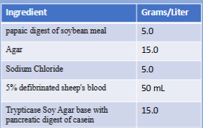
Blood agar
- Alpha hemolysis: Partial breakdown; greenish zone.
- Beta hemolysis: Complete breakdown; clear zone.
- Gamma hemolysis: No hemolysis; no clearing.
Starch agar
- Tests whether bacteria hydrolyze starch.
- Iodine is added after incubation.
- Clear halo around growth = positive starch hydrolysis.

Staphylococcus epidermidis (Gram-positive)
Gram-positive cocci in clusters; typically mannitol-negative; gamma hemolysis

Staphylococcus aureus (Gram-positive)
Gram-positive cocci in clusters; mannitol fermentation; beta hemolysis

Catalase test
2H2O2 —— 2H20+O2
Positive: Bubbles appear
Negative: No bubbles appear
MacConkey Agar
Type: Selective and differential.
- Bile salts and crystal violet inhibit most Gram-positive bacteria.
- Differentiates bacteria based on lactose fermentation.
- Pink/red colonies = lactose-positive.
- Colorless or pale colonies = lactose-negative.
- Example: E. coli typically forms pink colonies.

EMB Agar (Eosin Methylene Blue)
Type: Selective and differential.
- Eosin Y and methylene blue inhibit most Gram-positive bacteria.
- Differentiates lactose fermenters.
- Metallic green sheen: Strong acid production from lactose fermentation, classically E. coli.
- Other lactose fermenters may appear pink or purple.
- Non-lactose fermenters are usually pale or colorless.

Triple Sugar Iron Agar (TSI)
TSI differentiates bacteria by glucose, lactose, and sucrose fermentation, sulfur reduction, and gas production.
