BIO 223 A&P I LAB PRACTICAL 1
 Call Kai
 Learn
Learn Practice Test
 Spaced Repetition
 Match
Match Flashcards
 Knowt Play
1/48

There's no tags or description
Looks like no tags are added yet.
Name | Mastery | Learn | Test | Matching | Spaced | Call with Kai | Chat |
|---|
No analytics yet
Send a link to your students to track their progress
49 Terms
What is the general chemical equation for an enzymatic reaction?
S + E → ES → P + E
Where S is a substrate, E is an enzyme, ES is enzyme-substrate complex, and P is product.
What is the specific chemical equation for the catalase enzymatic reaction?
2H2O2 + catalase → H2O2 / catalase complex → 2H2O + O2 + catalase
In the reaction catalyzed by catalase, what molecule acts as the substrate?
Hydrogen peroxide (H2O2)
What are the products formed when catalase decomposes hydrogen peroxide?
Water (H2O) and Oxygen gas (O2)
Because the enzyme (E) appears on both sides of an enzymatic reaction equation, what does it imply about the enzyme?
Enzymes are not used up or permanently changed during the reaction, they can be reused.
What primary cellular function does the enzyme catalase perform in living organisms?
It detoxifies hydrogen peroxide (H2O2), a toxic metabolic byproduct, by breaking it down into harmless water and oxygen.
How was the rate of oxygen (O2) production experimentally measured in the catalase lab?
By recording the time (in seconds) it takes for a catalase-soaked paper disc to float to the surface of a 1% hydrogen peroxide solution due to oxygen bubbles accumulating on the paper.
How does paper disc surface time relate to catalase enzyme activity?
A faster float activity (faster rate of O2 production), whereas a shower float time (or not movement) indicates reduced activity.
What happens to the molecular structure of catalase when placed in an extreme acidic environment like pH 2.
The enzyme denatures (unfolds/unwinds), altering the active site shape so the substrate (H2O2) can no longer properly bind.
Predict the experimental outcome if catalase is boiled (100oC) prior to adding it to hydrogen peroxide.
The enzyme denatures completely, stopping O2 production (0 rate). The filter disc will remain at the bottom of the beaker and fail to surface.
Based on lab experimental findings, what is the optimal pH and optimal temperature for catalase activity?
Optimal pH= 7
Optimal Temperature = 37∘C
Why are 7 pH and 37∘C the optimal operating conditions for catalase?
They mimic the native internal environment of the human body (blood/cytoplasm), where catalase naturally functions at peak efficiency.
Define a solution in terms of its components.
A homogeneous mixture composed of a solvent (dissolving medium, present in greatest amount) and a solute (substance dissolved, present in lesser amount).
What defines passive transport in cellular processes?
Movement of substances down a concentration gradient without cellular energy (ATP) input.
Define diffusion.
The net passive movement of solute particles from an area of high concentration to an area of low concentration.
Define osmosis.
The passive diffusion of water (solvent) across a semipermeable membrane toward an area of higher solute concentration.

What happens to red blood cells (RBCs) when placed in an isotonic solution, and why?
The cells maintain their normal, stable shape. Solute concentrations inside and outside the cell are equal, resulting in equal, constant movement of water in and out.

What happens to red blood cells placed in a hypertonic solution, and what is this process called?
Water moves out of the cell into the higher solute concentration, causing the cell to shrink and look spiky. This process is called crenation.

What happens to red blood cells placed in a hypotonic solution, and what is this process called?
Water moves into the cell toward the higher intracellular solute concentration, causing the cell to swell and burst. This process is called lysis (or hemolysis).
Which reagent is used to test for the presence of glucose, and what procedure must be followed?
Reagent: Benedict's Solution.
Procedure: Add 5 mL test fluid and Benedict's drops to a test tube, then boil in a water bath for 1 minute.

Describe a positive versus a negative result for Benedict's glucose test.
Positive: Solution changes color from green to yellow+, orange++, or deep red/rusty+++ precipitate.
Negative: Solution stays original blue.

Which chemical reagent tests for Sodium Chloride (NaCl), and what indicates a positive result?
Reagent: Silver Nitrate (AgNO3).
Positive result: Formation of a cloudy white precipitate.

Which reagent tests for starch, and what indicates a positive result?
Reagent: Lugol's Iodine.
Positive result: Appearance of a dark blue or black color.

In the cell permeability dialysis sac experiments, what property determines whether a solute can cross the membrane?
The relative molecular size (diameter) of the solute particle compared to the pore size of the semipermeable membrane.
Order the three tested solutes—Glucose, Starch, and NaCl—from largest to smallest molecular size based on dialysis diffusion results.
Starch > Glucose > NaCl
Largest: Starch (unable to diffuse out of sac)
Middle: Glucose (diffuses slowly)
Smallest: NaCl (diffuses rapidly)
In Experiment 1 (40% glucose in sac, distilled water in beaker), why did the sac gain weight over 45 minutes?
Distilled water moved passively into the sac via osmosis because the hypertonic glucose solution inside the sac created a higher solute concentration.
Why was starch detected inside the dialysis sac after 45 minutes, but never in the beaker water?
Starch molecules are too large to pass through the microscopic pores of the semipermeable dialysis membrane.
What are the four primary tissue types found in the human body?
Epithelial (covering/lining)
Connective (support/protection)
Muscle (movement)
Nerve (communication/control)
List three hallmark functional or structural characteristics of Epithelial Tissue.
High cellularity (mostly cells joined by junctions)
Avascular (no direct blood vessels; nourished by underlying connective tissue)
Innervated
Polarity (apical vs. basal surfaces)
Rapid regeneration
How are epithelial tissues classified according to number of cell layers?
Simple: A single cell layer thick
Stratified: Two or more cell layers thick
How are epithelial cells classified according to cell shape?
Squamous: Flattened, scale-like cells
Cuboidal: Cube- or square-shaped cells
Columnar: Tall, column-shaped cells

What are the key structural features and anatomical locations of Simple Squamous Epithelium?
Features: Single layer of flat, scale-like cells with central nuclei.
Functions: Rapid diffusion and filtration.
Locations: Air sacs of lungs (alveoli), kidney glomeruli, lining of blood vessels (endothelium).
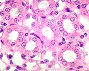
What are the structural characteristics and primary anatomical location of Simple Cuboidal Epithelium?
Features: Single layer of cube-like cells with large, spherical central nuclei.
Key Indicator: Forms clear rings/tubules with round central lumens.
Location: Commonly found in kidney tubules and small gland ducts.
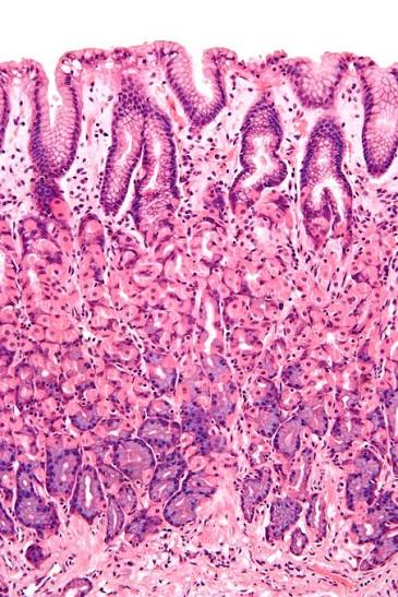
What are the key identifying features and primary anatomical location of Simple Columnar Epithelium?
Features: Single layer of tall cells with oval nuclei lined up in a row near the basal surface.
Key Indicator: Large open space in the middle.
Locations: Lining of the gastrointestinal (GI) tract (stomach, small intestine, large intestine).

What are the identifying features of Pseudostratified Ciliated Columnar Epithelium?
Features: Single layer of cells of varying heights (nuclei at different levels, creating a false stratified look); contains cilia on the apical surface and mucus-secreting goblet cells.
Where is Pseudostratified Ciliated Columnar Epithelium primarily located in the human body?
Lining the human trachea and upper respiratory tract.
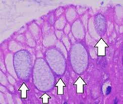
What is the function of goblet cells within columnar and pseudostratified epithelia?
To synthesize and secrete mucus, which traps foreign particles and lubricates mucosal surfaces.

What structural feature distinguishes Stratified Squamous Epithelium from simple squamous epithelium?
Multiple layers of cells, with flattened squamous cells at the superficial apical layer and cuboidal/columnar cells at the basal layer.
How do keratinized and non-keratinized stratified squamous epithelia differ in location and structure?
Keratinized: Outer layers are dead, filled with keratin protein, and lack nuclei; forms skin epidermis.
Non-keratinized: Surface cells remain alive and retain nuclei; lines moist cavities (esophagus, mouth, virginia).

What are the unique identifying features and location of Transitional Epithelium?
Features: Stratified epithelium with dome-shaped "umbrella cells" at the apical surface that flatten when stretched.
Location: Urinary system (urinary bladder, ureters).
What are the main components that make up Connective Tissue?
Extracellular Matrix: Composed of ground substance and fibers (collagen, elastic, reticular).
Cells: Fibroblasts, chondrocytes, osteocytes, etc.
List the two major subclasses of Connective Tissue Proper.
Loose Connective Tissue: Areolar, Adipose, Reticular
Dense Connective Tissue: Regular, Irregular, Elastic

What are the structural features and identifying indicators of Adipose Loose Connective Tissue?
Features: Large empty-looking cells ("white blobs") where a central lipid vacuole pushes the single nucleus to the outer membrane margin.
Indicator: One nucleus per vacuole/fat droplet.

How can you distinguish Adipose Connective Tissue from Simple Squamous Epithelium under a microscope?
Adipose tissue shows large fat vacuoles with sparse nuclei pushed to cell edges (one nucleus per vacuole), whereas simple squamous shows many closely packed dots/nuclei across a flat sheet.

What are the key structural components visible in Areolar Loose Connective Tissue?
A soft gel-like matrix containing all three fiber types (thick pink collagen fibers, thin dark elastic fibers, reticular fibers) and scattered fibroblast nuclei.

How do Dense Regular and Dense Irregular connective tissues differ in fiber orientation?
Dense Regular: Parallel collagen fibers oriented in the same direction (tendons, ligaments).
Dense Irregular: Interwoven collagen fibers oriented in different/multiple directions (dermis of skin).

What are the identifying features and locations of Hyaline Cartilage?
Features: Amorphous, glassy, firm matrix with scattered chondrocytes in lacunae; fibers are not obvious.
Locations: Tracheal rings, costal cartilages of ribs, nose, embryonic skeleton.

What is the key microscopic visual indicator for Elastic Cartilage?
Matrix filled with very prominent, dark, "hairy" elastic fibers surrounding closely packed chondrocytes in lacunae (found in external ear/pinna, epiglottis).

What are the structural features and locations of Fibrocartilage?
Features: Matrix filled with thick, parallel, wavy collagen fibers with rows of chondrocytes in lacunae.
Locations: Intervertebral discs, pubic symphysis, knee joint menisci.