10 Cardiac 1,2, and 3
 Call Kai
 Learn
Learn Practice Test
 Spaced Repetition
 Match
Match Flashcards
 Knowt Play
1/72

Earn XP
Description and Tags
W4 S.Lahmers
Name | Mastery | Learn | Test | Matching | Spaced | Call with Kai | Chat |
|---|
No analytics yet
Send a link to your students to track their progress
73 Terms
What is the function of the cardiovascular system?
circulation of blood to deliver what the cell/tissue/body needs and remove what the cell/tissue/body doesn't need
What are arteries?
they deliver blood from the heart to the tissues
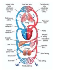
What are capillaries?
where the exchange occurs; the business end of things
they pick up waste and deliver oxygen (AKA where all the functions are happening)

What are veins?
They return blood to the heart
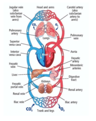
What is the heart?
central pump that moves blood forward
Just pumps spontaneously
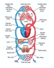
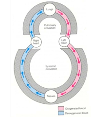
What occurs in pulmonary circulation?
Deoxygenated blood returns to the right heart to take up oxygen and drop off carbon dioxide (at the pulmonary capillaries)
right heart --> pulmonary artery --> pulmonary capillaries --> pulmonary veins --> left heart

What occurs in systemic circulation?
oxygenated blood leaves the left heart to go to systemic circulation to deliver oxygen to tissues and take up carbon dioxide (at the tissue capillary bed)
left heart --> aorta --> systemic capillaries --> systemic veins --> back to heart

True/false- all arteries carry oxygenated blood
FALSE- all arteries carry blood AWAY from the heart... it has nothing to do with oxygenation

What are the main components of the heart?
1. vena cava
2. 4 chambers
3. pulmonary artery/aorta
What is the vena cava?
the venous return to the heart
it carries deoxygenated blood
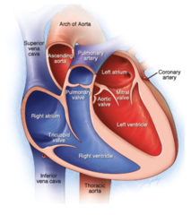
What are the four chambers of the heart?
2 atria: conduit and booster filling functions
2 ventricles: pumping to pulmonary and systemic circulations

What do the pulmonary artery and aorta do?
provide arterial delivery to circulations

true/false- atrial walls are thicker than ventricular walls
FALSE- ventricular walls are ticker than atrial walls
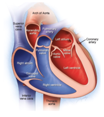
True/false- the right ventricular wall is thinner than the left ventricular wall
TRUE- the left ventricle pumps blood to the systemic circulation = higher pressure!!!

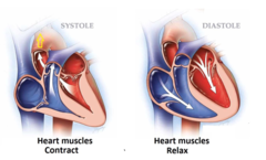
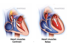
What is systole?
When the ventricles contract

What is diastole?
When the ventricles relax and fill with blood
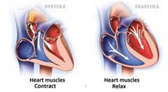
Does systole of the atria and ventricles occur at the same time or sequentially?
Sequentially
The valves in the heart are _________________
UNIDIRECTIONAL
What are the atrioventricular valves?
They separate the atria from the ventricles and open for ventricular filling (diastole)

What are the two AV valves?
Tricuspid (right AV valve) and mitral valve (left AV valve)

What are the semilunar valves?
they separate the ventricle from the great vessel and open during ventricular contraction (systole) to deliver blood to the pulmonary and systemic circulations

What are the two semilunar valves?
pulmonic and aortic

Which valves are open/closed for systole?
OPEN = pulmonic and aortic semilunar
CLOSED = tricuspid and mitral valves

What valves are open/closed for diastole?
OPEN = tricuspid and mitral valves
CLOSED = pulmonic and aortic semilunar valves
spontaneous depolarization at the ______________________ initiates events that results in mechanical pumping
sinus node

What are the two types of muscle cells (Myocytes) present in the heart?
1. specialized cardiac myocytes
2. working cardiac muscle myocytes
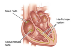
What do specialized cardiac myocytes do?
They make up the conduction system which controls the spread of depolarization through the heart. This depolarization initiates contraction
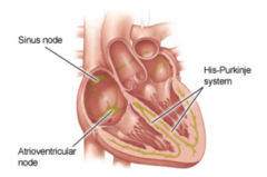
What do the working cardiac muscle myocytes do?
they are responsible for muscle contraction and "pumping" of blood and they make up most of the atria and ventricles

What are the steps of the cardiac cycle?
1. sinus node depolarization initiates the cardiac cycle
2. atrial depolarization and contraction enhances ventricular filling (diastole)
3. AV node (delay)- a pause between atrial depolarization to optimize ventricular filling before ventricular pumping starts
4. ventricular depolarization and contraction results in ventricular pumping (systole)

What is the purpose of the delay at the AV node?
the delay allow for sequential contraction of the atria and ventricles

What would happen if there was a blockage in the right heart?
-it wouldn't be able to pick up oxygen at the lungs
-there would be less flow to the systemic circulation
-less going FWD and more backing up = CONGESTION
Are right and left ventricles contracting at the same time or sequentially?
At the same time- both atria and both ventricles contract at the same time
True/false- blood flows from low pressure to high pressure
FALSE- blood flows from region of higher pressure to region of lower pressure

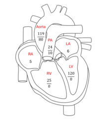
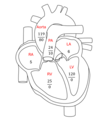
What is the pressure in the right atrium?
5 mmHg

What is the pressure in the right ventricle?
25/0 mmHg

What is the pressure in the pulmonary artery?
24/10 mmHg
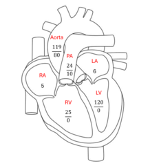
What is the pressure in the left atrium?
6 mmHg
What is the pressure in the left ventricle?
120/0 mmHg

What is the pressure in the aorta?
119/80 mmHg
systolic pressure is always _____________ than diastolic
HIGHER

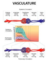
The systemic circulation is a ________________ pressure system than the pulmonary circulation
HIGHER

arteries have ____________ pressure than veins
HIGHER
What causes the unidirectional vales to open?
when the pressure in one chamber exceeds that of the other (ex. pressure in the ventricles exceeds that in the pulmonary artery and aorta during systole. The pressure in the atria is greater than the ventricles during diastole)
what part of the vasculature provides the greatest resistance?
arterioles

true/false- there are no valves in arteries
TRUE
How are veins able to get blood back to the heart when they have such a low pressure?
Veins have unidirectional valves that help favor forward blood flow
What does Ohm's law tell us about the cardiovascular system?
The cardiac output (Q) (mL/min) is directly related to the pressure between two points and is inversely related to resistance
note: *R = resistance = systemic vascular resistance = SVR = peripheral vascular resistance = PV

What characteristic of the vasculature helps to maintain diastolic pressure (AKA how can you have an intermittent pump with uninterrupted blood flow)
When the heart pumps blood into the arteries during systole (the contraction phase),
the elastic walls of the arteries expand to accommodate the surge of blood.
This expansion stores potential energy in the arterial walls
During diastole (the relaxation phase), the heart is not actively pumping blood.
The elastic arteries then recoil, releasing the stored energy.
This recoil helps to maintain a continuous pressure on the blood, ensuring that it keeps flowing even when the heart is at rest

what sensors are present in the cardiovascular system and what do they do?
baroreceptors "sense" blood pressure and chemoreceptors (pH, oxygen, and carbon dioxide) "sense" the tissue needs

What are the effectors of the cardiovascular system and what do they do?
1. sympathetic nervous system (fight or flight)
2. parasympathetic nervous system (rest and digest)
3. local and humoral modulators
They modulate heart rate, cardiac muscle contraction, blood flow, vascular resistance, and blood pressure
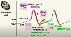
How does the sinus node spontaneously depolarize?
pacemaker cells in the sinus node do not have a resting phase.
These cells begin to depolarize automatically after an action potential ends.

what are the internodal pathways?
Transfer impulses from the SA node throughout the atria to the AV node

What are the interatrial pathways
a set of specialized muscle fibers that connect the right and left atria of the heart

how is the sinus node always depolarizing?
Due to the inflow of positive ions into specialized myocytes
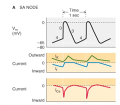
how does depolarization of the sinus node occur?
When the membrane potential reaches the threshold potential, the cell rapidly depolarizes.
This is due to the opening of calcium channels, which allow calcium to flow into the cell.
The cell repolarizes by closing calcium channels and opening up potassium channels that cause the K+ to flow out of the cell.

What is the main role of the SA node?
it sets the heart rate

what is the main role of the AV node?
it allows for the sequential atrial then ventricular contraction (AKA it creates the pause from atria to ventricles)

What is the main role of the internodal pathways/purkinje fibers?
rapid spread of depolarization for near simultaneous contraction of the chamber
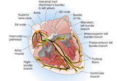
What is the difference in the structures of working myocytes and specialized myocytes?
working myocytes are striated with a central nucleus, intercalated disc. They have myosin and action = SARCOMERE
specialized myocytes don't have sarcomeres (THEY DON"T CONTRACT)
What important structure do specialized myocytes have?
GAP JUNCTIONS
they have increased gap junctions for transmission of impulse except for AV node
which has reduced gap junctions to slow transmission of impulse
(AKA the AV node does not have a lot of gap junctions because it is it's job to slow down the transmission)
What is the fastest component of the conduction system of the heart?
purkinje fibers- you need the ventricles to contract at the same time

What is the second fastest component of the cardiovascular conduction system?
atrial internodal pathways (need atria to contract at the same time (there are less myocytes to depolarize so they don't have to be quite as fast as the purkinje fibers)

What is the slowest component of the conduction system of the heart?
AV node (obviously....)

Explain excitation-contraction coupling?
The coupling activity of specialized myocytes with working myocytes
Specialized cardiomyocytes initiate and propagate the wave of cellular depolarization that starts at the SA node (excitation)
Working myocytes contract (contraction) in response to cellular depolarization

Which comes first in the heart, electrical or mechanical activity?
Electrical!!!
What is the functional significance in the difference in action potential of SA node and ventricular myocyte?
The action potential in the SA node is characterized by a slow depolarization phase (Phase 4), which is due to the gradual influx of calcium (Ca2+) ions.
This slow depolarization is crucial for the rhythmic generation of impulses
The action potential in ventricular myocytes has a rapid depolarization phase (Phase 0) due to a fast influx of sodium ions, followed by a plateau phase (Phase 2) where calcium ions enter the cell.
This plateau phase is essential for the prolonged contraction of the ventricles

What does the P wave represent in an ECG?
atrial depolarization
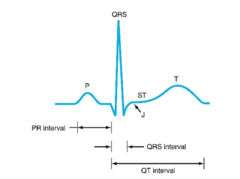
What does the flat line before the QRS wave represent in an ECG?
AV node delay
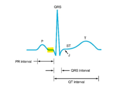
what does the QRS complex represent in an ECG?
ventricular depolarization

What does the ST wave represent in an ECG?
end of QRS (it is not flat in animals)

what does the T wave represent in a ECG?
ventricular repolarization

What are the phases of a ventricular action potential?
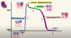
What are the phases of the sinoatrial node potential?