Unit 4, Chapter 7A-7C: Pathogens and the Innate Immune System
 Call Kai
 Learn
 Practice Test
 Spaced Repetition
 Match
 Flashcards
 Knowt Play
1/61

62 Terms
Define Pathogens
Pathogens are agents that cause disease.
Define Antigens and their Role with the Immune System
Antigens are molecules that interact with the immune system and are able to trigger an immune response. They act as markers that signal to the immune system whether or not something is foreign, and can be used by immune cells to identify specific pathogens.
Define Self-Antigens
Self-antigens are located on the surface of cells and mark the cells of an organism as ‘self’ so that the immune system does not attack them.
Define Major Histocompatibility Markers (MHC) Markers
NOTE: MHC markers is an accepted abbreviation
MHC markers are a group of proteins present on the surface of all self-cells in vertebrates, that enables the immune system to distinguish them from non-self material.
NOTE: red blood cells are an example of non-nucleated cells
Define MHC Class I Markers
MHC Class I markers are expressed on all nucleated cells in the body. They mark cells as ‘self’ so that the immune system doesn’t attack them, and differ between individuals.
Define MHC Class II Markers
MHC Class II Markers are expressed on antigen-presenting cells that allow them to present foreign antigens to T helper cells, initiating immune responses.
Define Non-Self Antigens
Non-self antigens are molecule that are recognised as foreign by the immune system - not belonging to an individual - and initiate an immune response when detected.
Define an Autoimmune Disease
An autoimmune disease occurs when errors in the immune system result in the recognition of self-antigens as non-self, which initiated as immune response against self cells.
Define Allergens
Allergens are non-pathogen antigens that trigger an immune response referred to as allergic reaction.
Define an Allergic Reaction
An allergic reaction is an unwarranted overreaction of the immune system to a non-pathogen antigen (allergen).
Explain how Red Blood Cells are Labelled as Self
As non-nucleated cells, red blood cells have glycoproteins to label them as self instead of MHC markers. Type A blood cells have A antigens, Type B cells have B antigens, Type AB have both, and type O have neither A or B antigens.
Define a Cellular Pathogen
A pathogen that has a cellular structure and exhibits the proceeses of a living organism.
Define Non-Cellular Pathogens
A pathogen that neither has a cellular structure nor exhibits the processes of a living organism. They cannot replicate on their own, typically requiring a host cell.
Differentiate Intracellular and Extracellular Threats
While both threats can interfere with cell functioning, intracellular threats are theats found within a cell such as viruses or non-pathogenic threats such as DNA mutations, while extracellular threats are founf outside of a cell, such as bacteria.
Identify 6 Pathogen Types and Whether they are Cellular on Non-Cellular
Bacteria (cellular)
Fungi (cellular)
Prions (non-cellular)
Protozoa (cellular)
Viruses (non-cellular)
Worms (cellular)
Define Bacteria as Pathogens
Bacteria are unicellular prokaryotes that can infect almost any part of the body and can cause disease through the production of toxins and enzymes which either affect cell functioning or kill them. They reproduce asexually (binary fission).
Define Fungi as Pathogens
Fungi are eukaryotes that include yeasts and moulds and contain long branching filaments (hyphae), which help them absorb nutrients. They reproduce both asexually and sexually via spores.
Define Prions as Pathogens
Prions are abnormally folded proteins only found in mammals that have the ability to induce normal proteins to become misfolded. They only affect the brain and other neural structures.
NOTE: they are currently the only known infectious agents that don’t contain nucleic acids!
Define Protozoa as Pathogens
Protozoa are single-celled eukaryotes that can be free-living or parasitic. They have many different mechanisms of action and reproduce both sexually and asexually.
Define Viruses as Pathogens
Viruses are non-cellular infectious agents composed of genetic material inside a protein coat (capsid) which is sometimes surrounded by a lipid envelope. They are unable to independently reproduce and rely on inserting genetic material into host cells to replicate.
Define Worms as Pathogens
Worms are multicellular invertebrate parasites which vary in length and size. They reproduce sexually via a complex life cycle (including egg, larval and adult stages).
Briefly Outline the Innate Immune System
The innate immune system is a component of the immune system that is composed of generalised and non-specific defences and/or responses to pathogens. It is includes the first and second lines of defence, both of which respond quickly and non-specifically.
Define the First Line of Defence
The first line of defence is a component of the innate immune system characterised by the presence of non-specific physical, chemical and microbiological barriers to keep pathogens out of the host.
Define Physical Barriers
Physical barriers are a component of the first line of defence that features solid or fluid obstacles that aim to block pathogen entry.
Define Chemical Barriers
Chemical barriers are a component of the first line of defence that features the use of enzymes, toxins and acids to protect against pathogen invasion by inhibiting their growth or development and/or destroying them.
Define Microbiological Barriers
Microbiological barriers are a component of the first line of defence in which the presence of normal flora limits the growth of pathogenic bacteria by taking up space and resources.
Identify the Components of the Immune System in Plants
Plants rely exclusively on the inntae immune system, prioritising the prevention of pathogen entry over responding to infection. They have physical and chemical barriers.
List 5 Physical Barriers in Plants
Thick bark
Waxy cuticles of leaves
Formation of galls
Presence of thorns and trichomes (small surface ‘hairs’)
Closing of stomata
Explain how Waxy Cuticles Deter Pathogens
Waxy cuticles are waxy protective films covering the surface of a plant leaf that act as a hydrophobic barrier. They can repel pathogens and prevent standing water from pooling on leaves and providing breeding grounds for certain pathogens.
Explain how Galls Respond to Pathogens
Galls are abnormal outgrowths of tissue in plants designed to isolate/trap and limit the spread of an invading pathogen.
List 3 Physical Barriers in Animals
Intact skin between external and internal environments
Mucous secretions in the respiratory tract
Cilia in the respiratory tract
Explain how Cilia deters Pathogens
Cilia are thin hair-like projections that protrude from eukaryotic cells. In the respiratory tract, they sweep pathogens away from airways and into the throat where they are swallowed and destroyed by the gastrointestinal tract.
List 5 Chemical Barriers in Animals
Lysozymes in tears and saliva
Acidic sweat
Stomach acid
Antibacterial compounds in earwax
Low pH in the vagina
Describe the Microbiological Barriers in Animals
Microbiological barriers refer to the presence of normal flora on the skin, in the lower gastrointestinal tract and in the vagina. This naturally occuring bacteria outcompetes pathogenic bacteria by taking up space and resources, defending these areas from bacterial infections.
Define the Second Line of Defence
The second line of defence is a component of the innate immune system characterised by the non-specific and immediate repsonse to injury or pathogens that have entered the body to prevent the spread of infection.
Define Leukocytes
Leukocytes are white blood cells, and are responsible for protecting the body against invading pathogens and foreign material. They make up the cellular components of the second line of defence.
List the 6 Cellular Components of the Second Line of Defence
Eosinophil
Mast cells
Neutrophil
Macrophages
Dendritic cells
Natural killer cells
Define Phagocytes and Name 3
Phagocytes are cells that engage in phagocytosis, a process in which they consume and destroy foreign or dead material by engulfing them in vesicles and breaking them down with lysozymes. Macrophages, dendritic cells and neutrophil are phagocytes.
Define Macrophages
Macrophages are a type of leukocyte found in the body that engage in the phagocytosis of pathogens, foreign material and cell debris, the release of cytokines, and antigen presentation.
Define Neutrophil
Neutrophil is the most common type of leukocyte in the body. They engage in the phagocytosis of pathogens, foreign material and cell debris, as well as the release of cytokines.
Define Dendritic Cells
Dendritic cells are a type of leukocyte found in the body that engage in the phagocytosis of pathogens, foreign material and cell debris, the release of cytokines, and antigen presentation.
Define Cytokines
Cytokines are signalling molecules released by certain cells which aid in communication between immune cells, helping to protect against pathogens.
Define Antigen-Presenting Cells and Name 2
Antigen-presenting cells are a phagocytes that display antigens from consumed pathogens on their surface MHC Class II markers and interact with the adaptive immune system. Dendritic cells and macrophages are antigen-presenting cells.
Define Natural Killer Cells
Natural killer cells are large granulated cells which recognise and destroy abnormal or virally infected cells.
Define the Killer Inhibitory Receptor of Natural Killer Cells
The killer inhibitory receptor examines the surface of cells for MHC Class I markers, checking if they have sufficient numbers of markers.
Define the Killer Activation Receptor of Natural Killer Cells
The killer activation receptor binds to certain molecules which appear on cell undergoing cellular stress.
Explain how Natural Killer Cells Destroy Abnormal Cells
If the killer inhibitory receptor detects a sufficient number of MHC Class I markers, then it overrides the killer activation receptor, preventing cell death.
When an infected or abnormal cell has missing MHC Class I markers, the killer inhibitory receptor is unable to bind to a sufficient number of markers and the killer activation receptor is activated. The natural killer cell then releases granules to target and kill the cell via apoptosis or lysis.
Define Mast Cells
Mast cells reside in connective tissues throughout the body and are responsible for releasing histamines during allergic and inflammatory responses. This occurs when they detect injury to surrounding cells or are stimulated by antigens or allergens.
Define Histamine
Histamine is a chemical messenger released by mast cells that plays a key role in inflammatory responses.
Define Eosinophils
Eosinophils are large granulated cells containing various toxic chemical mediators which help destroy invading pathogens by degranulating on contact. They typically target pathogens which are too large to be phagocytosed.
Define Interferons
Interferons are cytokines released by virally infected cells that increase the viral resistance of uninfected neighbouring cells. This helps prevent the spread of infection.
Define Complement Proteins
Complement proteins are a number of different types of proteins found in the blood that opsonise, cause lysis and attract phagocytes to invading pathogens.
Describe Opsonisation with Complement Proteins
Complement proteins attach to the surface of pathogens, making them easier to recognise as foreign and phagocytose.

Describe Chemotaxis with Complement Proteins
Complement proteins gather near a pathogen and attract phagocytes to it, making it more likely to be destroyed.

Describe how Complement Proteins cause Lysis of Cells
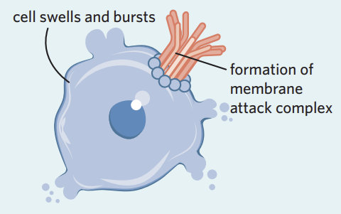
Complement proteins can join together on the surface of pathogens, forming a membrane attack complex which creates pores in the cell membrane. This destroys the pathogen by causing lysis via the sudden influx of fluid.
Outline Fever as an Innate Immune Response
A fever is a temporary increase in body temperature following a complex series of responses that result in raising the set temperature point of the body. This is an innate response to potential infection as many pathogens cannot survive the elevated temperatures.
Identify 4 Aims of the Inflammatory Response
Eliminate the effects of an injury
Defend against potential pathogens
Clear out cells that may be damaged or destroyed
Initiate repair
Identify 4 Symptoms of the Inflammatory Response
Swelling
Pain
Redness
Heat
Outline the First Stage of the Inflammatory Response
HINT: Initiation
In response to injury and/or introduced bacteria, mast cells in the tissue release histamine
Macrophages activate in the tissue to release cytokines
Damaged cells also release cytokines
Outline the Second Stage of the Inflammatory Response
HINT: Vasodilation
Histamine triggers vasodilation by binding to receptors of nearby blood vessels
This increases blood flow to the site
The formation of gaps in the vessel wall increase its permeability for immune cells
Outline the Third Stage of the Inflammatory Response
HINT: Migration
Vasodilation and gaps in the blood vessel walls allow for a number of immune system components to enter the site from the bloodstream
Phagocytes are guided by cytokines
Complement proteins are attracted to pathogens
Summarise the Stages of the Inflammatory Response
