MMED 8115 exam 3
 Call Kai
 Learn
 Practice Test
 Spaced Repetition
 Match
 Flashcards
 Knowt Play
1/394

There's no tags or description
Looks like no tags are added yet.
395 Terms
What is the circulation system composed of?
heart, blood vessels and blood
What are the circulatory systems of blood?
systemic and pulmonary
What does the systemic circulation carry blood between?
heart and organ systems
What/where does the systemic circulation carry in the blood in terms of CO2 and O2?
moves O2 to tissues and CO2 back to heart
Which circulatory circulation system maintains appropriate blood pressure?
systemic circulation
Where does the pulmonary circulation carry blood?
between heart and lungs
what is the pulmonary circulation relation to O2/CO2 in the blood?
moves O2 from inhaled air into blood and removes CO2 from blood and releases it to the atmosphere
Roles of circulatory system
transports O2, CO2, nutrients, waste products, hormones, excess heat; and regulates blood pressure
how many chambers is the heart?
four
How does the two-sided pump in the heart function?
in parallel
Direction of blood through the heart
deoxyngenated blood from the body enters the right atrium through the vena cava, then moves to the right ventricle, then gets pump to the lungs in the pulmonary artery to exchange CO2 for O2, then moves through the pulmonary vein to the left atrium, then moves to the left ventricle, then moves the oxygenated blood to the body through the aorta

What is the black pointing to?
pulmonary artery

What is the dark blue pointing to?
deoxygenated blood from the body

What is the dark green pointing to?
vena cava

What is the light green box?
right atrium

What is the pink box?
right ventricle

What is the dark purple box?
lungs

Ehat is the light purple box?
left atrium

What is the light blue box?
left ventricle

What is the brown pointing to?
Atrial septum

What is the dark pink pointing to?
venticular septum

What is the grey pointing to?
pulmonary vein
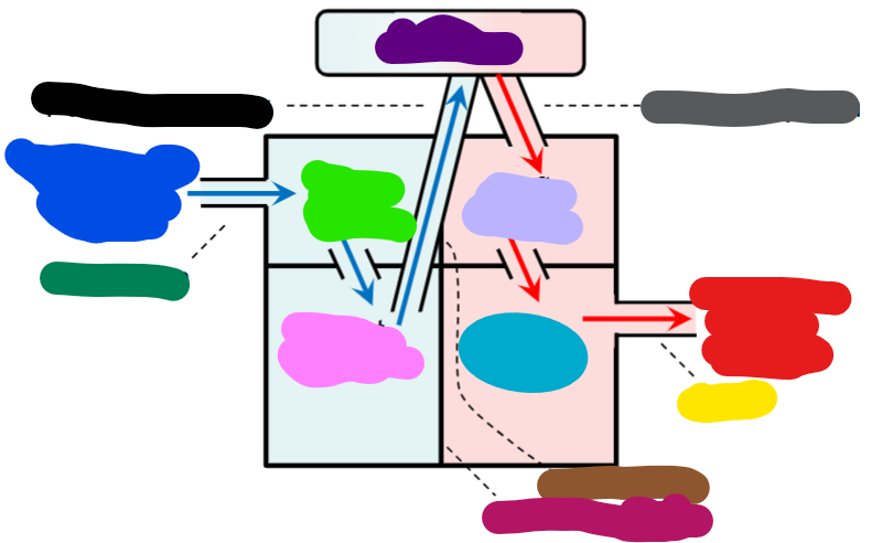
What is the red indicating?
oxygenated blood to the body

What is the yellow pointing to?
aorta
What heart chambers does deoxygenated blood pass through?
right atrium and right ventricle
What heart chambers does the oxygenated blood pass through?
left atrium and left atrium
arteries
carries blood to other organs
veins
carries blood to heart from other organs
base of the heart
top part of the heart above left and right atrium
Superor vena cava
returns blood from head and upper limbs
right pulmonary veins
returns blood from right lung
pulmonary semiluar valve
valve between right ventricle and pulmonary arteries
right atrioventricular valve
valve between right atrium and right ventricle
inferior vena cava
returns blood from trunk and legs
left pulmonary veins
returns blood from left lung
aortic semilunar valve
valve between left ventricle and aorta
left atrioventicular valve
valve between left atrium and left ventricle
Ventricular septum
separates right and left ventricles
apex of heart
bottom of heart (below the ventricles)
full name of right trioventricular valve
right atrioventricular ticuspid valve
full name of left atrioventricular valve
left atrioventricular mitral/bicuspid valve
Which ventricular muscles are thicker/stronger?
left
Why are the left ventricular mucsles thicker/stronger?
left pumps blood out into the arota against a much higher systemic blood pressure and the right pumps blood to pulmonary circulation with a lowr pressure
pericarditis
inflamation/viral
cardiac tamponade
fluid in cavity/trama

What is the yellow box pointing to?
fibrous pericardium

What is the red box pointing to?
parietal pericardium

What is the dark pink/red box pointing to?
pericardial cavity

What is the grey box pointing to?
visceral pericardium

What is the green box pointing to?
pericardium

What is the blue box pointing to?
mycoardium
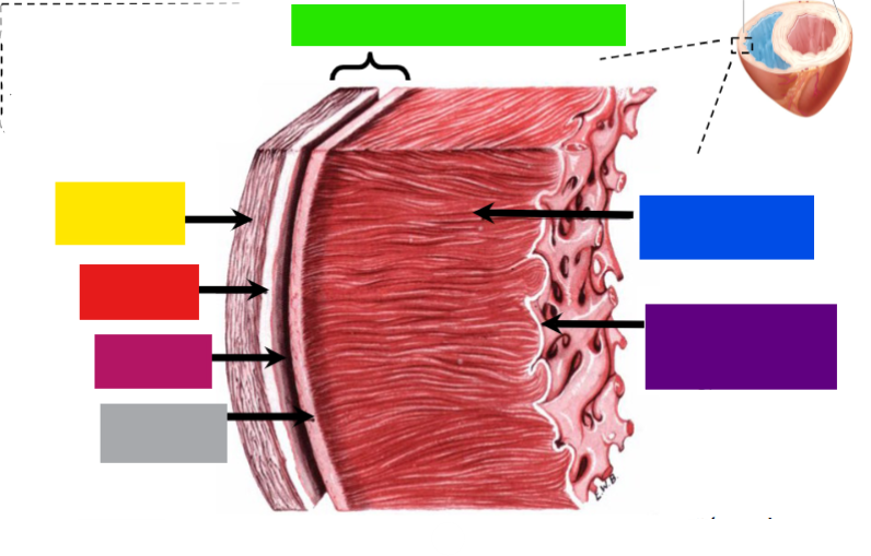
What is the purple box pointing to?
endocardium
What is the pericardium of the heart?
the heart sac
myocardium
cardiac muslce layer/tissue
myocardial infaraction
heart attack; happens when a part of the myocardium fails to receive enough oxygen
situs inversus totalis
left-right inverted organs
Where is the heart beat felt the most by hand (point of maximal impulse (PMI))?
near apex, on the mid-clavicular line
Which direction do pressure-operated valves allow blood flow?
only one direction; from chambers of higher pressure to thos of lower pressure
What happens to atrioventricular valves when the ventricles relax ?
blood enters the atria, pushing the atrioventricular valve cusps down ino the ventricles, opening the valves
What happens to atrioventricular valves when the ventricles contract?
blood presses up against the atrioventricular valve cusp, forcing the valves closed. Contraction of the papillary muscles tightens the chordae tendineae, preventing the valve cusps from being pushed into the atria

What is in the purple box?
atrioventricular valve cusp

What is the green box?
chordae tendineae
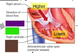
What is the brown box?
pulpillary muscle
Does the chordae tendineae pull the valve open?
no, they serve as an anchor to prevent eversion of valve during ventricular contraction
What happens to semilunar valves when ventriculars contract?
blood presses up against the semilunar valve cusps, forcing the valves open and allowing blood to flow into the aorta and pulmonary artery
What happens to the semilunar valves when the ventricles to relax?
the blood in the aorta and pulmonary artery presses down against the valve cusps, forcing them to close
Properties of “cardiac skeleton”
composed of rings of dense connective tissue around 4 valves; forms a single spatial plane that contains 4 valves
roles of “cardiac skeleton”
anchors valves; anchors atrial muscles above and ventricular muscles below; acts as electrical insulator between atria and ventricles
what is the electrical role of “cardiac skeleton” important for?
controlling the timing and direction of cardiac muscle contractions
Echocardiogram
real-time view of heart, using ultrasound
Heart valve diseases
stenosis, regurgitation, atresia
stenosis (heart valve disease)
the valve flaps/leaflets become thick or stiff and possibly fuse together; results in a narrowed valve opening and reduced blood flow through the valve
regurgitation (heart valve disease)
the valve flaps don’t close properly, causing blood to leak backward in heart; this commonly occurs due to valve flaps bulging back (prolapse)
prolapse
valve flaps bulging back
Atresia (heart valve disease)
the valve isn’t formed and a solid sheet of tissue blocks the blood flow between the heart chambers
directional waves in the heart involve what?
electrical excited (surface) and muslce contraction
Directional waves of electrical ecitation and muscle contraction order
from top of atrium to its bottom; intermettent period (delay); from the bottom of the ventricle to its top
Which heart chambers contract simultaneously?
left and right atria contract simultaneously; left and right ventricles contract simultaneously
Cardiac functions made possible by directional waves of electrical excitation and muscle contraction
atria push blood downward to ventricles; delay is necessary to fill ventricles with blood; ventricles push the blood upeard to either pulmonary artery or aorta
What do irregular electrical activities lead to?
abnormal contractions; in extreme cases, ventricles twitch uselessly, leading to sudden cardiac death
ventricular fibrillation
ventricles twitching uselessly
Intercalated discs
specialized structures that allow cardiac muscle cells to be joined end-to-end
What are the two main structures of intercalated discs?
desmosomes and gap junctions
desmosomes
a type of adherence junction which holds the cells together
gap junctions
areas of low electrical resistance that allow action potentials to spread from cell to cell
functional syncytium
if an action potential is initiated in any cell, it will spread throughout the connected muslce cells
initiation of contraction in cardiac muscle
myogenic/pacemaker potentials (has the ability to initiate its own muscle contractions)
What happens once electrical activity is initiated?
will spread throughout the myocardium; but cardiac skeleton electrically separates artia and ventricles and creates two subgroups of syncytia
what of the two subgroups of syncytia in the heart?
atrial and ventricular; which contract sequentially with a delay
What is the atrio-ventricular node (AV node)?
a single, narrow connecting path between atrial and ventricular syncytia
What does the AV node allow for?
atrial excitation conduction to ventricles with a delay
What is the cardiac conduction system composed of?
cardiac pacemakers
cardiac pacemakers
specialized myocardial cells that are devoted to generation and conduction of auto-rhythmic, pacemaker potentials
Where are pacemaker potentials initiated and where does it spread to?
initiated in the SA node in the right atrium and spreads to the left atrium
What is normally the only way for a depolarization wave to get to the atria to the ventricles?
the AV node (due to the insulating cardiac skeleton)
AV nodal delay
conduction through AV node is slow, which creates a delay between atria and ventricles
wave of depolarization from the AV node
does not spread uniformly over the venricular myocardium
wave of depolarization from the AV nodes pathway
spreads down the ventricular septum through the Bundle of His, left and right bundle branches; then spreads from apex upward through the Purkinje fibers
what do Purkinje fibers enable?
ventricular contraction beginning primarily at the apex and pushing the blood upward toward pulmonary and arotic valves

what is the brown box?
SA node