spore stain
 Call Kai
 Learn
Learn Practice Test
 Spaced Repetition
 Match
Match Flashcards
 Knowt Play
1/17

There's no tags or description
Looks like no tags are added yet.
Name | Mastery | Learn | Test | Matching | Spaced | Call with Kai | Chat |
|---|
No analytics yet
Send a link to your students to track their progress
18 Terms
The endospore stain is a differential stain used to distinguish ….
the presence of endospores in bacteria. Certain gram positive bacteria from the genera Bacillus and Clostridium are able to produce endospores in response to hostile environmental conditions (lack of nutrients, heat, cold, radiation, etc.).
The formation of these resting bodies is a protective mechanism and is not a means of reproduction. In the proper environment, an endospore will germinate to produce a new vegetative cell, which can grow and reproduce.
The formation of what resting bodies is a protective mechanism and is not a means of reproduction.
endospores
In the proper environment, an endospore will germinate to produce a new …..
vegetative cell, which can grow and reproduce.
The endospore first develops within the vegetative cell when nutrients such as carbon and nitrogen become limiting. Once the endospore develops…
the vegetative cell disintegrates, resulting in a free spore.
____ are not metabolically active and, due to the thick outer covering made of protein, are highly resistant to desiccation, extreme temperatures, and toxic chemical agents such as disinfectants, acids, and staining reagents .
Spores
To penetrate the spore with the primary stain, malachite green, what must be applied?
vigorous heating
. Once the spore is stained, decolorizer will not easily remove the stain. Any vegetative cells present will decolorize easily with water and will be counterstained with safranin. The endospore stain is differential and results in a preparation containing red vegetative cells and green spores.
What are the results of spore staining?
red vegetative cells and green spores.

Image of the spore stain results
Image of the spore stain results

The position and size of the endospore within the vegetative bacterial cell are important diagnostic features. See Figure 2 for examples of endospore locations and shapes.
The position and size of the endospore within the vegetative bacterial cell are important diagnostic features. See Figure 2 for examples of endospore locations and shapes.
Spore Staining Procedure
1. Prepare a heat-fixed smear of Bacillus megaterium in the center of a clean slide.
2. Place a square piece of blotting paper slightly smaller than your slide over your smear.
3. Saturate the smear with malachite green.
4. Heat the smear by placing the paper-covered slide on a slide warmer for 10 minutes. The heat provided is necessary to drive the stain into the spore. DO NOT ALLOW THE PAPER ON THE SLIDE TO DRY. Add additional stain as needed. DO NOT BOIL THE STAIN ON THE SLIDE.
Note: Make sure you use forceps to remove your slide from the slide warmer, and NEVER your fingers after steaming.
5. Once you have finished heating your slide, use a pair of forceps to place it on the slide rack over a staining tray and allow it to cool. Remove the blotting paper with forceps or a strong stream of water and dispose of the filter paper in the trash.
Note: All subsequent steps should be performed over a staining dish.
6. Gently rinse the slide with water using your squeeze bottle to remove excess stain.
7. Counterstain with safranin for at least one minute.
8. Rinse thoroughly with water and then gently blot the smear dry with bibulous paper.
9. Examine the finished slide under the oil immersion lens on your microscope. On your slide, vegetative cells will stain red and the spores will appear green from the malachite green stain that is trapped inside of the various layers, coating the spores. Remember, the location of an endospore can also be used as a means of identification.
10. do a drawing to indicate which cells are positive or negative for the presence of endospores. Indicate the location of the endospores with respect to the cell and any swelling of the cell walls.
The staining reagents used and the order they are used in
malachite green, safranin
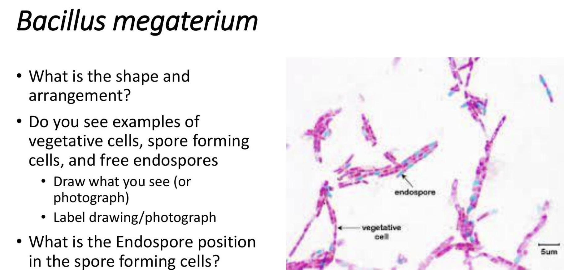
bacillus megaterium
image of bacillus megaterium
What common errors could occur during the staining technique and how they would affect the final results
ALLOWING THE PAPER ON THE SLIDE TO DRY.
BOILING THE STAIN ON THE SLIDE.
forgetting to use the coutnerstain= everything is green
What is the purpose of the malachite green in the spore stain technique?
Stains endospores green if present
Which staining reagent is used to stain the vegetative parts of the cells in the spore stain?
Safranin
How would spore-forming cells from the genus Bacillus, appear if you performed a Gram stain instead of a Spore stain on them when observed under oil immersion?
Vegetative cells would be purple, Endospores would be colorless
Why is heating the smear with malachite green on the slide required for a successful Spore stain
To force the malachite green through the thick walls of the endospores.
What possible errors could have happened during the endospore stain if the endospores inside the spore forming cells and the free endospores have remained colorless?
Possible errors that resulted in colorless endospores inside the spore forming cells and the free endospores could be not heating the slide long enough to allow the malachite green penetrate the spore, or simply forgetting to use the malachite green could be another error.