Test 3 Anatomy
 Call Kai
 Learn
Learn Practice Test
 Spaced Repetition
 Match
Match Flashcards
 Knowt Play
1/119

There's no tags or description
Looks like no tags are added yet.
Name | Mastery | Learn | Test | Matching | Spaced | Call with Kai | Chat |
|---|
No analytics yet
Send a link to your students to track their progress
120 Terms
thoracic mammae
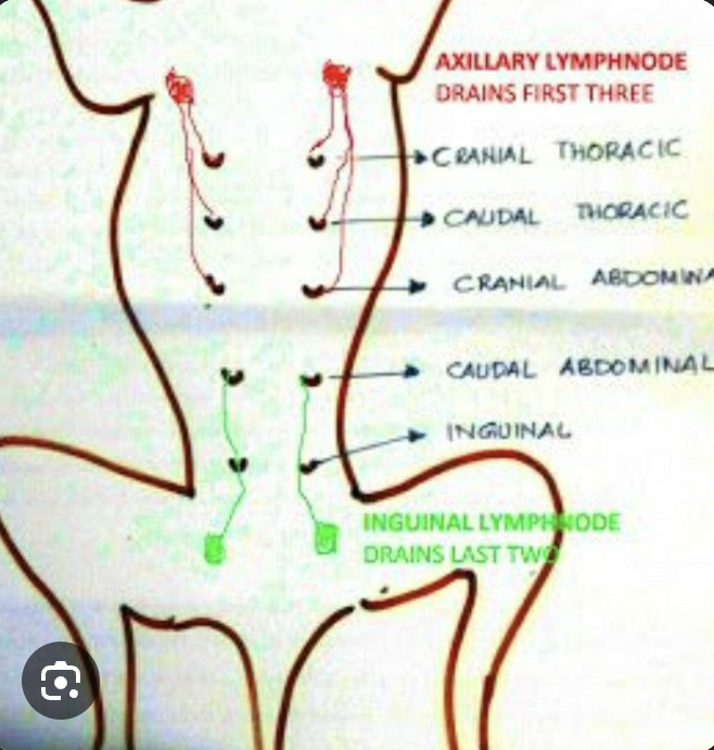
abdominal mammae

inguinal mammae

Clinical note: Cats have how many mammae
4 pairs, with cranial thoracic being absent
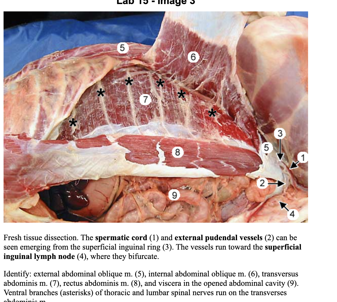
external abdominal oblique muscle (EAO)
1

superficial inguinal ring
arrow

vaginal process (females)
arrow


external pudendal vessels
2
inguinal ligament
??
internal abdominal oblique muscle (IAO)
6
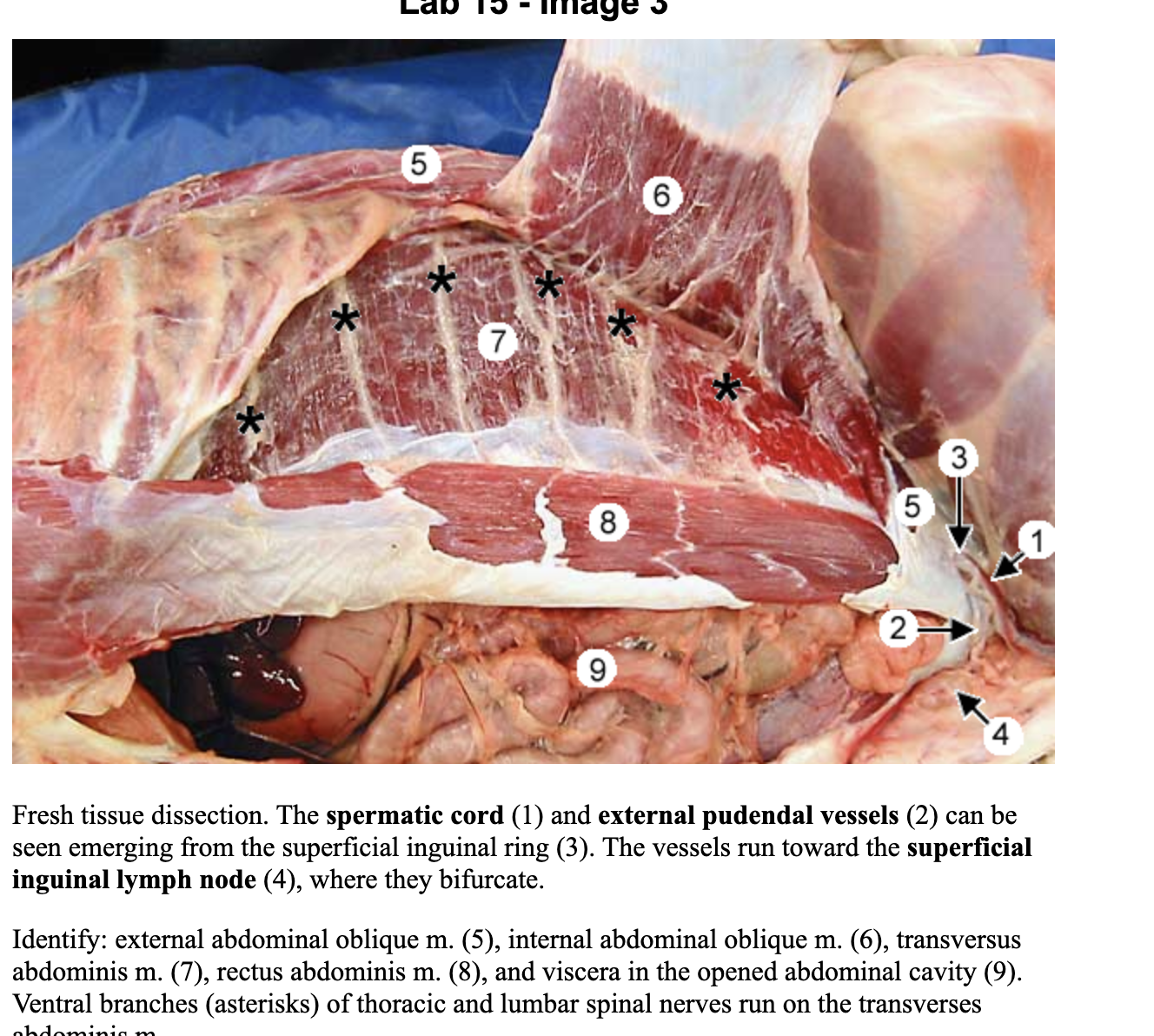
transverse abdominis muscle (TA)
7

rectus abdominis muscle (RA)
8
linea alba

rectus sheath
thick fascia that encapsulates rectus abdominis and is formed by EAO,IAO, T.
has superficial and deep leaves.

Clinical note: when an incision into the abdomen deviates to the side of the linea alba during celiotomy, closure begins with the closure of the _ leaf of the rectus sheath.
Superficial leaf.
it is the holding layer for those sutures as it is present throughout the length of the abdomen
cranial epigastric aa.
1

cranial superficial epigastric aa.
3

caudal superficial epigastrics
??
caudal epigastric vessels
??
cranial abdominal a.
?? ik where its at
deep circumflex iliac a.
8

lateral cutaneous femoral nerve
??
diaphragm: sternal, costal, and lumbar portions
costal 2
sternal 3

right and left crura of diaphragm
right crus 4
left crus 5

caval foramen, esophageal hiatus, aortic hiatus of diaphragm
caval foramen 6
esophageal hiatus 8
aortic hiatus 9

parietal, visceral, and connecting peritoneum
connecting is where the blood vessels are

peritoneal cavity
nothing located in there expect a small amount of fluid

Clinical note: in disease states, fluid can accumulate within the peritoneal cavity which can be removed through a procedure called _
abdominocentesis
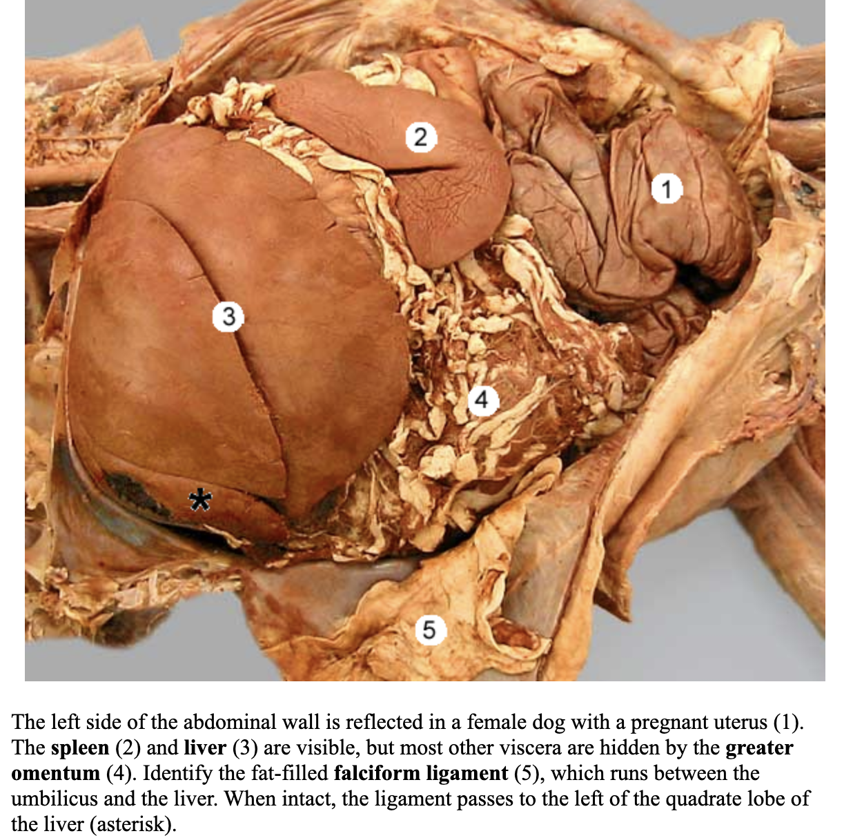
falciform ligament
5
round ligament of the liver

median ligament of the urinary bladder
6

lateral ligaments of the uinary bladder
12

Clinical note: The falciform ligament and the median ligament of the bladder may be incised from the _ during celiotomy
linea alba
mesoductus deferens
3
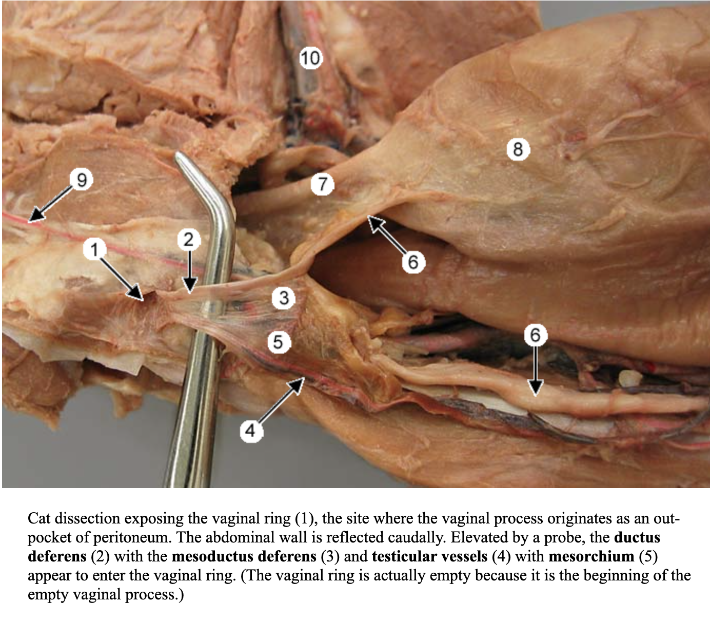
greater omentum
has superficial and deep leaves
4
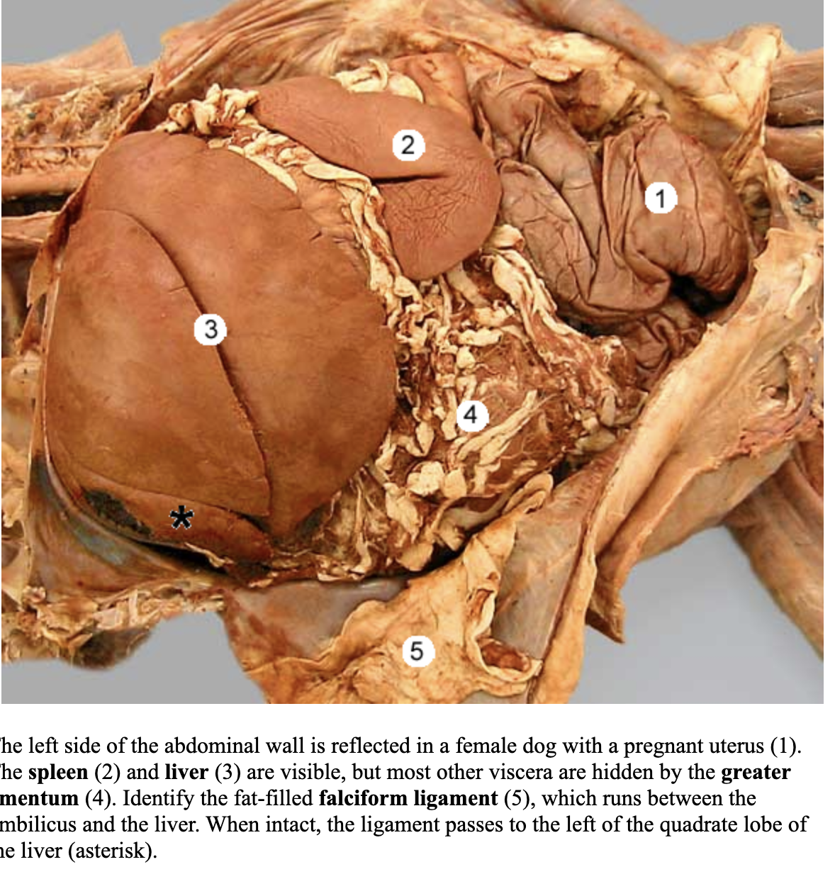
omental bursa

epiploic foramen

Clinical note: The _ is an important organ defense for the abdominal cavity. When inflammation or injury occurs at the peritoneum, the omentum adheres to the site for protection. This attribute can be used in surgery by suturing to weakened viscera (enterotomy sites) to provide blood supply and facilitate sealing of leaks
greater omentum
lesser omentum
connects lesser curvature of stomach to the liver
5

spleen
2
has head, body , and tail

gastroplenic ligament
asterisk

Clinical note: In what condition does the stomach twists on its long axis, pulling the spleen with it. This may cause enough vascular compromise to the spleen such that is must be removed when the torsion is corrected
gastric dilation and volvulus (GDV)
liver & its lobes
caudate process of caudate lobe
right lateral lobe
right medial lobe
quadrate lobe
left lateral lobe
left medial lobe
papillary process of the caudate lobe
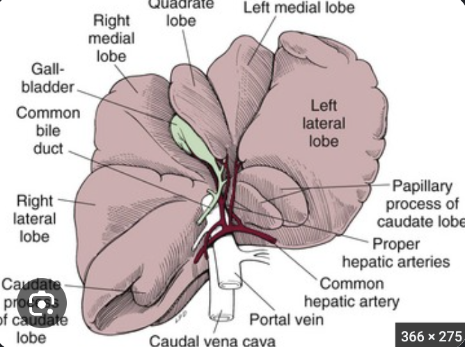
gall bladder
between right medial and quadrate lobe of liver

ligaments of the liver
coronary ligament
right and left triangular ligaments
falciform ligament

stomach lesser and greater curvature

cardia of stomach

fundus of stomach

body of the stomach

the pyloric region of the stomach is made up of what
antrum, canal, pylorus (sphincter)

Clinical note: _ is a serious condition where the stomach fills with gas and may lead to torsion. Consequently, circulatory failure may occur. This is an emergency requiring surgery to replace the stomach to its normal position and restore blood flow. A gastropexy is usually also performed to secure the pyloric part of the stomach to the right body wall or last rib
GDV
small intestine consists of what
duodenum, jejunum, and ileum
duodenum
ascending duodenum 8
descending duodenum 6

cranial and caudal duodenal flexure
cranial near stomach
caudal 7

mesoduodenum
5
holds duodenum in place on right side of the abdomen

duodenocolic fold
connects ascending duodenum with the descending mesocolon

Clinical note: The _ is held such that the mesoduodenum serves as an anatomical retractor to retract viscera to the left in order to explore the right lumbar area during a celiotomy,. The duodenocolic fold is a landmark used during “running the bowel” during celiotomy
duodenum
pancreas
left lobe 1
right lobe 3
body 5

major & minor duodenal papilla
major 1
minor 2 (more caudal)

bile duct
major papilla
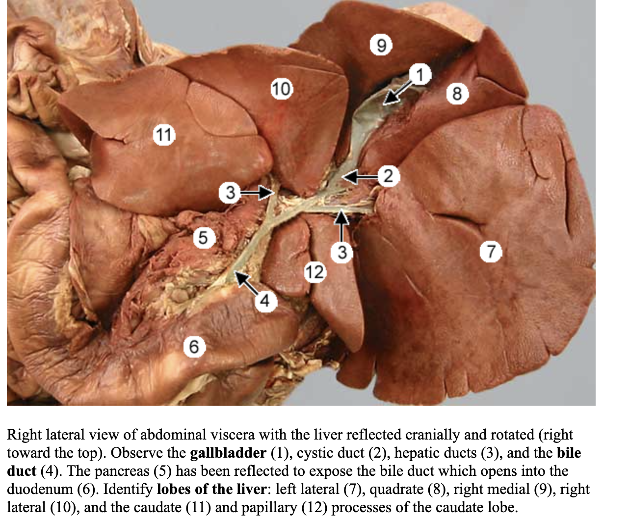
pancreatic duct
major papilla

accessory pancreatic duct
minor papilla
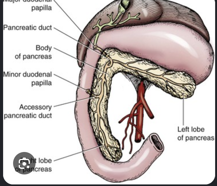
Clinical note: One should take care when doing an exploratory celiotomy not to disturb the or roughly handle it. This may inadvertently cause _
pancreas, pancreatitis
jejunum
4

root of the mesentery
5
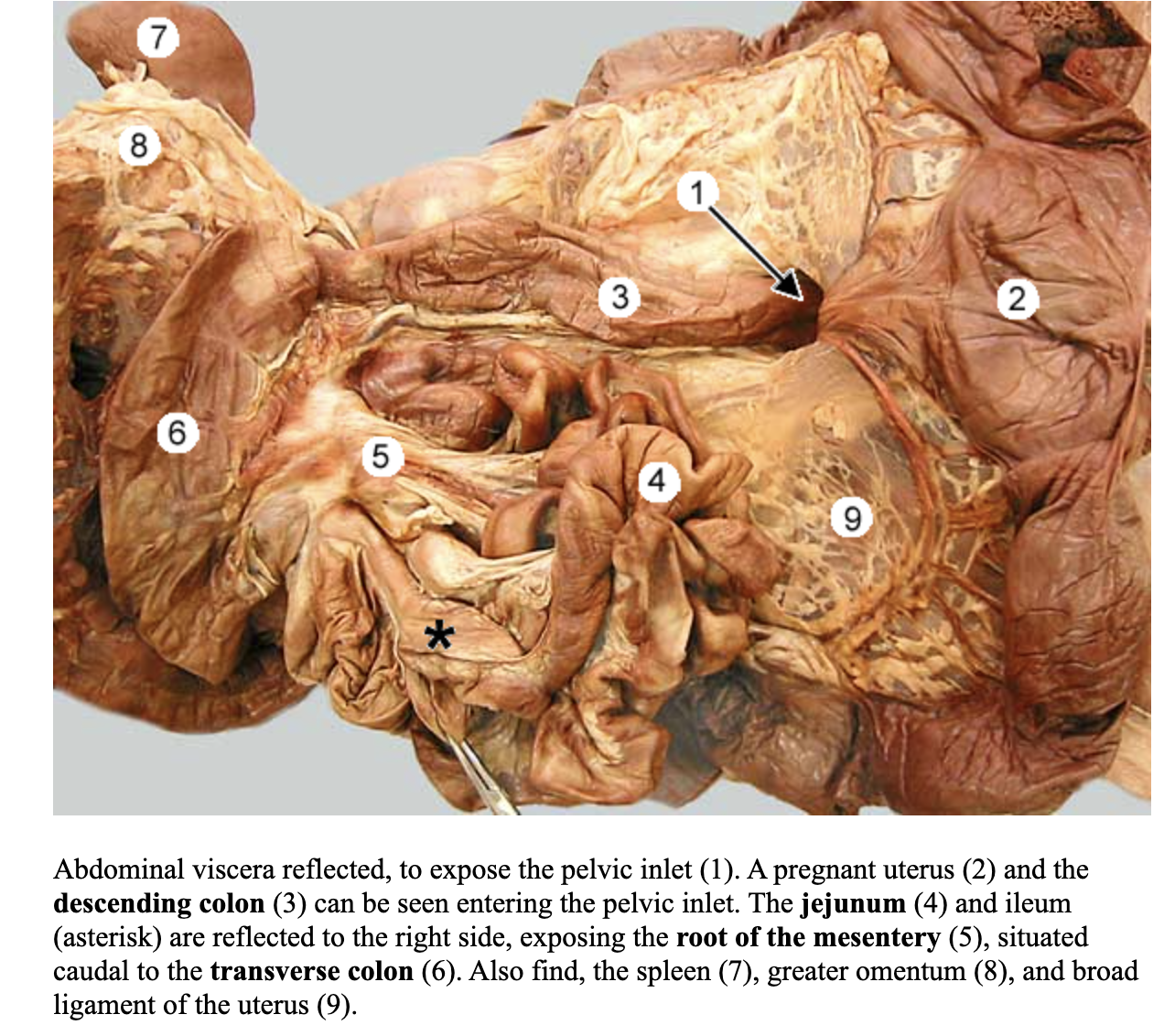
mesojejunum
??
jejunal lymph nodes
??
Clinical note: String foreign bodies tend to lodge and erode through the mesenteric border of the jejunum. Enterotomies for this and other foreign body removals are performed along the _ border of the jejunum to preserve intestinal blood supply
anti-mesenteric border
ileum
1

antimesenteric a.
2

ileocolic orifice

Clinical note: The _ is the most common part of the small intestine involved in intussusception (ileocolic or cecocolic)
ileum
The large intestine consist of
colon, cecum, rectum
ascending colon
2

right colic flexure

transverse colon
3
always cranial to the root of mesentary

left colic flexure

descending colon
4
lies on left side of the abdominal cavity

mesocolon
peritoneal layer that supports three segments of large colon

Clinical note: During celiotomy, the descending colon is held so that descending mesocolon an be used as an __ to retract viscera to the right for exploration of the left lumbar area
anatomical retractor
cecum
asterisks

ileocecal fold
3

cecocolic orifice

Cat difference: In cats and are short
the large intestine and cecum (comma shaped). as a result, the ileocecal fold is short as is the ileum
rectum
arrow

kidneys
1
left and right
right lies caudal to the caudate process of the caudate lobe of the liver
left is “left behind” more caudal than right

hilus of kidney

renal cortex of kidney

renal medulla of kidney
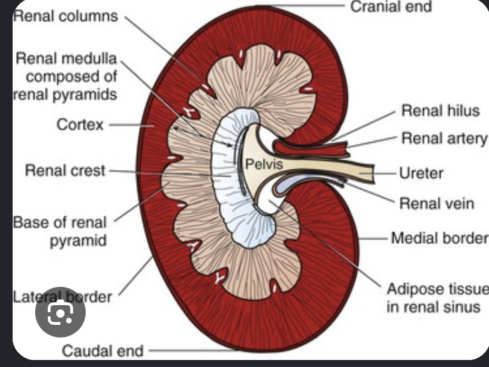
arcuate arteries and veins of kidney

interlobar aa. of kidneys

renal sinus of kidneys

renal pelvis of kidneys

renal crest of kidneys

renal pyramids of kidneys

pelvic recesses of kidneys

ureters
2

Clinical note: It is important to understand the anatomic location of the _ in relation to the ovary and uterus, because accidental ligation of the ureter during ovariohysterectomy would be BAD
ureter
Cat difference: the kidneys are more pendulous and therefore easier to palpate In addition there are large subscapular veins on the cortical surface that converge to the renal vein at the hilus
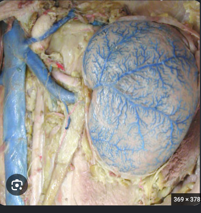
renal arteries and veins
11
