quiz 4 SHN, breast lymph
 Call Kai
 Learn
Learn Practice Test
 Spaced Repetition
 Match
Match Flashcards
 Knowt Play
1/63

There's no tags or description
Looks like no tags are added yet.
Name | Mastery | Learn | Test | Matching | Spaced | Call with Kai | Chat |
|---|
No analytics yet
Send a link to your students to track their progress
64 Terms
vitiligo
absence of melanin pigment in patchy areas (more common in people with dark skin)
freckles/ephelides
small, flat, brown macules
Pigmented nevi (moles)
inspect for changes
pallor
white or lighter coloration; ashen gray with brown/black skin
Erythema
flushed appearance
Cyanosis
bluish, grayish): due to decreased perfusion of tissues (tissue hypoxia)
Central cyanosis
2o to cardiopulmonary problems (inspect lips, tongue, oral
mucosa, and conjunctiva)
■ Dark skin may appear blue, dull
Peripheral cyanosis
d/t vasoconstriction
Jaundice
yellow, icteric
● Bilirubin is a by product of RBC breakdown & is normally excreted through the GI tract.
● Jaundice results from rising amounts of bilirubin in the blood with reabsorption into the skin.
Hypothermia
d/t to decreased circulation)
● generalized [shock]
● localized [peripheral arterial insufficiency]
Hyperthermia
Generalized (increased metabolic rate) - hyperthyroidism, fever, heavy exercise
● Localized (inflammation, infection, dvt)
Perspiration
normal sweating
Diaphoresis
profuse sweating

Cherry angiomas
genetic bright red papular lesion, 1-5 mm
located on trunk, upper chest, extremities
increases with age Benign

Telangiectasia
dilated superficial arteriole blood vessels
spider like shape or linear
legs or anterior chest
Does not blanch with pressure
Secondary to superficial dilatation of venous vessels and capillaries
associated with increased venous pressure states similar to varicosities

Spider angioma
central arteriole with capillary radiations; blanches (turns white) with pressure
located on face, neck, arms, upper trunk; usually not below waist
compression of the center completely blanches the lesion
seen w/ liver disease, pregnancy, Vit B deficiency, estrogen excess. A few may be normal
Venous star
bluish spider angioma; non blanching with pressure
associated with increased venous pressure (seen with varicose veins) -
primarily located on legs

Petechiae
1 - 3 mm round purple or red macule
results from superficial capillary bleeding / blood
extravasation
caused by bacteremia, bleeding disorders (thrombocytopenia or emboli to the skin)
Does not blanch with pressure
Ecchymosis
larger patch of capillary bleeding
r/t trauma, bleeding disorders or liver dysfunction
Purple/purplish-blue fading to green, yellow, brown over time
Hematoma
localized collection of blood (raised bruise)
characteristics of skin lesions
color
elevation
configuration
size
number
location and distribution
discharge or exudate
primary lesion
initial lesion
secondary lesion
results from changes in the primary lesion such as scratching, infection, scarring
example of flat lesion
macule, patch
macule
flat, circumscribed, discolored, <1 cm (ephelides, solar
lentigines, flat nevi, petechiae)
patch
flat, irregular, >1 cm (vitiligo)
example of raised lesion
papule, plaque, nodule, tumor, wheal, urticaria, vescile, bulla, pustule, cyst

papule
solid, elevated, circumscribed, < 1 cm (raised nevus, wart
[verruca])
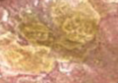
plaques
coalesced papules, > 1 cm (psoriasis)
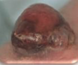
nodule
solid, elevated, 1-2 cm (lipoma = fatty growth)
tumor
larger than a few centimeters, firm or soft (lipoma)

wheal
superficial, raised, erythematous, irregular (allergic
reaction, PPD, mosquito bite) - causes interstitial edema

urticaria (hives)
wheals coalesce to form extensive reaction; intensely pruritic
example of fluid flled lesions
vesicle, bulla, pustule, cyst

vesicle
elevated cavity with clear fluid, <1 cm (herpes simplex, varicella [chickenpox], herpes zoster [shingles], contact dermatitis [e.g., poison ivy])
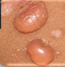
bulla
elevated cavity with fluid, >1 cm (blister, burns)

pustule
contains pus; filled with leukocytes, not
necessarily infected (acne)
cyst
encapsulated fluid filled cavity in dermis or sub-q (sebaceous cyst) If deep, may be hard to differentiate from a nodule or tumor (e.g., breast cyst versus tumor)

malignant melanoma
Highly Metastatic
● Grows Deep, not wide
● Increased Risk: Fair skin, Sun Exposure
● Characteristics: ABCD: Asymmetry, Border is
Irregular, Color: Varied, Diameter > 6 mm
Vernix Caseosa
white coating at birth
Lanugo
fine downy hair darker than Vellus hair. Can be present at birth or shed in utero
puberty
Increased secretion of apocrine glands - results in body odor
Increased secretion of eccrine sweat glands – increased perspiration
Increased secretion of sebaceous glands (oily skin/acne)
Open comedones (blackheads)
Closed comedones (white heads)
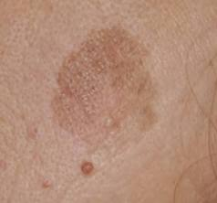
Solar Lentigines
flat or slightly raised, well-demarcated dark spots on the skin caused by cumulative ultraviolet (UV) light exposure

Actinic Keratosis
common, precancerous skin condition caused by long-term damage from ultraviolet (UV) light
Seborrheic Keratosis
Waxy, rough, scaly, or slightly raised bumps that look like they were "stuck on" the skin

Koilonychia
(spoon nails/concave) - iron deficiency anemia

Paronychia
inflammation/infection of skin around nail bed

Onycholysis
loosening of nail plate (fungal infection)

Leukonychia:
white hairline markings from trauma or picking at cuticle (normal)
Clubbing
nail angle straightens out to ≥ 180 degrees & nail bed becomes spongy
r/t chronic hypoxia - COPD, lung CA, Chronic Cardiopulmonary problems
stage 1 pressure ulcer
non-blanchable erythema of intact skin
stage 2 pressure ulcer
partial–thickness loss of skin with exposed dermis. Pink bed of skin, top layer of skin may look like it peeled off.
stage 3 presure ulcer
full thickness skin loss-adipose is visible
stage 4 pressure ulcer
full thickness skin and tissue loss with exposed fascia, muscle, tendon, ligament, or bone
Unstageable
full thickness skin and tissue loss, obscured by
slough or eschar.

breast review
most cancer in upper outer quadrant and axillary tail of spence
subjective data for breast pain
localized or general
CA can cause pain but not all breast CA is painful
character
cyclic or constant
trauma
associated factor
fibrocystic:
cyclical changes
caffeine intake may worsen sypmtoms
associated skin changes
objective clinical breast exam: inspection
CBE indicated if pt C/O lump on SBE
inspect:
expose both breasts for comparison
note size nad shape
slight asymmetry may be normal for patient
retraction maneuvers
inspect skin:
smooth, even color
localized area of redness, bulging, dimpling, edema
redness w heat
peau d orange
peau d orange
caused by blocked lymph ducts, common in CA, edema may exaggerate hair follicles causing ubmpy textue like orange peel
objective data: examination
Nipples are normally symmetric and on same plane
Deviation could indicate CA
May be everted, flat, or inverted
Inverted nipples: suspicion for CA (if recent change & unilateral)
Note any dry scaling, fissures, ulcerations, or bleeding
Supernumerary nipple – 2 nipples along the mammary ridge at MCL
how to feel pectoral nodes
grasp the anterior axillary fold between your thumb and fingers and with your fingers palpate the inside border of the pectoral muscle

where are epitrochlear nodes
along the upper part of arm
breast lump characteristics
Location
● Size
● Shape
● Consistency
● Tenderness
● Borders
● Retraction/dimpling
● Mobility
● Cancer characteristics: painless lump, ulcerations, retractions, nipple d/c
pregnnacy related changes
montgomery glands and mastitis