A2 Exam 1
 Call Kai
 Learn
Learn Practice Test
 Spaced Repetition
 Match
Match Flashcards
 Knowt Play
1/64

There's no tags or description
Looks like no tags are added yet.
Name | Mastery | Learn | Test | Matching | Spaced | Call with Kai | Chat |
|---|
No analytics yet
Send a link to your students to track their progress
65 Terms

Frontal bone

Maxilla (left) bone

Mandible bone

Temporal Bone (right)

Sphenoid bone

Occipital Bone

Vomer bone

Ethmoid bone

Zygomatic bone

Parietal bone (right)

Nasal bone (left)

Foramen Magnum:
Part of occipital bone
Opening for continuity of brain stem and spinal cord

fibrous, synarthrosis joints

Hyoid bone

Temporomandibular joint (TMJ)
Bilateral modified synovial hinge joint
Mandibular condyle articulates with temporal bone through articular disc of TMJ
Depression / elevation, protrusion / retrusion, and lateral deviation

Movements?
Superior joint cavity: translation
Inferior joint cavity: rotation

Mandibular condyle / head of mandible
Forms the floor of the inferior TMJ cavity.
Articulates with the temporal bone’s Mandibular fossa.

TMJ articular disc
Sits between the temporal bone’s Mandibular fossa and the Mandibular condyle, separating the TMJ into superior and inferior cavities

Ramus of mandible

Body of Mandible

Coranoid process of mandible

Angle of mandible

Neck of mandible

TMJ
Zygomatic Arch (temporal + zygomatic bones ; landmark for finding TMJ)
Ramus of Mandible
Inferior border of mandible
Hypoid bone

Lateral TMJ Ligament
Reinforces the lateral capsule and resists excessive posterior displacement of the Mandibular condyle
Connects temporal bone to neck of mandible

Stylomandibular Ligament
Limits excessive protrusion (little contribution to TMJ stability
Connects Styloid process of the temporal to angle of mandible

Sphenomandibular Ligament
Primary passive support ; acts like ropes guiding Mandibular movement
Connects spine of sphenoid to lingula of mandible
Explain TMJ disc pathology reduction and without reduction
Click on opening + click on closing + full ROM (disc is recaptured) = anterior displacement with reduction
Disc stays anterior + difficulty opening = with out reduction

what vertebrae are considered upper and lower cervical spine and what are the functional joints
Upper cervical spine: C0 (occipital condyles) -C2
C0-C1 (AO joint) and C1-C2 (AA joint)
Lower cervical spine: C2-C3 → C6-C7 (first intervertebral disc is at C2-C3)

Atlanto-occipital (AO) Joint
Occipital condyles articulating with superior facets of C1 (Atlas)
Synovial condyloid
Flexion / Extension. Nodding your head “yes”

Atlantoaxial (AA) Joint
50% of cervical rotation (shaking “no”) is at AA joint
Median AA joint
anterior surface of the dens (C2) ←→ posterior surface of the anterior arch of the atlas (C1)
Synovial pivot
Lateral AA Joint
Inferior facets of C1 ←→ superior facets of C2
Synovial plane

Tectorial membrane
Continuation of the PLL (changes around C2)
Resists Cervical flexion

Alar ligaments:
Dens ←→ Medial occipital condyle regions
Each primarily resists contralateral rotation and lateral flexion
Transverse ligament of atlas:
Resists anterior translation of C1 on C2
Contacts posterior surface of dens but does not attach to it
Cruciform / Cruciate Ligament of atlas
Transverse ligament + sup and inf longitudinal bands (weaker than transverse)

Posterior AO membrane
connects posterior arch of C1 to the occiput
provides stability and limits excessive flexion at the AO joint
Ligamenta flava
Strongest resistor of flexion

Anterior Longitudinal Ligament (ALL)
Resists extension
Nuchal Ligament
Resists flexion and major muscle attachment site
The supraspinous and interspinous ligaments blend into the nuchal ligament at approximately C7
Explain coupled motion in the lower vs upper cervical spine
In the LOWER cervical spine: rotation and lateral flexion couple to the SAME side. (Right side bending → Right rotation)
In the UPPER cervical spine: rotation and side bending couple to the OPPOSITE sides. (Right side bending → Left rotation)
Explain Atlantodental interval (ADI)
Space between posterior C1 anterior arch and anterior dens. <3mm in adults is normal, widening raises instability concerns


Sternoclavicular joint
Synovial Saddle joint (elevation / depression and protraction / retraction)
Coracoid process
Acromioclavicular joint
Synovial Plane joint
Acromion
Glenohumeral Joint
Synovial Ball and Socket joint (F/E, ABD/ADD, IR/ER, Horizontal ABD/ ADD)

Acromioclavicular joint (synovial plane)
Posterior angle of the acromion
Superior angle of scapula
Spine of scapula
Roof of spine of scapula
medial border of scapula
Lateral border of Scapula
Inferior angle of scapula

Explain the movements of the Pectoral Girdle (Clavicle + Scapula) in Overhead Reaching
Clavicular movements:
Elevation
Retraction
Posterior Rotation
Scapular movements:
Upward Rotation
Posterior Tilt (superior border away from thorax)
Internal Rotation
Explain Scapulohumeral Rhythm
120 degrees from glenohumeral (GH) elevation + 60 degrees scapulothoracic (ST) upward rotation = 180 degrees of shoulder abduction in scapular plane (30-45 deg ant to frontal plane). 2:1 ratio the whole way up.


Coracohumeral Ligament
Extrinsic
Resists superior humeral head translation

Intrinsic Ligaments
Superior Glenohumeral (GH) Ligament
Resists ER when the arm is ADD
Middle GH Ligament
Resists ER when arm is “neutral”
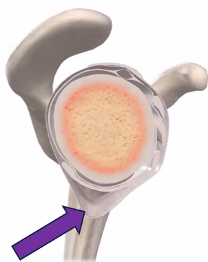
Inferior GH Ligament
Anterior band: Resists ER when arm is ABD
Posterior band: Resists IR when arm is ABD
Axillary fold is between ant and post fibers (purple arrow)

Coracoclavicular (Conoid): Resists superior displacement of the clavicle relative to the scapula
Coracoclavicular (Trapezoid): Resists separation and displacement between clavicle and scapula, including superior clavicular displacement.
Coracoacromial: Resists superior translation of the humeral head
Transverse Humeral Ligament: Holds long head of biceps tendon in the intertubercular groove

Costoclavicular Ligament: Resists elevation of the clavicle
Interclavicular Ligament: Resists depression of the clavicle
Anterior Sternoclavicular Ligament: Resists retraction of the clavicle
Posterior Sternoclavicular Ligament: Resists protraction of the clavicle

Acromioclavicular Ligament: Resists excessive horizontal displacement, especially anterior / posterior movement at the AC joint

What structure is this and explain the difference between a SLAP and Bankart tear.
This is the Glenoid Labrum
SLAP tear is located at the superior labrum and extends anterior to posterior
Bankart tear is located at inferior anterior labrum

Acromion
Greater tuberosity
Intertubercular groove (ER/IR the shoulder)
Lesser tuberosity (ER/IR the shoulder)
Coracoid process
Medial epicondyle
Olecranon fossa (flex the elbow)
Lateral epicondyle
Ulnar groove

Together form the elbow joint (Hinge Joint)
Humeroulnar joint: synovial hinge joint allowing for flexion and extension
Humeroradial joint: synovial hinge joint allowing for flexion and extension

Radial / Lateral Collateral ligament
Resists varus stress: excessive ADDuction of the forearm relative to the arm
intrinsic

Annular Ligament
Holds the radial head against the radial notch of the ulna, while allowing is to spin during pronation / supination
Helps prevent displacement of the radial head during distraction (“Nursemaid’s elbow”)
intrinsic

Oblique Cord
Connects the proximal ulna to radius, just below the radial tuberosity
Becomes taut during distraction / traction (think hanging or carrying a load), helping resist the radius being pulled distally.
Transfers distraction forces from radius → ulna to share the pulling load

Interosseus Membrane
Forms the middle radioulnar joint (a fibrous syndesmosis)
holds the radius and ulna together while permitting pronation / supination
Becomes taut during compression, transferring force from the radius → ulna
Example: during a pushup, load enters mainly through the radius and is shared with the ulna

Medial / Ulnar Collateral Ligament
Resists Valgus stress: excessive ABDuction of the forearm relative to the arm
3 bands (anterior, posterior, oblique) anterior is the strongest
Injury cue: baseball pitcher from repeated valgus loading
intrinsic
Quadrate Ligament
stabilizes the proximal radioulnar joint and limits excessive pronation and supination

Proximal and Distal radioulnar joints
Both are synovial pivot joints allowing pronation and supination

Olecranon
Humeroradial joint ( flex the elbow to 90 degrees, pronate / supinate)
Radial head
Radial neck
Lateral shaft of radius
Posterior shaft of ulna
Head of ulna
Styloid process of ulna
Dorsal (Lister’s tubercle)
Styloid process of radius

Styloid process of radius
Radiocarpal joint
Scaphoid
CMC - 1 joint
Triquetrum
MCP - 1 joint
IP joint
MTP joints
PIP joints
DIP joints


Joints of the Hand
Radiocarpal (RC) Joint:
— Between Radius and scaphoid / lunate / triquetrum
— Synovial Condyloid Joint: wrist FLX/EXT and ABD/ADD (radial/ulnar deviation)
Carpometacarpal (CMC) Joints:
— 5 total, between carpals and metacarpals
— 1st CMC (thumb) is a Synovial Saddle joint allowing thumb FLX/EXT, ABD/ADD, OPP/ REP, and circumduction
—2-5 CMC are synovial plane joints
Metacarpophalangeal (MCP) Joints:
— Knuckles
— 1st MCP (thumb) is a Synovial Condyloid joint BUT only FLX/EXT
— 2-5 MCP are Synovial Condyloid joints and allow FLX/EXT, ABD/ADD, and combined circumduction
PIP, DIP, IP Joints:
— All Synovial Hinge Joints, allowing FLX/EXT
—The thumb has a IP instead of PIP or DIP


Wrist Ulnar/Medial Collateral Ligament (UCL/MCL):
Resists radial deviation (ABD)
Wrist Radial/Lateral Collateral Ligament (RCL/LCL):
Resists ulnar deviation (ADD)
Posterior/Dorsal radiocarpal ligament:
Resists excessive wrist flexion
Dorsal Intercarpal Ligaments (also some on palmar side just not highlighted in picture)
Anterior/Palmar radiocarpal ligaments:
Resists excessive wrist extension
Anterior/Palmar ulnocarpal ligaments:
Resists excessive wrist extension

Flexor retinaculum / transverse carpal ligament: forms the roof of the carpal tunnel

Finger Collateral ligaments
Stabilize the sides of MCP/IP joints against excessive sideways movement
Thumb MCP LCL resists excessive thumb ABD

What is the suspected result of a FOOSH
Scaphoid or distal radius fracture
The shape of the distal radius favors:
Flexion > Extension and Ulnar deviation > Radial deviation
