Histology Identification Exam 1
 Call Kai
 Learn
Learn Practice Test
 Spaced Repetition
 Match
Match Flashcards
 Knowt Play
1/201

There's no tags or description
Looks like no tags are added yet.
Name | Mastery | Learn | Test | Matching | Spaced | Call with Kai | Chat |
|---|
No analytics yet
Send a link to your students to track their progress
202 Terms

Mitotic figure

Mitotic figures

nucleus of fibroblast cell

cytoplasm of fibroblast cell

1
Nucleus
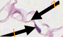
2
Cytoplasm

What cellular structure is outlined in yellow?
Cytoplasm

What cellular structure is outlined in red?
Nucleus

What cellular structure is the arrow pointing to?
Nucleolus

1
Nucleolus

2
Nucleus

3
Cytoplasm

Mitotic figure

How would you describe the metabolic state of this cell?
Metabolically active (euchromatic)

How would you describe the metabolic state of this cell?
Metabolically inactive (heterochormatic)

How would you describe the metabolic state of this cell?
Metabolically inactive (heterochromatic)

How would you describe the metabolic state of this cell?
Metabolically active (euchromatic)

Golgi apparatus

Lipofuscin

What type of tissue is this?
Epithelium

What type of tissue is this?
Muscle tissue

What type of tissue is this?
Connective tissue

What is the green highlighted structure?
basement membrane

What is the blue highlighted surface?
Apical surface

What is the red highlighted surface?
Basal surface

What surface is the arrow pointing to?
Apical surface

What surface is the arrow pointing to?
Basal surface

What tissue is the arrow pointing to?
Epithelial tissue

What is the area of basophilic staining near the apical surface?
Ribosomes
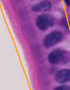
What is the pink layer on the apical surface?
Microvilli

What is the large structure embedded in the epithelium?
Goblet cell

What type of epithelium is shown?
Simple squamous epithelium

What type of epithelium is shown?
Simple squamous epithelium

What type of epithelium is shown?
Simple columnar
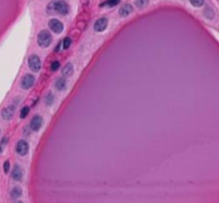
What type of epithelium is shown?
Simple cuboidal

What type of epithelium is shown?
Stratified columnar

What type of epithelium is shown?
Non-keratinized stratified squamous

What type of epithelium is shown?
Keratinized stratified squamous

What type of epithelium is shown?
Pseudostratified columnar

What type of epithelium is shown?
Transitional

What apical surface modification is the arrow pointing to?
Microvilli

What apical surface modification is the arrow pointing to?
Cilia

Which type of glandular epithelium is circled?
Endocrine

Which type of glandular epithelium is pointed at by the arrows?
Exocrine

What type of gland is shown in this picture?
Serous gland

What type of gland is shown in this picture?
Mucous gland

What type of gland is shown in this picture?
Mixed gland

Goblet cell
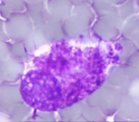
Goblet cell

What type of neoplasia is shown?
Epithelial neoplasia

What type of neoplasia is shown?
Epithelial

What type of cell is shown in the image?
Mesenchymal

What type of neoplasia is shown?
Mesenchymal

What type of neoplasia is shown?
Mesenchymal

What type of tissue is shown?
CCT

What type of cells do the arrows point to?
Fibroblasts (mesenchymal)

What is the cytological classification of the cells in this image?
Epithelial

What is the cytological classification of the cells in this image?
Mesenchymal

What type of tissue is shown?
Unilocular adipose tissue (white fat)

What type of cell is shown?
Adipocyte

What type of tissue is shown and what type of mass would you expect this cytology came from?
Unilocular adipose tissue; Lipoma

What section of bone is number 1 pointing to?
Compact bone
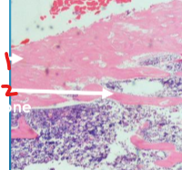
What section of bone is number 2 pointing to?
Cancellous bone

What type of cell is shown?
Osteoblast

What type of cell is shown?
Osteocyte

What type of connective tissue cell is shown?
Osteoblast

What type of cell is circled?
Osteoclast

What type of connective tissue cell is shown?
Osteoclast

What type of muscle did this cytology come from?
Smooth m.
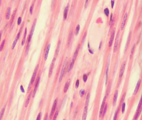
What type of muscle is shown on this histology?
Smooth m.

What type of muscle is shown on this histology?
Skeletal m.

What type of muscle did this cytology come from?
Skeletal m.

1
Epithelium

2
Lamina propria

3
Muscularis mucosae

4
Mucous membrane/mucosa

5
Submucosa

6
Tunica muscularis

7
Serosa

What layer of a tubular organ is shown?
Submucosa
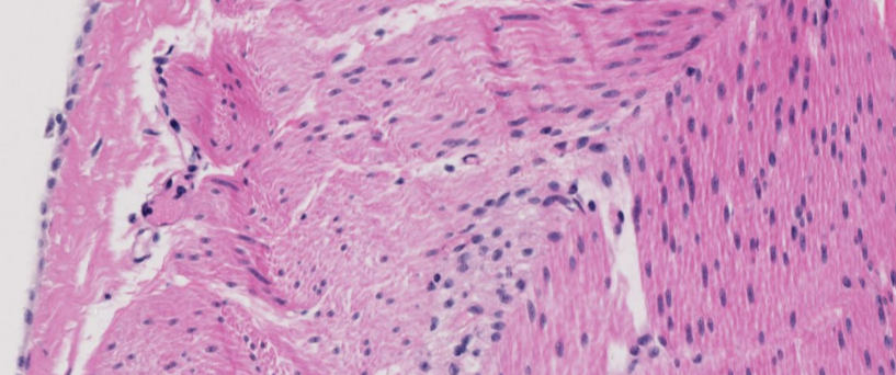
What layers of a tubular organ are shown in the image
Serosa (Far left); tunica muscularis (right - majority of photo)

1
Lumen

2
Muscosa
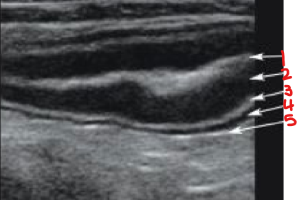
3
Submucosa

4
Muscularis

5
Serosa
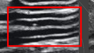
What intestinal layer is significantly thickened in this ultrasound?
Muscularis

What cell in the blood is shown?
Erythrocyte

What cell in the blood is shown?
Neutrophil

What cell in the blood is shown?
Eosinophil

What cell in the blood is shown?
Basophil

What cell in the blood is shown?
Monocyte

What cell in the blood is shown?
Neutrophil

What cell in the blood is shown?
Eosinophil
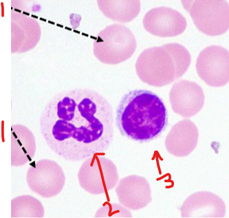
What cell in the blood is number 1?
Erythrocyte

What cell in the blood is number 2?
Neutrophil

What cell in the blood is number 3?
Small mature lymphocyte

What type of cell in the blood is shown?
Camelid erythrocytes

What type of cell in the blood is shown?
Avian, reptilian, amphibian, or fish erythrocytes

What type of cell in the blood is shown?
Neutrophils