Biology - Cells and cell transport
 Call Kai
 Learn
 Practice Test
 Spaced Repetition
 Match
 Flashcards
 Knowt Play
1/20

There's no tags or description
Looks like no tags are added yet.
21 Terms
What are the differences between electron microscopes and light microscopes? (7)
Electron microscopes use a beam of electrons focused by electromagnets, light microscopes use light focused by glass lenses
Electrons have a much shorter wavelength (~0.005 nm) than light (400-700 nm)
Electron microscopes have a much greater resolution (~0.05 nm) than light microscopes (~200 nm)
Electron microscopes have a higher maximum useful magnification (over 1 million ×) than light microscopes (~1500 ×)
Electron microscopes can reveal the ultrastructure of cells in much greater detail
Electron microscopy requires dead, dehydrated specimens in a vacuum, whereas light microscopy can be used with living specimens
Electron microscope images are black and white (often false-coloured), while light microscope images are colour
Compare transmission electron microscopes (TEM) and scanning electron microscopes (SEM) (7)
Transmission electron microscopes (TEM) - passes a beam of electrons through a thin specimen stained with heavy metal salts
Advantages of TEM - produces images of internal cell structures and has a higher resolution than SEM
Limitations of TEM - equires very thin sections, does not produce a 3D image, specimens may deteriorate in the electron beam
Scanning electron microscopes (SEM) - scans a specimen coated in a thin layer of heavy metal (e.g. gold) with an electron beam
Electrons reflected from the surface produce an image of the specimen's surface features
Advantages of SEM - gives a 3D appearance and can examine thicker specimens
Limitation of SEM - lower resolution than TEM and only shows the surface of specimens

What are the equations used in magnification calculations and unit conversions?
Magnification = Image size ÷ Actual size
Actual size = Image size ÷ Magnification
Image size = Actual size × Magnification
Conversion of mm to µm - multiply by 1000
Conversion of µm to nm - multiply by 1000
mm —> nm = x106

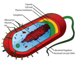
What are the features of prokaryotic cells? (6)
Prokayote - a small, single-celled organism that lacks a nucleus and membrane-bound organelles e.g bacteria
A typical bacterial cell contains a cell wall (murein/peptidoglycan), cell membrane, circular DNA, ribosomes, cytoplasm and food reserve granules
Plasmids - small circular DNA molecules carrying non-essential genes
Pili - used for attachment and genetic exchange between cells
Flagella - used for locomotion
Mesosomes - associated with respiration and binary fission
What are the characteristics of eukaryotic cells? (6)
Eukaryotes - make up the vast majority of living organisms, including plants and animals
Eukaryotic cells contain a nucleus surrounded by a nuclear membrane
The cytoplasm contains numerous membrane-bound organelles
Examples of organelles includes: mitochondria, endoplasmic reticulum, Golgi apparatus and lysosomes
Eukaryotic cells contain 80S ribosomes for protein synthesis
Eukaryotic cells are generally larger and more complex than prokaryotic cells

What structures are found only in a plant cell? (4)
Cellulose cell wall - provides support and maintains cell shape
Starch grains - store carbohydrate reserves
Chloroplasts - contain chlorophyll and are the site of photosynthesis
Vacuole - large, permanent, contains cell sap (soluble sugars, salts and pigments)

What are the differences between prokaryotic and eukaryotic cells? (6)
Prokaryotes have no nucleus, whereas eukaryotes have a nucleus
Prokaryotes lack membrane-bound organelles such as mitochondria, Golgi apparatus and endoplasmic reticulum, which are present in eukaryotes
Prokaryotes contain 70S ribosomes, whereas eukaryotes contain 80S ribosomes
Prokaryotic DNA is circular, whereas eukaryotic DNA is linear
Prokaryotic cells are smaller (0.5-10 µm) than eukaryotic cells (10-100 µm)
Prokaryotic flagella are simple and lack microtubules, eukaryotic flagella contain a 9+2 arrangement of microtubules
What is differential centrifugation and how are cell organelles prepared for it? (5)
Differential centrifugation - separates cell structures based on their density by centrifuging at different speeds
Cells are first homogenised (broken open) in an ice-cold, isotonic buffer solution
The isotonic solution prevents organelles from gaining or losing water by osmosis so they don’t burst or shrivel up
The low temperature prevents enzyme activity and autolysis (self-digestion) of organelles
The homogenate is centrifuged at increasing speeds to separate and isolate organelles according to their density

What are the stages of differential centrifugation? (6)
The homogenate is filtered to remove debris, intact cells and tissues
The filtrate is centrifuged at a low speed, causing the heaviest particles to form a pellet (sediment)
The liquid above the pellet, called the supernatant, is removed and centrifuged at a higher speed for longer
Nuclei form the next pellet, while lighter organelles remain in the supernatant
The process is repeated using increasing speeds and centrifugation times to separate organelles by density
Organelles are typically isolated in the order: nuclei → chloroplasts → mitochondria → endoplasmic reticulum → ribosomes

What are the structures and functions of the nucleus? (5)
Nucleus - contains the cell's genetic material (DNA), which controls the development, structure and function of the cell
Nuclear envelope - a double membrane that surrounds the nucleus
Nuclear pores - allow communication between the nucleus and cytoplasm
Nucleoplasm - a substance in the nucleus which contains chromatin (DNA and proteins) and nucleoli
The nucleus controls protein synthesis, cell division and the production of ribosomes

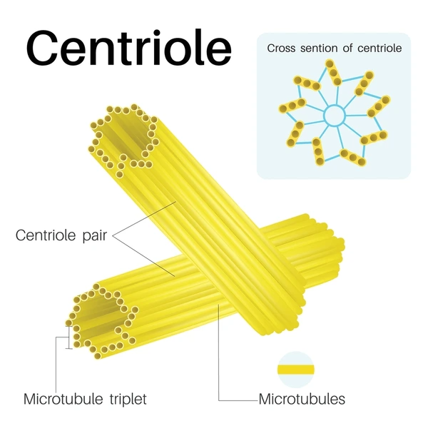
What are the functions and the structure of centrioles? (3)
Centrioles - occur as a pair within animal cells
Function: produce spindle fibres (microtubules) that separate chromosomes during mitosis and meiosis
Each centriole is a small, hollow cylinder composed of 9 triplets of microtubules
What are the structures in mitochondria and its functions? (8)
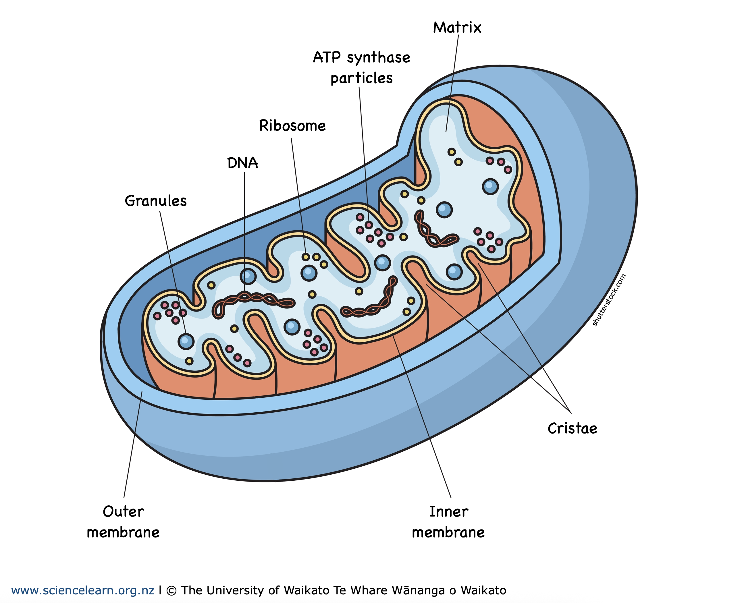
Mitochontria - the site of aerobic respiration and ATP production
Structure:
Double membrane - surrounds the mitochondria and consists of an outer membrane and an inner membrane
Intermembrane space - the space between the membranes
Cristae - folding of the inner membrane, providing a large surface area for oxidative phosphorylation
The cristae contain stalked particles with enzymes involved in the electron transport chain and ATP synthesis
Matrix - contains enzymes for the Krebs cycle, as well as mitochondrial DNA and ribosomes
Mitochondrial DNA - allows mitochondria to replicate and ribosomes enable protein synthesis
Cells with high energy demands, such as muscle cells, liver cells and cells carrying out active transport, contain many mitochondria
What is a ribosome and its main function? (4)
Ribosomes - small organelles (about 20 nm in diameter) made of a large and small subunit
Each subunit contains ribosomal RNA (rRNA) and protein
Location in the cell - free in the cytoplasm, arranged in polysomes, or attached to the rough endoplasmic reticulum (RER)
Main function - the site of protein synthesis, where amino acids are joined together to form polypeptides

What is the structure and functions of the endoplasmic reticulum? (6)
Endoplasmic reticulum (ER) - consists of flattened membrane sacs called cisternae, forming an internal transport system within the cell
Rough endoplasmic reticulum (RER) - has ribosomes attached to its surface
The RER synthesises and transports proteins, particularly secretory proteins that are exported from the cell
Proteins produced by the RER are often transported to the Golgi apparatus for modification and packaging
Smooth endoplasmic reticulum (SER) - lacks ribosomes
The SER is involved in the synthesis and transport of lipids

Describe the structure and functions of the Golgi body (8)
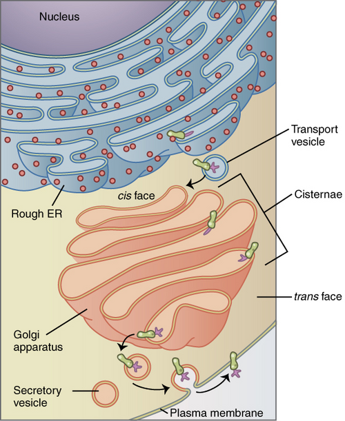
Golgi body - consists of stacks of flattened membrane-bound sacs called cisternae
The cisternae are continuously formed at one end and pinched off as vesicles at the other end
Cells specialised for secretion contain large Golgi bodies
Functions:
Produces glycoproteins e.g mucin
Modifies, packages and secretes proteins e.g enzymes
Formation of cell walls in plant cells
Formation of lysosomes
Lipid biosynthesis
Describe the structure and functions of lysosomes (7)
Lysosomes - small spherical organelles surrounded by a single membrane
They contain hydrolytic (digestive) enzymes such as proteases, nucleases and lipases, which digest proteins, nucleic acids and lipids
The enzymes are enclosed within lysosomes to prevent damage to the cell
Enzymes are synthesised on the rough ER, processed by the Golgi apparatus, and packaged into lysosomes
Main functions:
Digestion of material - lysosomes fuse with vesicles formed by endocytosis and release enzymes to digest material, products are absorbed by the cytoplasm
Autophagy - lysosomes digest worn-out organelles and unwanted cell structures within autophagic vacuoles
Enzyme release - enzymes may be released outside the cell

What are the structures found in chloroplasts and their main functions? (5)
Chloroplasts - found in photosynthetic plant cells and are the site of photosynthesis
They are surrounded by a double membrane envelope
Contains flattened membrane sacs called thylakoids, which are stacked into grana
Grana - provide a large surface area for chlorophyll and are the site of the light-dependent reactions
Stroma - surrounds the membrane system, contains enzymes, sugars and starch grains and is the site of the light-independent reactions

Describe cell differentiation (5)
Cell differentiation - the process by which cells become specialised to perform specific functions
Specialised cells are grouped together to form tissues
Tissue - a group of similar cells that perform a specific physiological function
Organ - consists of different tissues working together to perform a specific function
Several organs work together in an organ system, e.g. the digestive system or plant vascular system
How are palisade mesophyll cells adapted for photosynthesis? (5)
Palisade mesophyll cells - located beneath the upper epidermis of the leaf
Adaptations:
Contain numerous chloroplasts to maximise light absorption for photosynthesis
Thin cell walls allow rapid diffusion of carbon dioxide
The cells are long and columnar (cylindrical), providing a large surface area for gaseous exchange
There are few air spaces between cells, allowing maximum light to be absorbed

How are columnar epithelial cells adapted for absorption? (4)
Columnar epithelial cells - found in the lining of the small intestine and proximal convoluted tubule of the kidney
They are specialised for the absorption of small molecules, often by active transport
The cell surface membrane is folded into microvilli, increasing the surface area for absorption
They contain numerous mitochondria to provide ATP for active uptake of substances such as amino acids and glucose
