Anat/Phys lab exam 1
 Call Kai
 Learn
 Practice Test
 Spaced Repetition
 Match
 Flashcards
 Knowt Play
1/63

There's no tags or description
Looks like no tags are added yet.
Name | Mastery | Learn | Test | Matching | Spaced | Call with Kai | Chat |
|---|
No analytics yet
Send a link to your students to track their progress
64 Terms
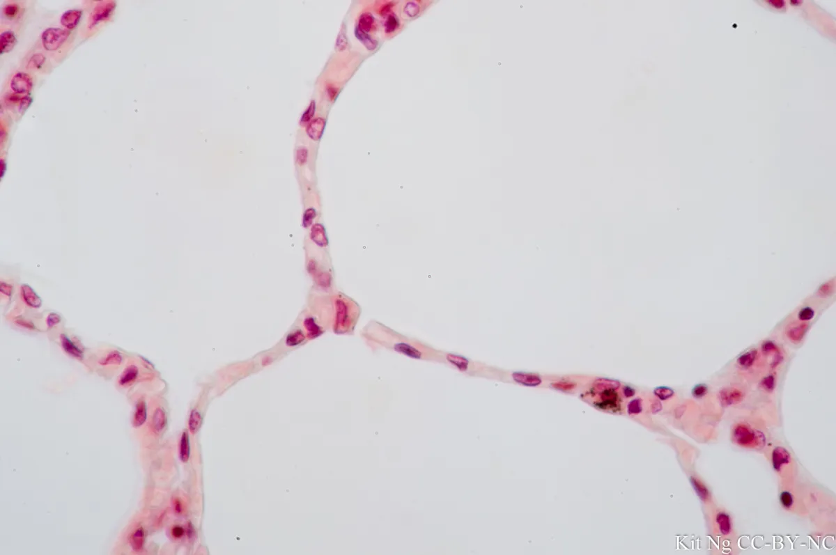
simple squamous

stratified squamous

simple cuboidal

simple columnar

pseudostratified columnar

transitional

connective tissue

adipose tissue

maxilla
zygomatic
mandible
lacrimal
palatine
incisive
nasal

parietal
frontal
temporal
occipital

spinous process
axis
transverse
articular
c3-c6
atlas

spinous process
transverse process
articular process
t1-t13

shaft
tubercle
neck
head (capitulum)

l1-l6
spinous process
articular process
transverse process

spinous process
articular process
sacral vertebrae
caudal vertebrae
transverse process

supraspinous fossa
spine of scapula
infraspinous fossa
subscapular fossa
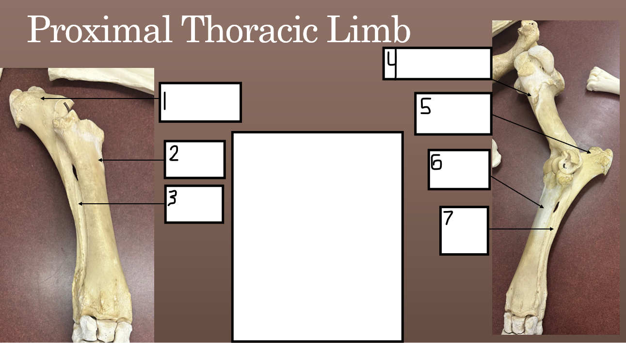
left olecranon
left radius
left ulna
left humerus
left olecranon
left radius
left ulna

carpals
3rd metacarpal
proximal phalanx
middle phalanx
distal phalanx

ischium
pubic bone
hooks
ilium
pins

left fibula
left tibia
left femur
left patella
left fibula
left tibia

tarsals
3rd metatarsal
proximal phalanx
middle phalanx
distal phalanx
right distal pelvic limb
left distal pelvic limb
calcaneus
tarsals
3rd metatarsal
Scapula justification
spine of scapula faces toward outside, supraspinous fossa positioned front, subscapular fossa on inside
Proximal and distal thoracic limb justification
with the olecranon pointing caudally, locate the radial tuberosity, if on the left, it’s a left limb and vice versa
Proximal pelvic limb justification
head of femur must face medially to articulate into hip socket of pelvis, the fibula will always sit on lateral side relative to tibia
distal pelvic limb justification
identify calcaneous, which will sit on lateral side, view limb from back, if calcaneous points toward the right it is right limb and vice versa
pelvis justification
the broad tuber coaxe of the ilium point cranially and laterally, the tuber ischii on the ischium point caudally
Masseter
origin- lower edge of the zygomatic arch
insertion- lateral surface of the mandible, up to its thickened lower edge
Masseter action
closes the mouth by lifting the lower jaw
Supraspinatus
origin- lateral surface of cranial part of scapula and its attached cartilage
insertion- cranial medial and lateral corners of the top of the humerus
supraspinatus action
stabilizes and extends shoulder
infraspinatus
origin- lateral surface of caudal part of scapula and its cartilage
insertion- lateral side of top of the humerus
infraspinatus action
stabilizes and rotates shoulder
deltoid
origin- spinal portion, spine of the scapula and surface of underlying infraspinatus muscle, caudal portion, upper caudal corner of the bony scapula
insertion- into the lateral surface of the humerus, third of the way down the bone
deltoid action
flexes shoulder, abducts limb
biceps brachii origin
from a bony bump on the lower cranial end of scapula
biceps brachii insertion
cranial medial corner of the top of the radius
ligament on the medial side of the elbow
by a long tendon that continues into tendon of extensor carpi radialis muscle, and indirectly into cranial surface of third metacarpal
biceps brachii action
flexes elbow, also extends shoulder
brachialis
origin- upper third of the caudal surface of the humerus
insertion- medial side of the radius near the top, slightly below and medial to insertion of biceps
brachialis action
flexes elbow
triceps brachii origin
long head: caudal border of the scapula
lateral head: upper lateral surface of the humerus
triceps brachii insertion
top and side of the olecranon of the ulna
triceps brachii action
extends elbow
longissimus dorsi origin
from medial and lateral surfaces of cranial part of pelvis, spinous process of thoracic and lumbar vertebrae, and cranial half of sacrum
longissimus dorsi insertion
attaches to sides of thoracic and lumbar vertebrae, upper ends of all ribs except first, and sides and tops of last four cervical vertebrae
longissimus dorsi action
extends/stabilizes vertebral column
latissimus dorsi
origin- surface of the spinal muscles from the top of the shoulder to lumbar region
insertion- medial surface of humerus, a little under halfway down the bone
latissimus dorsi action
draws humerus up and back
trapezius origin
continuous line along dorsal midline of the neck and chest
neck portion: starting at 2nd cervical vertebra down to top of shoulder
thoracic portion: from spinous process of thoracic vertebrae and connecting ligaments, from shoulder to middle of chest
trapezius insertion
neck portion: along full length of spine of scapula
thoracic portion: into bony expansion about one third of way down the spine of scapula
trapezius action
elevates/rotates scapula
gluteus medius
origin- top of longissimus, upper surface of ilium, strong ligaments that connect the sacrum to ilium and to tuber ischii
insertion- top and caudal side of upper end of femur
gluteus medius action
extends hip, abducts limb
quadriceps femoris origin
vastus lateralis: lateral surface of femur, just below the hip socket down about two thirds of bone
vastus medialis: medial surface of femur, just below the hip socket down about two thirds of bone
rectus femoris: two small areas on pelvis just cranial to hip socket
vastus intermedius: cranial surface of femur, beneath rectus femoris
quadriceps femoris insertion
all parts attach to cranial surface and upper edge of patella, through three patellar ligaments, quadriceps attach to cranial surface of top of tibia
quadriceps femoris action
extends stifle, rectus femoris also flexes hip
semimembranosus
origin- from caudal edge of sacrotuberal ligament and the tuber ischii
insertion: medial surface of lower end of femur
semimembranosus action
extends hip, adducts limb
semitendinosus
origin- first and second caudal vertebrae, tail fascia, and tuber ischii
insertion- fascia on medial side of leg, attaches to cranial edge of tibia and calcaneus
semitendinosus action
extends hip and hock, flexes stifle
gluteus superficialis
origin- tuber coaxe and lateral edge of ilium
insertion- third trochanter of femur, third of way down lateral side of bone
gluteus superficialis action
flexes hip, abducts limb
biceps femoris origin
long head: ligament connects to sacrum to ilium near third and fourth sacral spines, surface of gluteal muscles and tail
short head: tuber ischii
biceps femoris insertion
fuses into caudal surface of femur, lower ends form a wide tendinous sheet into leg fascia and stifle region
long head: patella and lateral patellar ligament
short head: lateral patellar ligament and cranial edge of tibia, leg fascia and calcaneus
biceps femoris action
extends hip/stifle/hock, can flex stifle