Ant&Post Abdominal Wall

0.0(0)
Studied by 0 people
call kaiCall Kai
learnLearn
examPractice Test
spaced repetitionSpaced Repetition
heart puzzleMatch
flashcardsFlashcards
GameKnowt Play
Card Sorting

1/29

encourage image

There's no tags or description

Looks like no tags are added yet.

30 Terms

1
New cards
<p>identify muscles of abdominal wall</p>

identify muscles of abdominal wall

1.External oblique (*inf and med direction)

2.Rectus abdominis (*R and L)

3.Tendinous intersection (*separates RA)

4.Linea alba (*fuses R and L RA)

5.Linea semilunaris

6.Internal oblique (*sup direction)

7.Transversus abdominis (*horizantal direction)

*obliques flatten out into tendon that forms sheath

*anterior and posterior rectus sheat (post for TA and part of IO)

2
New cards

boundaries of abdominal cavity

-superior boundary: diaphragm

-inferior boundary: no physical boundary

<p>-superior boundary: diaphragm</p><p>-inferior boundary: no physical boundary</p>
3
New cards

quadrants of the abdomen

-Upper Right: gallbladder, liver

-Upper Left: spleen, stomach

-Lower Right: appendix, ureter, ovary

-Lower Left: ureter, ovary

<p>-Upper Right: gallbladder, liver</p><p>-Upper Left: spleen, stomach</p><p>-Lower Right: <strong>appendix,</strong> ureter, ovary</p><p>-Lower Left: ureter, ovary</p>
4
New cards

layers of abdominal wall: fatty and membranous layer + muscular layer

-Fatty and membranous layers of superficial fascia (Not separable in lab; deep to skin)

-Muscles and investing fascia / aponeuroses (*deep to fascia)

  • Rectus sheath may be removed to show underlying muscles

*aponeroses=big flat tendon

<p>-<span>Fatty and membranous layers of superficial fascia (Not separable in lab; deep to skin)</span></p><p><span>-Muscles and investing fascia / aponeuroses (*deep to fascia)</span></p><ul><li><p style="text-align: left;"><span>Rectus sheath may be removed to show underlying muscles</span></p></li></ul><p>*aponeroses=big flat tendon</p>
5
New cards

deep layers of abdominal wall

knowt flashcard image
6
New cards

features of anterior abdominal wall part of past fetal circulation (what structures are contained in the umbilical ligaments)

-Urachus → Median umbilical fold

-Umbilical a → Medial umbilial fold

-Inf Epigastric a (supplies anterior ab wall, branch external illiac) → Lateral umbilical fold WHEN covered by periteinum

-Umbilical V → Ligamentum Teres

<p>-Urachus → Median umbilical fold</p><p>-Umbilical a → Medial umbilial fold</p><p>-Inf Epigastric a <mark data-color="purple" style="background-color: purple; color: inherit;">(supplies anterior ab wall, branch external illiac)</mark> → Lateral umbilical fold WHEN covered by periteinum</p><p>-Umbilical V → Ligamentum Teres</p>
7
New cards

Inguinal region + Inguinal Ligament + Inguinal Triangle

-transition abdominal wall and lower limb

-Inguinal Ligament: spans ASIS <-> Pubic Tubercle; inf part of EO

-Inguinal Triangle: inguinal ligament, rectus abdominis, inf epigastric vessels

<p>-transition abdominal wall and lower limb</p><p><strong>-Inguinal Ligament:</strong> <mark data-color="blue" style="background-color: blue; color: inherit;">spans </mark><mark data-color="red" style="background-color: red; color: inherit;">ASIS</mark><mark data-color="blue" style="background-color: blue; color: inherit;"> &lt;-&gt; Pubic Tubercle</mark>;<mark data-color="red" style="background-color: red; color: inherit;"> inf part of EO</mark></p><p><strong>-Inguinal Triangle:</strong> inguinal ligament, rectus abdominis, inf epigastric vessels</p>
8
New cards

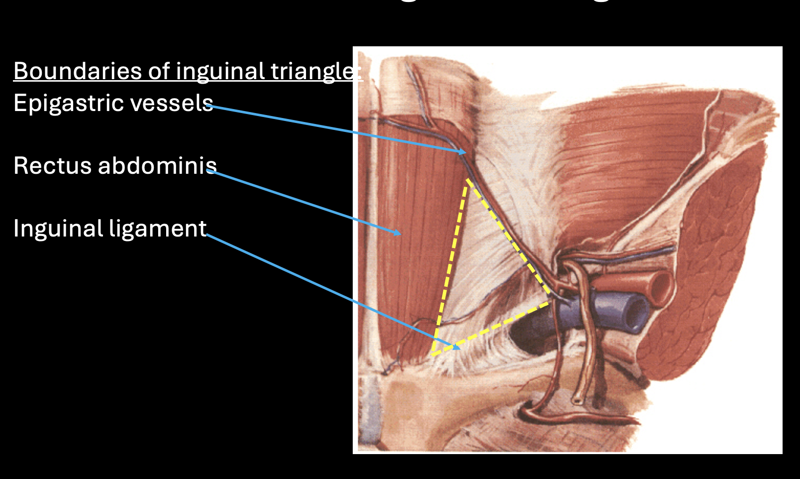
what are the boundries of the inguinal triangle?

-Inferior Epigastric vessels

-Rectus abdominis

-Inguinal ligament

*less thick part of body wall so high risk of hernia
<p>-Inferior Epigastric vessels</p><p>-Rectus abdominis</p><p>-Inguinal ligament</p><p>*less thick part of body wall so high risk of hernia</p>
9
New cards

inguinal canal: what is it?

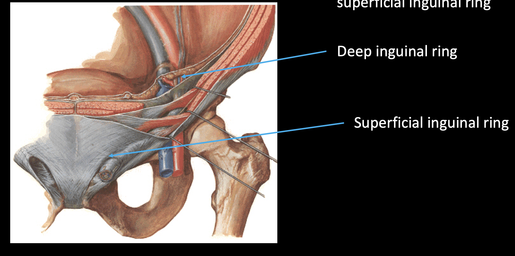
-Inguinal canal is a space (tunnel) that starts at deep inguinal ring and ends at superficial inguinal ring

  • deep=gap in fascia

  • superficial=opening/hiatus in EO


<p>-Inguinal canal is a space (tunnel) that starts at deep inguinal ring and ends at superficial inguinal ring</p><ul><li><p>deep=gap in fascia</p></li><li><p><mark data-color="purple" style="background-color: purple; color: inherit;">superficial=opening/</mark><strong><mark data-color="purple" style="background-color: purple; color: inherit;">hiatus in EO</mark></strong></p></li></ul><p></p>
10
New cards

Inguinal ring and nearby structures

knowt flashcard image
11
New cards

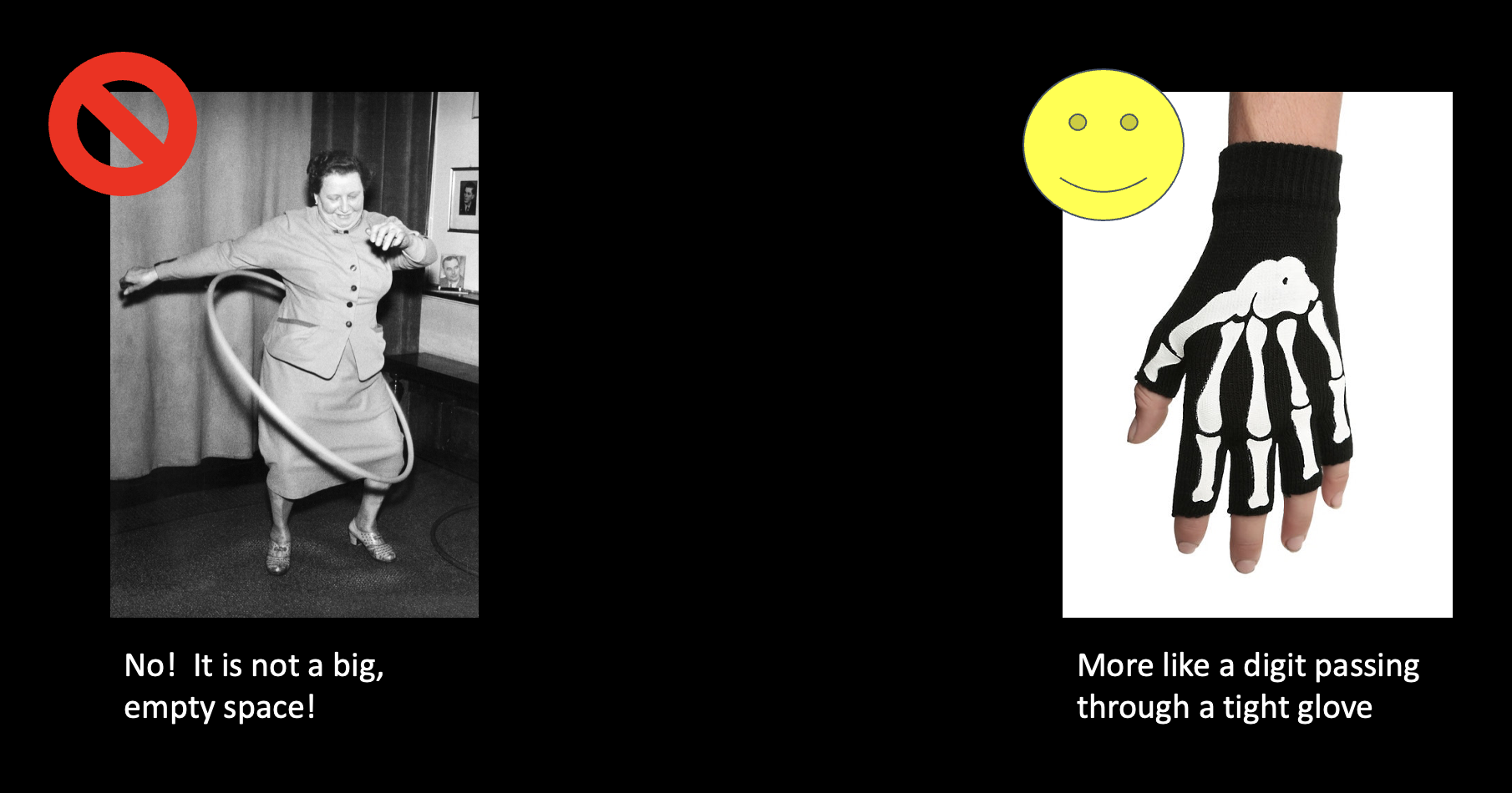
what does the inguinal ring resemble?

*if big would be a big risk for hernias
<p>*if big would be a big risk for hernias</p>
12
New cards

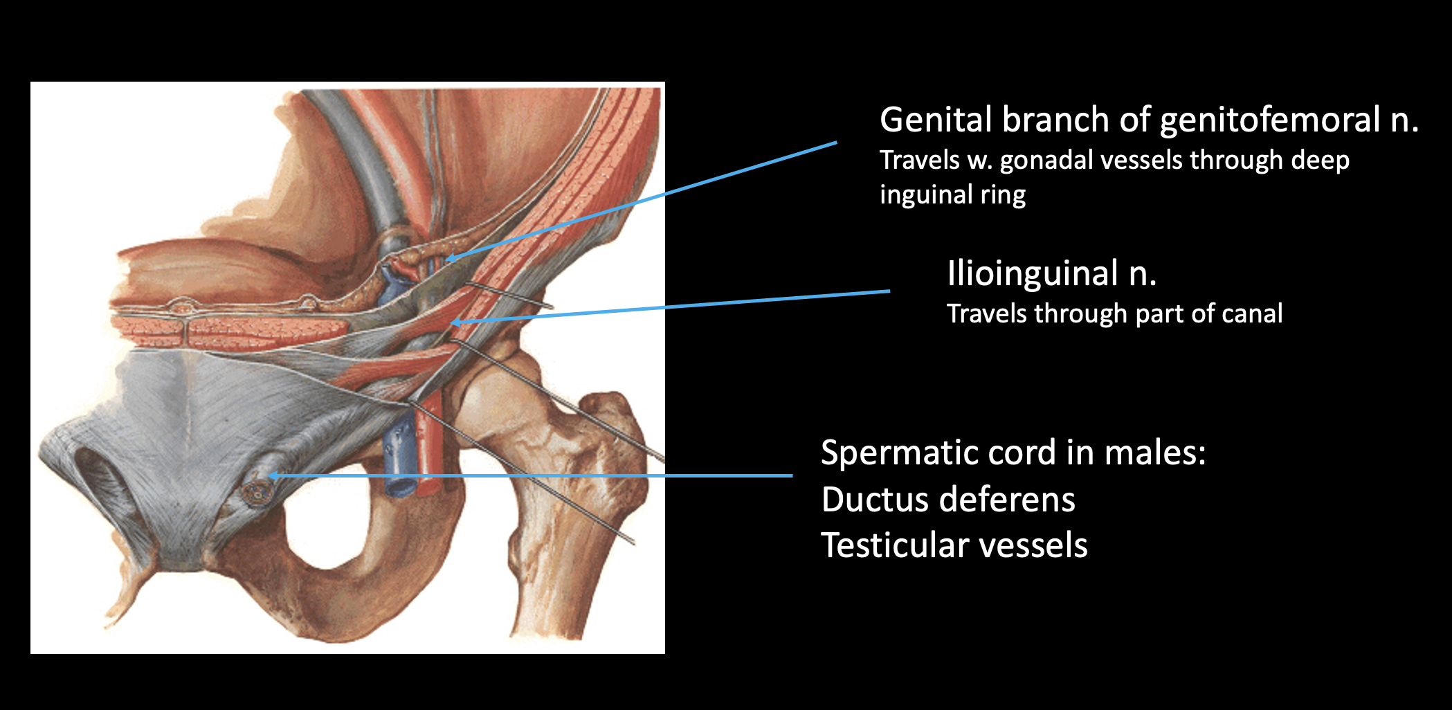
contents of the inguinal canal?

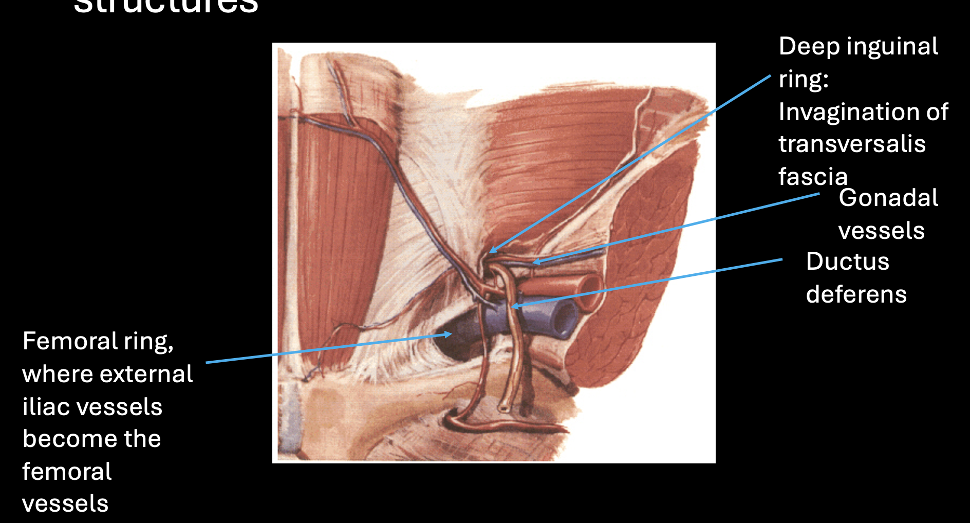
-Genital branch of genitofemoral n.: Travels w. gonadal vessels (in M) through deep inguinal ring

-Ilioinguinal n.: Travels through part of canal/just superficial ring

  • Ilioinguinal n. and genitofemoral n. are the same in all patients

-Spermatic cord in males: Ductus deferens + Testicular vessels

OR

-Round ligament of the uterus in females

  • The ovarian vessels do NOT travel in the inguinal canal (*in pelvis)

<p><strong>-Genital branch of genitofemoral n.</strong>: Travels <mark data-color="red" style="background-color: red; color: inherit;">w. gonadal vessels (in M) </mark>through deep inguinal ring</p><p><strong>-Ilioinguinal n.</strong>: Travels through part of canal/<em><mark data-color="purple" style="background-color: purple; color: inherit;">just superficial </mark></em><mark data-color="purple" style="background-color: purple; color: inherit;">ring</mark></p><ul><li><p style="text-align: left;"><mark data-color="red" style="background-color: red; color: inherit;">Ilioinguinal n. and genitofemoral n. are the same in all patients</mark></p></li></ul><p><strong>-Spermatic cord in males:</strong> Ductus deferens + Testicular vessels</p><p>OR</p><p><strong>-Round ligament of the uterus in females</strong></p><ul><li><p style="text-align: left;"><mark data-color="red" style="background-color: red; color: inherit;">The ovarian vessels do NOT travel in the inguinal canal (</mark>*in pelvis)</p></li></ul><p></p>
13
New cards

why an inguinal canal?

-The gonads begin their development in the abdomen (*~L1)

-The gonads then descend along a tract called the gubernaculum

<p>-The gonads begin their development in the abdomen (*~L1)</p><p>-The gonads then descend along a tract called the <strong>gubernaculum</strong></p>
14
New cards

gonadal position differs for males and females

Males:

-Gubernaculum pulls testis through the inguinal canal (*longer pathway)

-Testis located outside body cavity (*in scrotum)

-Thermoregulatory adaptation for optimal sperm production (*want lower temp than body)

Females:

-Gubernaculum pulls ovary from abdomen into pelvic cavity

-Ovary protected inside bony pelvis

-Protective adaptation for optimal ovum development

<p><u>Males:</u></p><p style="text-align: left;">-Gubernaculum pulls testis <mark data-color="blue" style="background-color: blue; color: inherit;">through the inguinal canal </mark>(*longer pathway)</p><p style="text-align: left;">-Testis located outside body cavity (*in scrotum)</p><p style="text-align: left;">-Thermoregulatory adaptation for optimal sperm production (*want lower temp than body)</p><p style="text-align: left;"><u>Females:</u></p><p style="text-align: left;">-Gubernaculum pulls ovary from <mark data-color="blue" style="background-color: blue; color: inherit;">abdomen into pelvic cavity</mark></p><p style="text-align: left;">-Ovary protected inside bony pelvis</p><p style="text-align: left;">-Protective adaptation for optimal ovum development</p>
15
New cards

how do the contents of the inguinal canal differ in males and females?

Males:

-The testis drags its blood vessels and the ductus deferens through the inguinal canal= SPERMATIC CORD

Females:

-Supportive ligaments of the uterus and ovaries derive from the gubernaculum= ROUND LIGAMENT

<p><u>Males:</u></p><p style="text-align: left;">-The testis drags its blood vessels and the ductus deferens through the inguinal canal= <strong>SPERMATIC CORD</strong></p><p style="text-align: left;"><u>Females:</u></p><p style="text-align: left;"><mark data-color="blue" style="background-color: blue; color: inherit;">-Supportive ligaments </mark>of the uterus and ovaries derive from the gubernaculum= <strong>ROUND LIGAMENT</strong></p>
16
New cards

list different types of abdominal hernias

-Inguinal: Direct and Indirect

-Umbilical

-Epigastric

-Spigelian

-Femoral

-Hiatal

17
New cards

inguinal hernias

-Inguinal region

-2 types:

Direct

-“Directly” through abdominal wall!

-Abdominal contents pass through the inguinal triangle

-Medial to the inf. epigastric vessels

Indirect

-The “scenic route” through the inguinal canal

-Abdominal contents pass through deep inguinal ring

-Lateral to the inf. epigastric vessels

<p>-Inguinal region</p><p>-2 types:</p><p><u>Direct</u></p><p>-“Directly” through abdominal wall!</p><p><mark data-color="red" style="background-color: red; color: inherit;">-Abdominal contents pass through the inguinal triangle</mark></p><p>-<strong>Medial</strong> to the inf. epigastric vessels</p><p><u>Indirect</u></p><p>-The “scenic route” through the inguinal canal</p><p><mark data-color="red" style="background-color: red; color: inherit;">-Abdominal contents pass through </mark><strong><em><mark data-color="red" style="background-color: red; color: inherit;">deep</mark></em></strong><mark data-color="red" style="background-color: red; color: inherit;"> inguinal ring</mark></p><p>-<strong>Lateral</strong> to the inf. epigastric vessels</p>
18
New cards

other hernias related to abdominal wall

-umbilical

-femoral

-epigastric

-spigelian

-umbilical: through umbilicus

-femoral: through femoral canal

-epigastric: through linea alba

-spigelian: through linea semilunaris

^bottom 2: if weak muscels/tendons (ex=pregnant, overweight); make sure RA still attached!

<p>-umbilical: through umbilicus</p><p>-femoral: through femoral canal</p><p><mark data-color="red" style="background-color: red; color: inherit;">-epigastric: through linea alba</mark></p><p><mark data-color="red" style="background-color: red; color: inherit;">-spigelian: through linea semilunaris</mark></p><p>^bottom 2: if weak muscels/tendons (ex=pregnant, overweight); make sure RA still attached!</p>
19
New cards

hital hernia

-stomach → diapgram → thorax

-can cause heartburn

*cases in lab!

<p>-stomach → diapgram → thorax</p><p>-can cause heartburn</p><p>*cases in lab!</p>
20
New cards

division of adbdominal cavity: which organs are intraperiteoneal vs retroperitoneal?

messentary=intraperiteneal

-Intraperitenal=stomach, liver and gallballder, spleen, first part duadenum, jejunum and ileum, cecum and appendix, transverse colon, sigmoid colon

-Retroperitenal(“SAD PUCKER”): adrenal glands, aorta and IVC, duodenum parts 2-4, pancreas, ureters, ascending and descending colon, kidney, esophagus, rectum

-alternalte intraparaternal and retroperiateails

  • helps aid in peristalsis


<p><mark data-color="blue" style="background-color: blue; color: inherit;">messentary=intraperiteneal</mark></p><p><strong>-Intraperitenal</strong>=stomach, liver and gallballder, <mark data-color="red" style="background-color: red; color: inherit;">spleen</mark>, first part duadenum, jejunum and ileum, cecum and appendix, transverse colon, sigmoid colon</p><p><strong>-Retroperitenal</strong>(“SAD PUCKER”): adrenal glands, aorta and IVC, duodenum parts 2-4, <mark data-color="red" style="background-color: red; color: inherit;">pancreas</mark>, <mark data-color="red" style="background-color: red; color: inherit;">ureters,</mark> ascending and descending colon, kidney, esophagus, rectum</p><p>-alternalte intraparaternal and retroperiateails</p><ul><li><p>helps aid in peristalsis</p></li></ul><p></p>
21
New cards

for future study…. match these names of mesentaris/ligaments to their organs

Falciform ligament

Ligamentum teres hepatis

Coronary ligament

Lesser omentum

  Hepatogastric ligament

  Hepatoduodenal ligament

Greater omentum

Gastrosplenic ligament

Mesentery proper

Transverse mesocolon

Sigmoid mesocolon


Falciform ligament

Liver to anterior abdominal wall and diaphragm

Ligamentum teres hepatis

Liver (in the free edge of the falciform ligament)

Coronary ligament

Liver to diaphragm

Lesser omentum

Liver to lesser curvature stomach and 1st part duodenum

Hepatogastric ligament

Liver to stomach

Hepatoduodenal ligament

Liver to duodenum

Greater omentum

greater curvature Stomach to transverse colon

Gastrosplenic ligament

Stomach to spleen

Mesentery proper

Jejunum and ileum (small intestine) + cecum to posterior abdominal wall

Transverse mesocolon

Transverse colon to posterior abdominal wall

Sigmoid mesocolon

Sigmoid colon to posterior abdominal wall

+Meso-appendix

22
New cards

muscles of the posterior abdominal wall

-Respiratory Disaphragm: anterior to other muscels

-Quadratus Lumborum: source low back pain

-Transverse Abdominis: lateral to QL

-Iliacus + Psoaas Major → Ilioposas (inf to IL)

-Psoas Minor

<p>-Respiratory Disaphragm: anterior to other muscels</p><p>-Quadratus Lumborum: source low back pain</p><p>-Transverse Abdominis: lateral to QL</p><p>-Iliacus + Psoaas Major → Ilioposas (inf to IL)</p><p>-Psoas Minor</p>
23
New cards

features of the respiratory diapram

-Median arcuate ligament

-Medial arcuate ligament: over psoas Major

-Lateral acurate ligament

-Aorta passing through aortic hiatus (@ T12 level)

<p>-Median arcuate ligament</p><p>-Medial arcuate ligament: over psoas Major</p><p>-Lateral acurate ligament</p><p><mark data-color="blue" style="background-color: blue; color: inherit;">-Aorta passing through aortic hiatus (@ T12 level)</mark></p>
24
New cards

Questions of Diapgragm features

-What passes posterior to each of the arcuate ligaments?

•Median

•Medial

•Lateral

-At what vertebral level does the aorta traverse the aortic hiatus?

•Median: aorta, thoracic duct, azygos and hemiazygous veins

•Medial: psoas major, smpathethic chain

•Lateral: quadratus lumborum, subcostal n/a/v

-T12

25
New cards
<p>identify lumbar plexus</p>

identify lumbar plexus

1.Subcostal (*inf to 12th rib)

2.Iliohypogastric

3.Ilioinguinal

4.Lateral femoral cutaneous: L2-3

5.Genitofemoral: w/ psoas major, L1-2

6.Obturator

7.Femoral

<p>1.Subcostal (*inf to 12th rib)</p><p>2.Iliohypogastric</p><p>3.Ilioinguinal</p><p>4.Lateral femoral cutaneous: L2-3</p><p>5.Genitofemoral: <mark data-color="purple" style="background-color: purple; color: inherit;">w/ psoas major, L1-2</mark></p><p>6.Obturator</p><p>7.Femoral</p>
26
New cards

nerve roots of lumbar plexus

*ventral ramus

<p>*ventral ramus</p>
27
New cards

what are the functions of the nerves of the lumbar plexus?

Provide motor and sensory innervation to the abdominal body wall and lower limb

*most both; LFC=sensory

28
New cards

muscle table: EO and IO

*intercostal n T8-12

<p>*intercostal n T8-12</p>
29
New cards

muscle table: Rectus Abdominis + Transverse Abdominus

knowt flashcard image
30
New cards

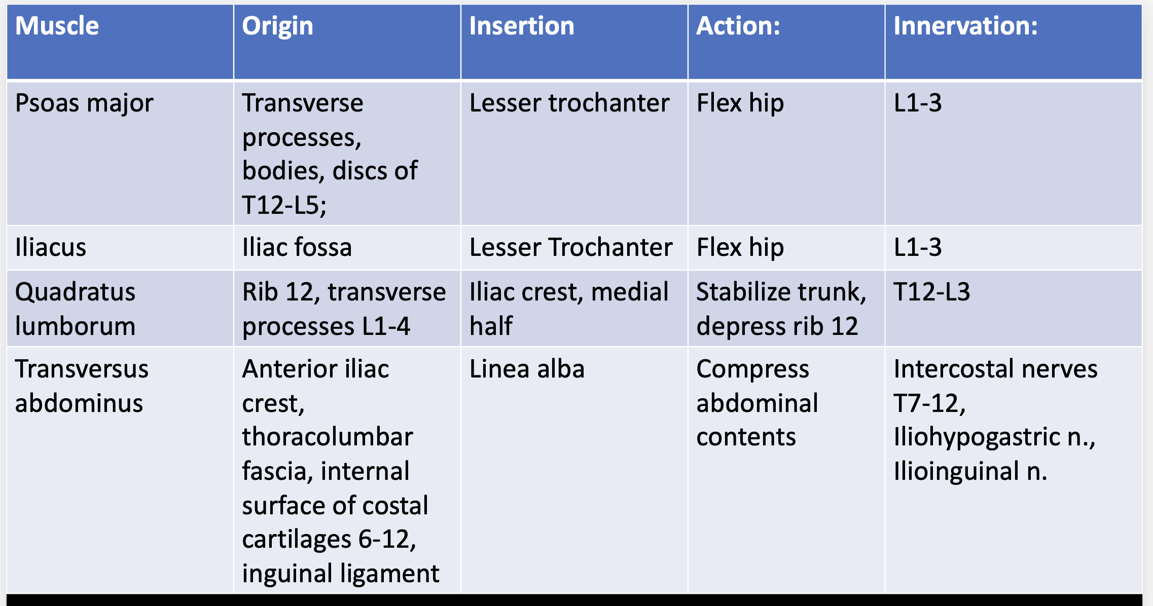
muscle table:

-psoas major

-iliacus

-quadratus lumborum

*QL in=T12-L3
<p>*QL in=T12-L3</p>