PATH 3810 Cardiovascular System
 Call Kai
 Learn
Learn Practice Test
 Spaced Repetition
 Match
Match Flashcards
 Knowt Play
1/86

There's no tags or description
Looks like no tags are added yet.
Name | Mastery | Learn | Test | Matching | Spaced | Call with Kai | Chat |
|---|
No analytics yet
Send a link to your students to track their progress
87 Terms
divisions of the cardiovascular system
blood circulatory system
lymph vascular system
functions of the cardiovascular system
transport of oxygen, nutrients, metabolic waste products, hormones, leukocytes
temperature regulation
atria
upper two chambers of the heart that receive and collect blood
ventricles
lower two chambers of the heart that pump blood out of the heart
septum
muscle that divides the heart into left and right halves
valves
4 heart valves that prevent backflow of blood
tricuspid valve
found between RA and RV
pulmonary semilunar valve
found between RV and pulmonary artery
mitral (bicuspid) valve
found between LA and LV
aortic semilunar valve
found between LV and aorta
deoxygenated blood from the body flows through the
inferior and superior vena cava
the vena cavae empty into…
…the right atrium
blood from the right atrium passes through…
… the tricuspid valve
blood passes through the tricuspid valve into…
…the right ventricle
blood from the right ventricle passes through…
…the pulmonary semilunar valve
blood flows through the pulmonary semilunar valve into…
…the right and left pulmonary arteries
pulmonary arteries take blood to…
…the lungs
the lungs send the blood to…
…the pulmonary veins
the pulmonary veins enter blood into…
…the left atrium
blood from the left atrium flows through…
…the mitral valve
blood flows from the mitral valve to…
…the left ventricle
blood from the left ventricle passes through…
…the aortic valve
blood flows from the aortic valve to…
…the aorta
pulmonary circulation
movement of blood from the heart to the lungs to the heart
systemic circulation
movement of blood from the body to the heart to the body
Which side of the heart is larger?
the left side
tunica intima
endothelium (simple squamous)
loose CT
+/- internal elastic lamina
tunica media
smooth muscle cells, elastin, collagen, ground substance
+/- external elastic lamina
tunica adventitia
outer connective tissue and smaller blood vessels (vasa vasorum), nerves (nervi vasorum), and lymphatic vessels
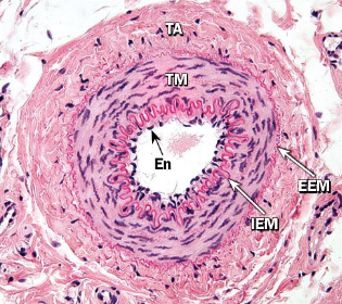
cross-section of an artery
tunica intima of the heart
endocardium
tunica media of the heart
myocardium
tunica adventitia of the heart
epicardium/visceral pericardium
order of circulatory layers from innermost to outermost
tunica intima → tunica media → tunica adventitia

layers of the heart

skeletal muscle cross-section
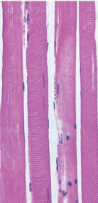
skeletal muscle longitudinal section

smooth muscle cross-section

smooth muscle longitudinal section

cardiac muscle
intercalated discs
bind cardiac cells, transmit forces of contraction, provide areas of low electrical resistance
cardiac conduction system consists of
sinoatrial node
atrioventricular node
bundle of His and Purkinje fibers
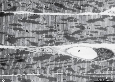
electron micrograph of cardiac muscle
the sinoatrial node is located…
…in the wall of the right atrium
impulse from the SA node passes through atria to…
…the atrioventricular node
the P-Q wave represents the
depolarization of the SA node, which causes atrial systole
the Q-R-S wave represents the
depolarization of the AV node, which causes ventricular systole
the S-T wave represents the
ventricles repolarizing
atrial repolarization is not seen on an EKG because
it happens during the AV impulse
P wave
atrial depolarization
QRS
ventricular depolarization
T wave
ventricular repolarization
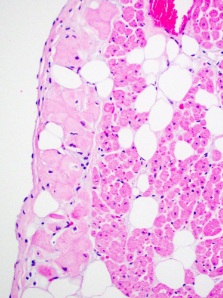
purkinje fibers
purkinje fibers
specialized cardiac fibers that lie beneath the endocardium, then penetrate the myocardium
appear larger and with paler sarcoplasm than typical cardiac fibers
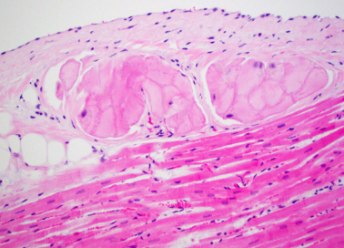
purkinje fibers
heart valves
leaflets of fibroelastic tissue (condensed endocardium)
covered by endothelium
prevent retrograde flow
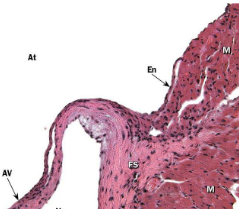
cardiac valve
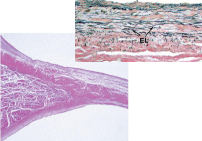
heart valve
arterial vessels
high-pressure distribution system from heart to capillaries
microcirculation vessels
capillary beds primarily concerned with gas, nutrient, waste exchange
venous vessels
low-pressure system collecting blood for return to the heart
endothelial cells
crucial in integrity and circulatory function of vessels
permeability barrier
secrete anticoagulants at rest and procoagulants when activated
secrete vasoconstrictive and vasodilative factors
anticoagulants produced by endothelial cells
prostacyclin
thrombomodulin
others
procoagulants secreted by endothelial cells
von willebrand factor
tissue factor
others
vasoconstrictive molecules secreted by endothelial cells
endothelin
vasodilative molecules secreted by endothelial cells
nitric oxide
arterioles
terminal branches of the arterial tree
arteries
elastic ± muscular, large diameter, thick tunica media with concentric layers of elastic laminae between collagen and smooth muscle
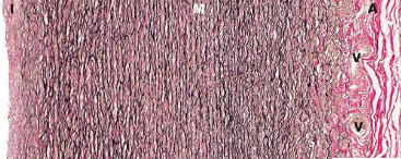
elastic artery under von giesin stain
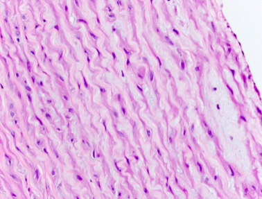
elastic artery under h & e stain
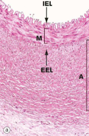
muscular artery
muscular arteries have…
…internal elastic lamina, which lies between tunica intima and tunica media
…may include external elastic lamina, less prevalent as vessels get smaller

muscular artery
arterioles
progression from small muscular arteries to arterioles is gradual, involving loss of IEL and reduction in smooth muscle layers
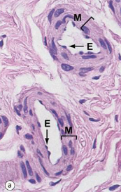
arterioles

electron micrograph of arteriole
microcirculation
involves arterioles, capillaries, and venules
capillaries
lumen diameter similar to that of red blood cells
single layer of endothelial cells joined by tight junctions and supported by basement membrane
no medial or adventitial layers
weibel-palade bodies
store von willebrand factor (aka factor VII)
found in capillary endothelial cytoplasm
pericytes
flattened cells surrounding epithelium with contractile properties
may or may not be found at capillaries
types of capillaries
continuous
fenestrated
sinusoidal
continuous capillaries
endothelial cells joined by tight junctions
exchange across endothelium by passive diffusion, pinocytosis, intercellular mechanisms
fenestrated capillaries
located in tissues with extensive exchange
continuous basement membrane
fenestrations allow enhanced permeability
sinusoidal capillaries
large fenestrations with discontinuous basement membrane
larger lumen
found in bone marrow, spleen, liver
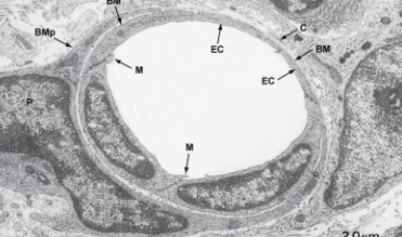
electron micrograph of capillary

electron micrographs of capillaries
venous system