Cell Bio Exam 2 Guide
 Call Kai
 Learn
Learn Practice Test
 Spaced Repetition
 Match
Match Flashcards
 Knowt Play
1/61

There's no tags or description
Looks like no tags are added yet.
Name | Mastery | Learn | Test | Matching | Spaced | Call with Kai | Chat |
|---|
No analytics yet
Send a link to your students to track their progress
62 Terms
membrane bound proteins
proteins attached to the cytosolic face of the ER and are engaged in synthesis of secretory proteins

cytosolic free ribosomes
proteins not attached to any cellular membrane and are engaged in synthesis of non-secretory proteins

Smooth ER
specializes in synthesizing lipids
Rough ER
has a series of flattened spaces and acts as a binding site for ribosomes during translation → specifically when proteins are being synthesized
Microsomes
can be either rough or smooth. A homogenizer is utilized to break down rough and smooth ER into a mixture → can be separated using density centrifugation. smooth have LOW densities and stop sedimenting at LOW sucrose concentrations, while rough have HIGH densities and stop sedimenting at HIGH sucrose concentrations

Cotranslational translocation
a ribosome begins reading mRNA to make a protein, this new protein will obtain a N-terminal signal sequence, a helper component Signal Recognition Particle (SRP) helps carry this protein to the rough ER, the protein then docks on the ER and feeds into the membrane while its still being generated
Cotranslational translocation factors
SRP, SRP receptor in membrane, a translocon (in the membrane) that allows protein to feed through ER, and GTP hydrolysis

Cotranslational translocation for soluble secretory proteins
Ribosome begins reading mRNA and generating a protein while free floating in the cytoplasm, the generated signal sequence binds to the SRP and directs where to go on rough ER. Here the SRP receptor binds and the ribosome attaches to a translocon to feed the protein through. GTP hydrolysis occurs as SRP detaches, and signal peptidase cleaves the off the signal sequence

Cotranslational translocation in yeast
Translocon in Sec61a, the SRP receptor is Sec63complex, BiP is a cofactor that binds to ATP for activation and creates a complex that stabilizes the growing protein chain into the ER lumen
Direction for protein synthesis
N terminus → C terminus (N terminus comes out of the ribosome first)
Type I terminal sequence
The only type of signal sequence that will cut (ie. insulin receptor)

Type II terminal sequence
A non-cutting terminal sequence that requires a single internal hydrophobic anchor sequence → this sequence serves as the ER targeting signal and becomes the hydrophobic transmembrane spanning sequence. Consequently, the N terminal extends to the cytosol

Type III terminal sequence
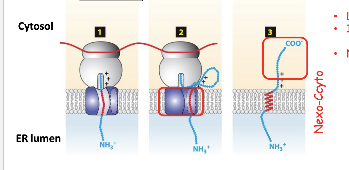
a non-cutting terminal sequence that requires a single internal hydrophobic anchor sequence. The N terminal is in the ER lumen, and the C terminal is in the cytosol
Insertion of Tail-anchored proteins
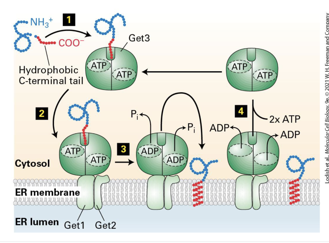
a non-cutting terminal sequence that does not utilize SRP. The hydrophobic tail sequence acts as SRP - it needs ATPase to bind. The tail extends into the cytosol (meaning the N terminus is in the cytosol)
Type IV terminal sequence - multipass
a non-cutting terminal sequence. The first transmembrane segment is C terminal facing the cytosol. Each subsequent segment takes the opposite orientation as the one before - an even number of transmembrane anchored proteins will then mean that the N terminus and C terminus point in same directions

GPI Anchored Proteins
Composed of a fatty acid tail with an opposing polar end and it links to a protein → polar end connects to the C terminus of a protein in the ER lumen

Protein modifications before final destinations
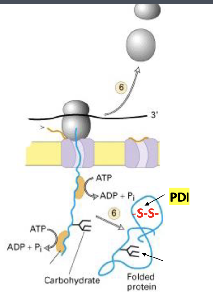
glycosylation in the ER and Golgi (adding carbohydrate chains), formation of disulfide bonds to the ER, proper folding of polytide assembly to the subunits, and proteolytic cleavage in the ER, Golgi, and vesicle
Resident ER proteins
Proteins that aid in translocation and folding
Binding Protein (BiP)
the “chaperone” protein that aids in folding and multimeric assembly that prevents back sliding and unfolded protein response - also assists in translocation

Protein Disulfide Isomerase (PDI)
catalyzes oxidation of free SH groups to S-S bridges - helps proteins achieve correct folded structures
Sugars and Glycoproteins
____ on _____ get modified as they move through the Golgi
Endosome
Receives material that has entered the cell via endocytosis and decides where molecules should go
Lysosome
acidic organelles that receive materials intended for degradation - cell digestion
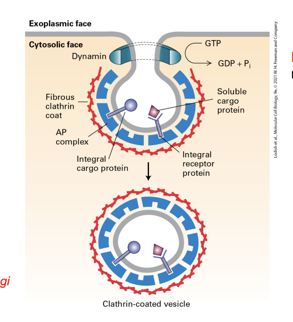
Dynamin
Required for clathrin coated vesicles → it pitches the neck of the bud and required GTP hydrolysis
S-S Formation and Rearrangement
the ionized S- of the substrate protein reacts with S-S bond in oxidized PDI to form and intermediate. Then the second ionized S- of the substrate protein reacts with intermediate to form a S-S within the protein

Glycosyltransferases
An enzyme that adds carbohydrate chains attached to the -OH in serine and threonine OR carbohydrate chains to the amide nitrogen (N2H) of asparagine
Biosynthesis of N-linked Oligosaccharaide precursor
the building of a large sugar chain that will attach to the protein in the ER - 3x glucose, 9x mannose, and 2x N-acetylglucosamine → all of it is assembled on dolichol phosphate and gets flipped to ER lumen

Protein glycosylation (N-linked oligosaccharide) in the RER
once biosynthesis of precursor chain is assembled - three glucose present signal this, the ER trims its designated components, and is then transported to the cis-Golgi

Unfolded Protein response
Ire1 monomers form dimers. This causes endonuclease activity to splice Hac1 mRNA. That spliced Hac1 mRNA is translated and taken back to the nucleus

Modification of N-linked Oligosaccharides
Used to monitor folding and quality control. One glucose is retained to bind to CNX/CRT. To retain the misfolded protein, UGGT can add a glucose and reflow binding. However if the protein cannot be folded, ERa-mannosidase will reduce mannose residue and bind the protein to OS-9 for degradation

Calnexin and Calreticulin
components in protein folding that bind to N-linked oliggosaccharaides and prevent incorrect folding with adjacent proteins

Coated vesicles
Compartments that transport proteins between cellular compartments. Their coat proteins (COPI, COPII, or clathrin) help select cargo and direct vesicle trafficking to specific destinations
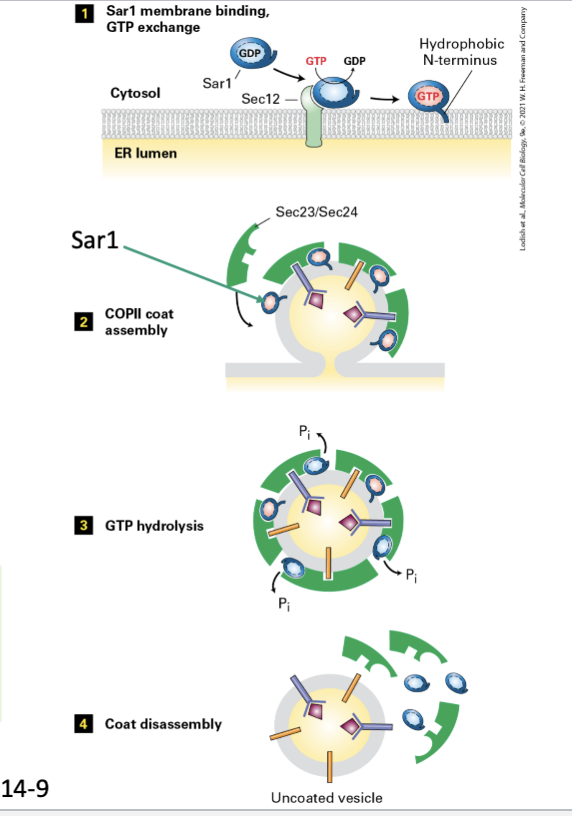
COPII
vesicle coat type that transports proteins FROM the ER to the cis-Golgi (Sar1) → Sar1 serves as binding site for the coat proteins, after the coat is complete, detachment of coat occurs. Signal sequence is Di-acidic (Asp-X-Glu)
COPI
vesicle coat type that transports proteins FROM the cis-Golgi, to the ER, and later to the Golgi cisternae. signal sequence is KEDEL and GTPase is ARF

Cathrin (and adapter proteins)
vesicle coat type that transports proteins FROM the trans-Golgi TO the endosome (ARF)

Rab GTPase
Small GTP-binding proteins that regulate vesicle targeting and docking. In their active GTP-bound state, they recruit tethering proteins that guide transport vesicles to the correct target membrane. This means v-SNARE proteins attach, formulate the SNARE complex with t-SNARE proteins on target membrane, and assimilate membrane fusion

Anterograde Transport by COPII
Forward vesicle transport from the ER to the cis-Golgi → requires di-acidic sorting signal for cargo protein
Retrograde Transport by COPI
Reverse transport from the cis-Golgi to the ER for particles that need to be returned. It retrieves v-SNAREs and other misc. items. The high pH in the golgi promote binding between KEDL and its receptor → it is taken back to a lower pH in the ER where the binding detatches
M6P
a carbohydrate marker added to lysosomal enzymes in the cis-Golgi apparatus. It is recognized by M6P receptors in the trans-Golgi network, which package the enzymes into clathrin-coated vesicles for transport to endosomes and ultimately lysosomes

Endocytosis
Receiving lysosomal enzymes from outside of the cell to the late endoscope (low pH) - involves budding, coating, and disassembly processes
Transocytosis
Involves endocytosis from blasolateral membrane and moves and fuses to the apical membrane (ie. move materials from one side to another side)

Internalization
Cells uptake macromolecules from surrounding outside intracellular destinations
Cell Polarity
ability of a cell to generate functionally distinct regions within itself
Cytoskeleton
composed of microfilaments, microtubules, and intermediate filaments
Microfilaments
rigid, high dynamic, and polarized protein fiber network of actin-binding proteins - motor protein is myosin → actin binds to ATP
Microtubules
rigid, high dynamic, and polarized tube shaped structures of tubulin → motor proteins are kinesis and dyneins
Intermediate Filaments
strong, less dynamic, and non polarized type of cytoskeletal structure protein that provides mechanical strength and structure to cells and tissues → composed of lamins and DO NOT have motor proteins
Critical Concentration (Cc)
the minimal concentration of monomers at which filaments form → when added short stable actin filaments as the starting point, elongation happens immediately

Microtubule Dynamics
Bulk tubulin versus individual microtubulin → bulk has its concentration above the critical point and polymerization assembly occurs while dynamic grows and shrinks overtime

Formats of actin
G-actin and F-actin
Dynamics of actin
nucleation, elongation, and steady state
Profilin
Binds to G-actin on the side opposite of the nucleotide binding cleft → binds to the (+) end and promotes filament growth by adding actin monomers to it
Cofilin
Binds to ADP-actin subunits with F-actin on the (-) end→ promotes depolymerization of filaments and actin disassembly - induces a small twist and allows more free (-) ends
CapZ
an actin capping protein that binds to the positive (+) end and prevents filament growth
Tropomodulin
an actin-capping protein that binds to the minus (−) end and prevents filament growth or shrinkage at that end
Thymosin-B4
STores actin filaments that cannot be added onto another monomer (buffer)
Arp 2/3 Complex
Creates a starting point for branched filament assembly. Nucleation promoting factors (NPF) bind to actin via WH2. This complex then changes its conformation as it binds to 2 more NPFs. After start is initiated NPF is released. This mechanism grows from the (+) end until it meets Zap (stop codon)
Listeria Monocytogenes
A bacteria obtaining ActA. This activates the host cell's Arp2/3 complex - since Arp2/3 generates branched actin filaments, it propells the bacterium through the cytoplasm and enables the cell to spread

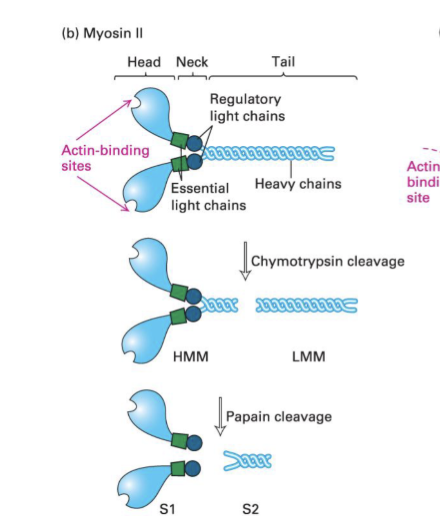
Myosin
Motor protein for actin filaments → the head domain interacts with actin filaments (S1 = the head). The distance filaments travel depends on their neck lengths
Class I Myosin
Step size is 10-14 nm, functions in membrane association + endocytosis, and moves in the (+) direction towards the barbed end

Class II Myosin
A (+) end directed actin motor protein with two motor heads and a long coiled-coil tail. Its step size is 8nm and it generates force for muscle contraction and cellular contractility

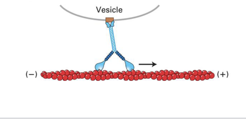
Class V Myosin
Step size is 36 nm, and it uses two motor heads to transport vesicles along actin filaments (cunty motor protein edit) for organelle support