Histology Lab Midterm
 Call Kai
 Learn
Learn Practice Test
 Spaced Repetition
 Match
Match Flashcards
 Knowt Play
1/100

There's no tags or description
Looks like no tags are added yet.
Name | Mastery | Learn | Test | Matching | Spaced | Call with Kai | Chat |
|---|
No analytics yet
Send a link to your students to track their progress
101 Terms

Transitional Epithelia

Simple Squamous Epithelia

Simple Cuboidal Epithelia
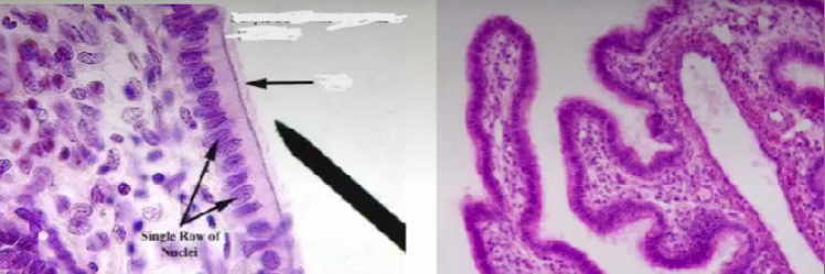
Ciliated Simple Columnar Epithelia
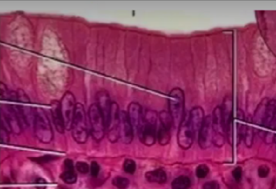
non-ciliated simple columnar epithelia

Non-keratinized stratified squamous epithelia
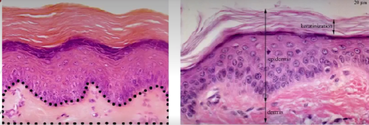
keratinized stratified squamous epithelia

stratified cuboidal epithelia

stratified columnar epithelia

pseudostratified ciliated columnar epithelia

non-ciliated pseudostratified columnar epithelium

brush border (microvilli)

cilia

Loose (areolar) CT

what type of CT is this and what are the numbers labeling?
Loose (areolar) CT
fibroblast
elastic fibers
collagen fibers
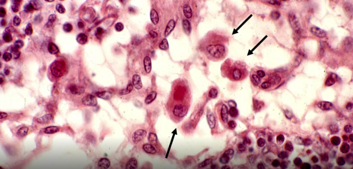
macrophages
capillaries

Dense Irregular CT
Dense irregular CT
fibroblast
collagen fiber
elastic fiber
arteriole
venules

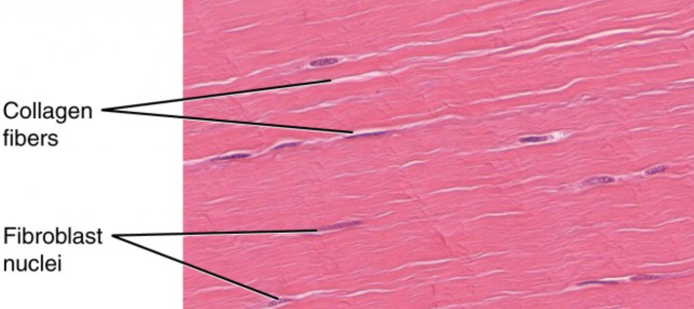
dense regular CT
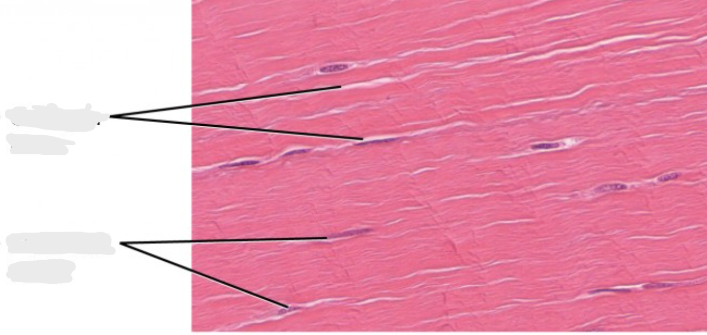
what type of CT is this and what are the lines pointing to?
Dense regular CT

adipose tissue
top - white (unilocular)
bottom - brown (multilocular)
lipid droplet
chondroblast
chondrocyte
lacunae

hyaline cartilage

what type of cartilage is this and label the structures
hyaline cartilage
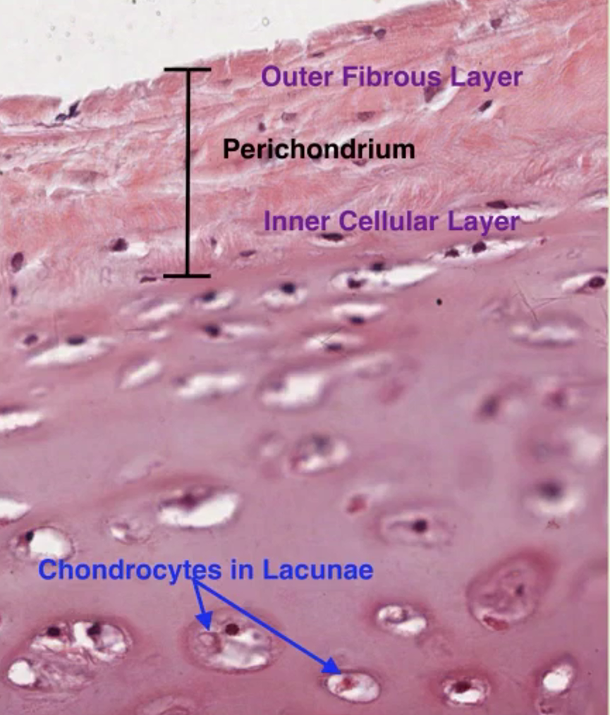

elastic cartilage

fibrocartilage

A & B - osteoblast
C - osteocyte
D - osteoclast
E - osteoid/bone matrix

A - canaliculi
B - osteocyte lacunae
C - Haversian (central) canal
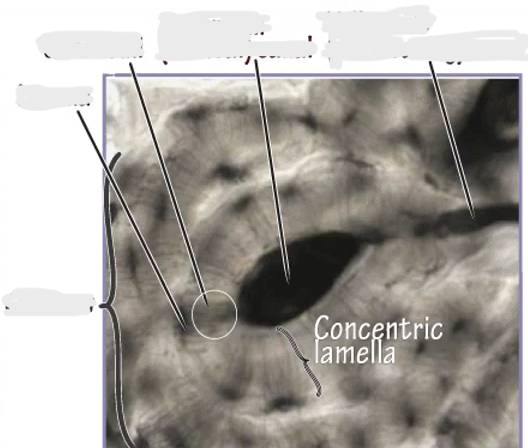
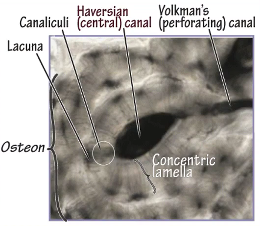

A - osteon
B - Haversian (central) canal
C - Interstitial lamella
D - osteocyte

epiphysis
metaphysis
diaphysis
periosteum
endosteum
bone marrow cavity
hypertrophied chondrocytes

bone development
proliferative zone chondrocytes
hypertrophic zone chondrocytes
vascular invasion front
secondary ossification center

Zone of reserve
zone of proliferation
zone of hypertrophy
zone of calcification
zone of ossification


skeletal muscle
skeletal muscle
endomysium
myofibrils
sarcomere
peripheral nuclei

sarcomere
A - Z disc
B - I band
C - A band
D - H band
*M line (may not be visible)

cardiac muscle

cardiac muscle
A - central nuclei
B - intercalated disk
C - cardiomyocyte

lipofuscin
purkinje fibers
cardiac muscle endomysium

smooth muscle

smooth muscle
central nuclei
fibers
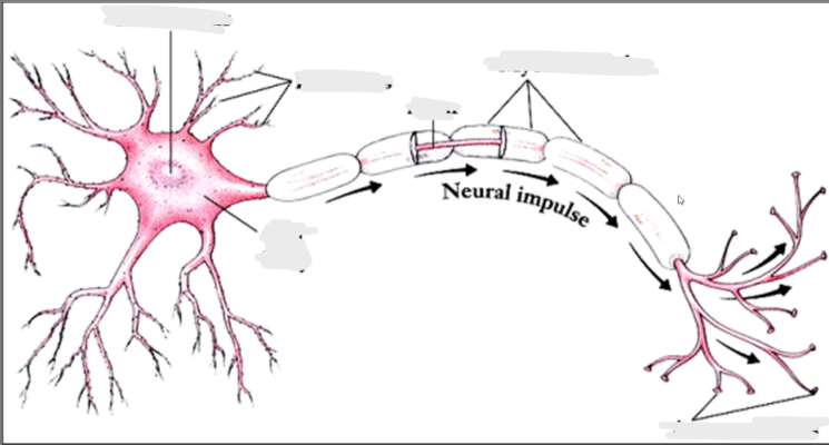

what is an axon hillock
the point where the cell body of the neuron merges into the axon
Glial cells
supporting cells of the CNS

microglia

fibrous astrocyte (white matter)

protoplasmic astrocyte (grey matter)
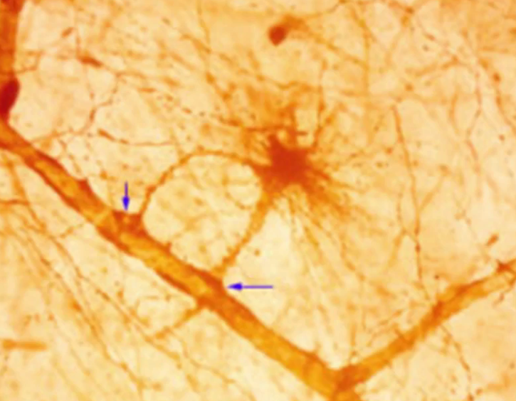
oligodendroglia/oligodendria


central canal
gray commisure
spinal cord
grey matter
white matter
anterior horn (ventral)
posterior horn (dorsal)
posterior median sulcus
anterior median fissure
spinal cord - motor neuron

pyramidal neuron
gyri
cerebrum
white matter
grey matter

satellite cells

A - Schwann cells
B - Axon
C - Myelin Sheath
cerebellum
folia

cerebellum
molecular layer
Purkinje neuron/cell
granular layer
grey matter
white matter
PNS ganglia
dorsal root/spinal ganglia
pseudounipolar neurons
multipolar neurons
peripheral nerve
fascicles of nerve fibers
myelin
node of Ranvier
peripheral nerve
endoneurium
perineurium
epineurium

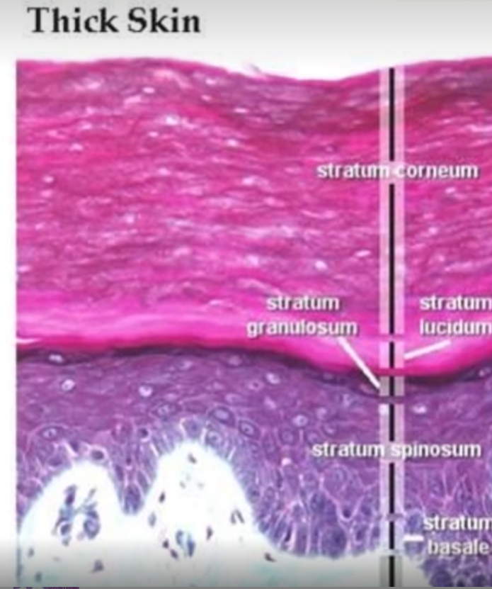

melanocytes

Merkel’s cell

keratinocytes

keratin layer
Langerhans cells

label number 4 and 5
dermal papillae
epidermal ridge

dermal layer
A - reticular layer
B - papillary layer

hair follicle
sebaceous gland

Pacinian Corpuscle
Meissner’s corpuscle
*(ID on image not a slide)
hypodermis (lies below dermis of integument and is not part of it)
artery
elastic artery
muscular artery
artery
tunica intima
tunica media
tunica adventitia
artery - tunica intima
internal elastic lamina
sub-endothelial layer of CT
endothelium
artery - tunica media
smooth muscle
elastic fibers
artery tunica adventitia - vasa vasorum
arteriole
capillary
capillary - pericytes
venule
vein
vein
tunica intima
tunica media (may be very thin)
tunica adventitia
vein - tunica intima
sub-endothelial layer of CT
endothelium
vein - tunica media
smooth muscle
vein - tunica adventitia
vasa vasorum
longitudinal smooth muscle (in large vein)
erythrocyte
reticulocyte
leukocyte
platelets
Granulocytes
neutrophil
eosinophil
basophil
agranulocytes - large lymphocyte
agranulocyte - small lymphocyte
agranulocyte - monocyte