Physiological Psychology Quiz 6
 Call Kai
 Learn
 Practice Test
 Spaced Repetition
 Match
 Flashcards
 Knowt Play
1/31

There's no tags or description
Looks like no tags are added yet.
32 Terms
Cochlear implants can help treat some forms of sensorineural deafness

Left: Image of a person with a cochlear implant, showing the external components (transmitter and microphone)
Right: Cochlear implant X-ray; each of the 29 surgically implanted beads is a stimulating electrode that excites the auditory nerve at positions along the organ of corti that correspond to successively lower frequencies from the base to the apex (tonotopy)
While hair cells are lost, the auditory nerve fibers are still there but there is nothing to stimulate them, sensorineural hearing loss
Transmitter → transmits electrical signals to cochlea
Microelectrodes implanted into the cochlea, they are stimulating electrodes, causes stimulation in response to sound based on the frequency
Electrodes are along the band, and
Tonotopic arrangement
Can help distinguish sounds that cannot be distinguished through lip reading alone
The earlier in life this is done, the better the outcome
Adults don't usually do this, under 1 year is optimal
Because of plasticity in the auditory cortex
Synaptic plasticity
Dendritic growth
If you can apply the right amount of stimulation early in life, it works best
As you get beyond this critical period, the gains become less
Cochlear implants do not fully replicate normal human hearing, they treat some forms of Sensorineural Deafness by essentially replacing inner hair cells

But over time the brain can adapt to the auditory signal
Takes training
People with cochlear implants struggle with music perception
Requires the ability to discriminate really small changes in pitch
They perceive music at flat and non-melody rich
With training + experience, people get better with differentiating sounds
A microphone detects sounds and directs the cochlear implant circuitry to stimulate the auditory nerve at frequency-specific locations along the cochlea corresponding to the frequency/pitch of the sound
This provides a somewhat crude simulation of hearing, but the brain can use this information to decipher speech and other sounds, and it gets better with training
Auditory System Summary/Review
The human ear consists of the outer, middle and inner ear, with each contributing to the sensation of sound
The inner ear includes the cochlea, which transduces sound waves into a tonotopic map of sound frequencies based on the changing biophysical properties of the basilar membrane
The cochlear apex is tuned to low frequencies (~20 Hz), while the base is tuned to high frequencies (~20 kHz)
Outer hair cells adjust the sensitivity of the basilar membrane, while inner hair cells are the auditory sensory receptors, responsible for transducing sound information to the brain
Hair cells have stereocilia which contain K+/Ca2+ channels that are mechanically opened when bent in the direction of the tallest stereocilium
Tonotopy is preserved from the level of the cochlea all the way to primary auditory cortex—this is thought to be important for the perception of “pitch” mainly through “place coding”
Brainstem auditory nuclei use different methods to localize sound depending on the frequency/pitch of the sound (low frequency = interaural time differences; high frequency = interaural intensity differences)
There are three main categories of hearing loss depending on the location of the auditory issue
Some forms of sensorineural deafness can be treated with cochlear implants which directly stimulate the auditory nerve—it’s a bit crude, but gets better with training!
VISION
Eye has evolved like 50 times
Eye pupil can take different shapes
Do we see the world the same way a traditional camera does?
Some things are the same
But there’s differences
To focus, camera lens moves
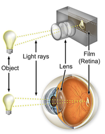
Lens
Refracts/bends light
Ours doesn’t move back and forth tho
High focuses in on a spot in the back of the eye
Sensory transduction in the visual system

Light rays converge on a single point
Retinal cells:
Cones
Rods
(Photoreceptors)
These two are responsible for sensation
Also taking light rays and turning them into action potentials
There are more than just rods and cones in retina
Retina is very thin, but it has 3 different layers
Sensory transduction
Sensory info makes it to primary visual cortex
Then higher cortices
Perception is about reconstruction
This process is subjective, each brain does it differently
Perception is all about interpretation by the brain!
Visual transduction: The process of converting living waves (sensory signal) from the external environment into an electrical signal (ex. action potentials) that the brain interprets–this process occurs in photoreceptors in the retina

Images are ambiguous

Brain is making a decision on which color is the foreground or background
Why is this static image moving?
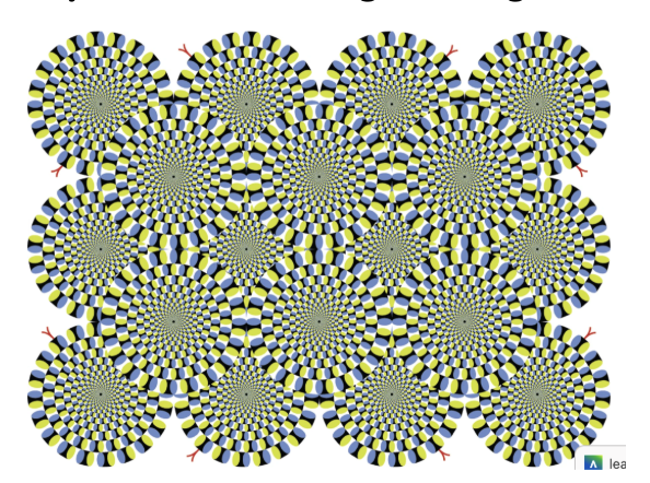
OUr eyes
The colors trick motion sensitive neurons in eyes
It all starts with light (electromagnetic waves)
Light is an electromagnetic wave and particle “photons”
We perceive “visible light” (400-700nm)
Long wavelength “hot” light (red/orange) is lower energy than “cool” (blue/violet) short wavelength light
Shorter wavelengths (faster frequencies) have more energy than longer wavelengths
We perceive different wavelengths as different colors
Colors are themselves a perception, subjectively “colored” by the brain
Electromagnetic spectrum

HIgher energy light repeats more often
→ We perceive as blue
Light travels very fast, lights is a particle (when you go to measure it)
Cones and color vision: The Trichromacy Theory

Cones
Absorb light
Responsible for color vision
Responsible for color vision
Many types of cones
Respond differently to light of varying wavelengths, providing us with color vision
We humans (usually) have three different types of opsin proteins in our cones “photopsins” that are preferentially sensitive to visible light in either blue, green, or red wavelengths (left image)
Equal mixing of the three primary colors causes equal activation of all three different cone types, resulting in a perception of “white” (right image)
Trimchromacy theory
Humans perceive color through combined activity, not just vision and direction
Colors
Brain assigns colors
Brain perceives colors
Individual expression differences:
Blue + Black dress
→ Brain uses color constancy to interpret colors under different lighting
→ Adjusts in diff brightness etc
If brain assumes warm yellow light, then subtracts yellow and makes the dress appear blue and black
Cross sectional anatomy of the eye

Cornea
Outer convex/dome shaped part of the eye
Convex tissue is transparent, light can pass through it, and refracts/bends light rays (allows light rays to enter the eye and provides most of the refractive “light-bending” power
Cornea doesn’t change in shape unless you have lasik done
The cornea of the eye (the curvature of which is fixed), bends light rays and is primarily responsible for forming the image on the back of the eye
Refraction = bending of light rays (basis of eyeglasses, telescopes, and microscopes)
Cornea: has specific shape, because its a dome, when light rays enters eye it bends light (light refraction)
Pupil
Dilation of the pupils is controlled by the sympathetic (autonomic) nervous system
Constriction is controlled by the parasympathetic division
Because usually both divisions are active, pupil size shows the result of the balance of both divisions
Like the lens opening
Opening in the opaque disc
In the middle of the iris
Iris
Shades
Lens opening
The opaque disc
Colored part of the eye
Can contract and expands
Controls the amount of light that enters the eye
Lens
A transparent structure that can bend, which also refracts light so a clear image is formed on the retina–helps with focusing “accomodation”
We can change the shape of our lens
We can contact the muscles in our eyes to do what we
Focuses light
Focus is adjusted by changes in the shape of the lens, controlled by the ciliary muscles inside the eye
Lens has to be more curved over a shorter working distance to see things
Reason for needing glasses to read a book for ex.
Retina
Transparent & thin (0.5mm) structure, layer of light sensitive cells at the back of the eye that processes and passes visual information to the brain
Where the first stages of visual-information processing occurs
Receptive surface inside the back of the eye
Thin, but has distinct layers of light sensitive cells
Bent or refracted by cornea,
Lens behind provides additional light rays, focuses views
Parallel rays converge
Air to liquid makes a difference, in a pool for example
Temporal retina - towards head
Nasal retina - towards nose
How we focus: Visual accommodation

light bent rays
If screen is too close to you, it produces a very wide angle
So the lens has to really curve the lens, has to be big and fat for the short amount of
The older we get, the worse it gets
Muscles are still fine, but
Your ability to focus gets worse
Old eye = presbyopia
One of the many consequences of aging is that the lens stiffens and becomes less flexible as we age, leading to reduced visual accommodation by the lens (less curvature) and ultimately, to “cheater” glasses to read the newspaper
The retina: CNS tissue in the periphery

Lots of blood vessels
Red eye in photos = reflection of blood vessels
There are neurons in retina
Retina develops as part of the brain
→ Macula lutea = yellow body, oval spot in center of retina, contains yellow photopigments, area where we have highest visual acuity (sharpness, ability to discriminate fine details/sharpness)
→ Fovea
Small “pit” at the center of the macula where visual acuity/sharpness is highest
Area where you see a depression in the thickness of the retina
Highest sharpness, resolution
In the center of the maculutea
High concentration of cones in the fovea provides high visual acuity in this region
→ Optic disc
Located to the nasal side of the fovea
Where blood vessels and ganglion cell axons leave the eye
Where all our blood vessels enter our retina
Where retinal nerve fibers (axons) leave the eye and travel to the brain via the optic nerve– this is your blindspot (no photoreceptors here!)
Some axons leave retina through the optic disc
Where axons leave retina
Where blood vessels enter and leave the eye
No photoreceptors here
So we have a blind spot in our vision
Binocural vision
One eye makes up for the other, both overlap
Blind spot

There are no photoreceptors at the optic disc so there is a blind spot here that we do not normally notice
There are many online sites that demonstrate your blind spot
Structure of the retina in cross section
Purple and green are the rods and cones
Photoreceptors
Phototransduction - initiated in the rods and cones
All the way in the back of the retina
Light comes in from the bottom, photons have to pass through the layers to get to the back
Rods and cones do not fire action potentials, but do release glutamate, they can depolarize or hyperpolarize, not considered neurons
Blue cells: called bipolar cells, excitatory, do not fire action potentials, have graded responses
Receive info from rods and cones and pass info down to yellow (retinal ganglion cells), project to middle of brain (thalamus)
All of these are excitatory
Only retinal ganglion cells fire action potentials
These three are the flow of visual information
Photons get absorbed in the rods and cones, info gets passed on to the brain
Horizontal cells
Make contacts among the receptor cells and bipolar cells
Amacrine cells
Contact both the bipolar and ganglion cells
These two are inhibitory, release GABA
*The rods, cones, bipolar cells and horizontal cells generate only graded local potentials, they do not produce action potentials
These neurons affect each other through release of neurotransmitters proportional to graded changes in electrical potentials
The ganglion cells do conduct action potentials
Because the ganglion cells have action potentials and are relatively large, they were the first retinal neurons to have their electrical activity recorded
From the receptor cells to the ganglion cells, enormous amounts of data converge and are compressed
The human eye contains a lot of rods compared to cones, but there are only a little bit of ganglion cells to transmit that information to the brain
A great deal of information processing is done in the eye itself

Path of visual information: (top to bottom)
Photoreceptors → Horizontal cells → Bipolar cells → Amacrine cells → Ganglion cells → Brain

Light: (bottom to top)
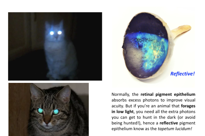
Pigment epithelial
Cells that support rods and cones
Recycle protein they use
DIternal = awake during the day
Ours is black in color, because it absorbs excess coors
Absorbs excess photons, because otherwise they’d be bouncing around everywhere
Nocturnal animals try to avoid being in environments where there is low light
Photons are reflected back out, these animals’ perception of the dark is maximizing every photons that come in
Overview of retinal cells
Retina: A photosensitive sheet in the back of the eye & part of the CNS
Contains (in order of light passage):
Retinal ganglion cells
Only source of output from the retina
Axons form the optic nerve and project to the thalamus
Only retinal cells that fire action potentials
Release glutamate, excitatory
Amacrine cells
Input from bipolar cells
Receive input from
Influence ganglion cells (release GABA–inhibitory, no APs
Bipolar cells
Input from the photoreceptors, output to the ganglion cells, on-center or off-center (release glutamate, excitatory, no APs)
Photoreceptors
The ONLY* light sensitive cells in the retina (rods & cones); output to the bipolar and horizontal cells (release glutamate, excitatory, no APs
What we know so far…

There’s a central part of the retina and a peripheral part of our retina
Central is where fovea is
Fovea is where the highest acuity is
Thinner, which is why we have better acuity
The fovea (central vision) in cross section
All other cell types Pushed out of the way]
When you orient your eyes on an object, you are directing light onto your fovea (central vision) which has the highest visual acuity (best “sharpness” or “resolution”)
This high acuity is in part due to the fact that in the center of the fovea, blood vessels, ganglion and bipolar cells are displaced laterally (pushed to the side) to allow light to directly strike cones (less light scattering by blood vessels and other cells

Acuity is a measure of the ability to resolve fine details from a given distance

The degree of convergence between photoreceptors and RGCs is different in central/foveal vs. peripheral vision

Convergence:
Describes how many photoreceptors and BPCs (bipolar cells) signal a single retinal ganglion cell (RGC)
In the image, the middle ones (blue) are bipolar cells and the upper ones (orange) are retinal ganglion cells
Center of the fovea: Low ratio of cones to RGCs(retinal ganglion cells) (1:1 in very center; good for acuity; preserves spatial information)
Central retina is mostly cones
Rods on outer periphery: High ratio “pooling” of rods to RGCs(retinal ganglion cells) (low acuity; good for detecting light in dim conditions, but spatial information is lost)
Peripheral retina is mostly rods
Mostly cones in the middle (central retina), rods around (peripheral retina)
Low convergence = high acuity
Low acuity = high convergence
High convergence is bad for acuity, but good for sensitivity

* Low convergence in the fovea is good for spatial acuity (precision), while high convergence in the peripheral retina is good for low light conditions (sensitivity), but bad for spatial acuity
So low convergence will give you more clear and sharp visuals in fovea
In the fovea, there is a 1:1 relationship where one cone connects to one bipolar cell, to one ganglion cell
You get maximal spatial acuity because there is just one at a time
And high convergence will give you worse spatial acuity but better in-the-dark visibility
In the periphery, you get spatial summation, amplifies tiny amounts of light to help you see better in the dark,
The trade-off is that the brain cannot tell which specific rod was triggered within that large group, resulting in blurry, low-acuity visuals.
The brain thinks that sound came from somewhere in the space, but it cannot distinguish from where
Which of these two retinal ganglion cells (A or B) would be able to distinguish between light projected on location 1 v.s location 2?
The leftmost retinal ganglion cell cannot tell, not good for acuity but yes for low acuity
Car line analogy:
You're at the light, turns green, you're distracted, and if there's only one behind you you know who beeped, if there’s multiple you don’t know who
*Central retina can tell diff between the two, the peripheral cannot because it’s receiving multiple signals
Rods
For seeing things in periphery (more numerous than cones in the periphery of the retina)
Long, narrow (look like rods)
Active in low levels of light
*From textbook:
*Rods are absent in the fovea, but are more numerous than cones in the periphery of the retina
*They are also more sensitive to dim light than the cones
Input from many rods converges on ganglion cells
There is greater convergence of information from photoreceptors to ganglion cells in rods than in cones, so acuity is greater in the cone-rich fovea
A cone here is more likely to have exclusive control over a single ganglion cell
Rods provide high sensitivity with limited acuity, while cones provide high acuity with limited sensitivity
Lecture 20 Thought Questions
What is a cochlear implant, what are the basics of how it works (e.g. how does it take advantage of cochlear tonotopy), and what form of hearing loss can it help treat? When in life should cochlear implant surgery be done in order to have the best results?
Where does sensation occur in the visual system? Where does perception occur? Be as specific as possible.
Describe the major anatomical components of the eye. What role do the cornea, lens, and retina play in vision?
What is meant by the term, “visual accommodation,” and how does this process help focus light rays onto the retina? Which structure(s) of the eye are responsible for refracting (bending) light? What shape does the lens take to focus on an object far away? What about up close? Why?
Lecture 21 Thought Questions
Describe the major cell types of the retina, their function, and how they’re connected/wired (cytoarchitecture) Which are excitatory and which are inhibitory? In which direction does light flow through the retinal layers? In which direction does visual information flow through the retinal layers?
Describe the concept of visual acuity, and provide two reasons why visual acuity is greater in the fovea/central vision vs. the peripheral retina. How does visual acuity differ from visual sensitivity? Where in the retina is visual sensitivity the greatest and why?