ANTR 350 - Learning Objectives Unit 3
 Call Kai
 Learn
 Practice Test
 Spaced Repetition
 Match
 Flashcards
 Knowt Play
1/391

There's no tags or description
Looks like no tags are added yet.
392 Terms


artery that supplies the anterior chest wall and breast tissue
internal thoracic and internal mammary
artery that supplies the heart
left and right coronary artery
artery that supplies the left head and neck
left common carotid artery
artery that supplies left upper limb
left subclavin artery
artery that supplies the posterior thoracic body wall
posterior intercostal
artery that supplies right head and neck
right common carotid artery
artery that supplies right upper limb
right subclavin artery
vein that drains the left head and neck
left internal jugular
vein that drains the left upper limb
left subclavin
veins that drain the right head and neck
right internal jugular
veins that drain the right upper limb
right subclavin
azygo system of veins
drains the posterior body wall
drainst o the superior vena cava


left and right renal arteries
kidney
left and right external iliac arteries
lower limb
left and right ovarian arteries
ovary
left and right testicular arteries
testis
left and right internal iliac artery
pelvic and gluteal region
celiac trunk artery
foregut abdominal organs
superior mesenteric artery
midgut abdominal organs
inferior mesenteric artery
hindgut abdominal organs
hepatic portal venous system
nutrient rich blood to liver
caval venous system
all other veins in the body
left and right reenal veins
kidney
left and right external iliac veins
lower limb
left and right ovarian veins
ovaries
left and right testicular veins
testes
left and right internal iliac vein
pelvic and gluteal region
splencic vein
foregut abdominal organs
superior mesenteric veins
midgut abdominal organs
inferior mesenteric vein
hindgut abdominal organs


hepatic portal vein
splenchcic vein where ther inferior mesesntieric drains into and hten the splenchic and superior mesenteric vein combine ot make the hepatic portal vein
hepatic vein
drains blood from the liver to the inferior vena cava
hepatic portal system veins
splenic
superior mesenteric vein
inferior mesenteric vein
heaptic (protal vein)
caval system veins
common iliac
gonadal
hpeatic
inferior vena cava
renal
mediastinum
centrally in thorax between right/left cavities
contains: heart, esophagus, trachea, thymus
systemic circulation
aorta
inferior vena cava
left atrium
left ventricle
superior vena cava
pulmonary circulation
pulmonary arteries
pulmonary trunk
pulmonary veins
right atria
right ventricles
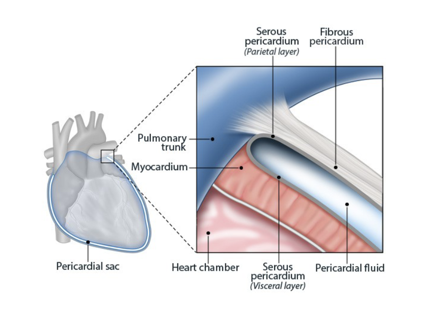
layers of pericardial sac from superifical to deep
fibrous pericardium
parietal layer of serous cavity
pericardial cavity
visceral layer of serous pericardium
heart
layers pericardial cavity is between
parietal layer of serous pericardium
visceral layer of serous pericardium
fibrous pericardium
anhcors the heart and prevents overfilling of hte chambers
serous pericardium
parietal and visceral layers create a sac which contain serous fluid which lubricates the heart

pericarditis
inflammation of the pericardium, cause fluid accumulaiton in pericardial cavity
pericardial effusion
slow accumulation of fluid wihtin pericardial cavity
cardiac tamponade
rapid, sudden accumulation of fluid that impedes hearts ability to pump
apex of the heart
tip of left ventricle
base of the heart
left atrium

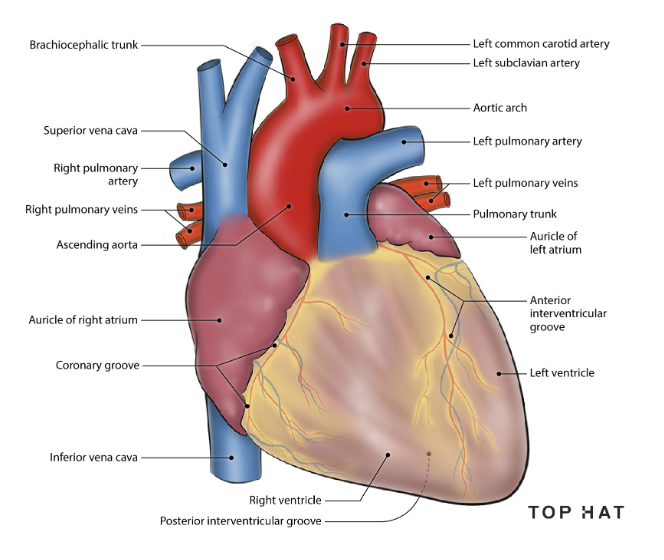
3 vessels that drain into right atrium and the regions of the body they drain
superior vena cava: drains all structures superior to thoracic diaphragm
inferior vena cava: drains structures inferior to thoracic diaphragm
coronary sinus: drains the cardiac muscle itself
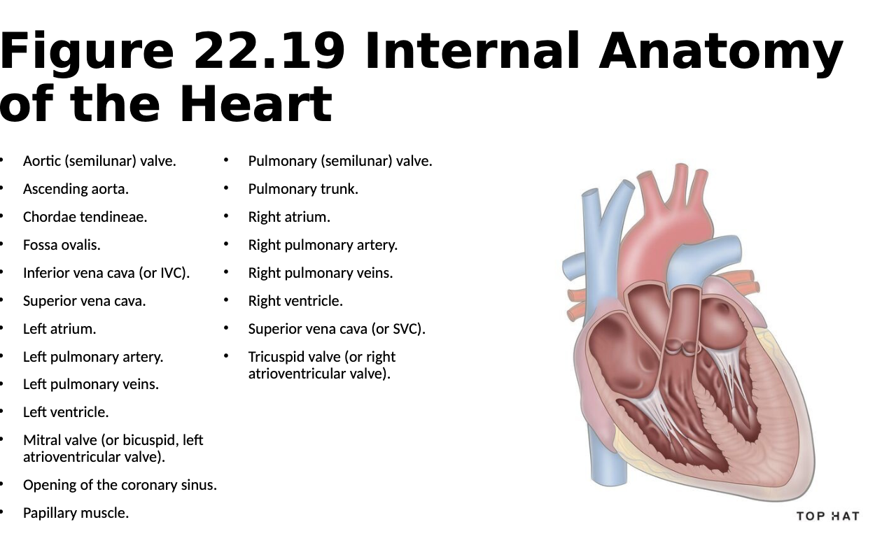

why is the left ventricle thicker
pumps blood to all structures of the body
tricuspid (right atrioventricular) valve
right atrium to right ventricle
pulmonary (semilunar) valve
right ventricle to pulmonary trunk
bicuspid (left anteroventricular) valve
left atrium to ventricle
aortic (semilunar) valve
left ventricle to aorta
chordae tendinae
prevents eversion of the cusps
cusps
prevent backflow of blood between atria and ventricle
paillary muscle
pull on chordae tendinae opening the valve
semilunar valve
ensure one way flow from ventricles to pulmonary/systemic circuit
aortic valve location of ausculation
right 2nd intercostal space, along sternal border
mitral valve (bicuspid) location of ausculation
left 5th intercostal space on midclavicular line
pulomonary semilunar valve location of ausculation
left 2nd intercostal space, along sternal border
tricuspid valve point of ausculation
left 5th intercostal space along sternal border
valve prolapse
excessviely floppy valve, improper closure leading to backflow of blood
valve stenosis
decrease in valve flexibility, limits amount of blood flow
blood flow starting in the superior vena cava
superior vena cava → right atrium → tricuspid valve → right ventricle → pulmonary valve → pulmonary trunk → pulmonary artery → lungs → pulmonary veins → left atrium → bicuspid valve → left ventricle → aortic valve → ascending aorta → aorta arch → body
high or low oxygen: aortic arch
high
high or low oxygen: ascending aorta
high
high or low oxygen: inferior vena cava
low
high or low oxygen: left atrium
high
high or low oxygen: left ventricle
high
high or low oxygen: pulmonary arteries
low
high or low oxygen: pulmonary trunk
low
high or low oxygen: pulmonary veins
high oxygen
high or low oxygen: superior vena cava
low
high or low oxygen: right atrium
low
high or low oxygen: right ventricle
low
foramen ovale
small hole in the intertrial septum in fetuses, allows blood ot flow from right ot left atrium, oxygenation by lungs is unneeded
fossa ovalis
closed foramen ovale after birth
patent foramen ovale
formane ovale remains open, leads to blood flwoing between atria and mixing oxygenated and deoxygenated blood
cardiac cycle
process of pumping blood through the heart
systole
contraction
diastole
relaxation
atrial systole + ventricular diastole
AV valves are open, semilunar valves are closed
atrial diastole + ventricular systole
AV valves are closed, semilunar valves are open
right coronary artery supplies…
right atrium
SA node
AV node
interatrial septum
right ventrincle
left coronary artery supplies…
left atrium
left ventricle
interventricular septum


occlusion
blockage of coronary arteries
ischemia
low to no blood supply to the heart
myocardial infarction
heart attack
3 veins that drain into the coronary sinus
great cardiac vein, middle cardiac vein, small cardiac vein


artery and vein on the: coronary groove
arteries: left and righ coronary arteries
veins: coronary sinus
artery and vein on the: right margin of hte heart
artery: right marginal artery
vein:small cardiac vein
artery and vein on the: posterior interventricular groove
artery: posterior interventricular artery
vein: middle cardiac vein